Review
. 2013 Sep;76(9):096601.
doi: 10.1088/0034-4885/76/9/096601. Epub 2013 Sep 4.

The physics of functional magnetic resonance imaging (fMRI)

Affiliations
Review

The physics of functional magnetic resonance imaging (fMRI)

Richard B Buxton. Rep Prog Phys. 2013 Sep.

Abstract

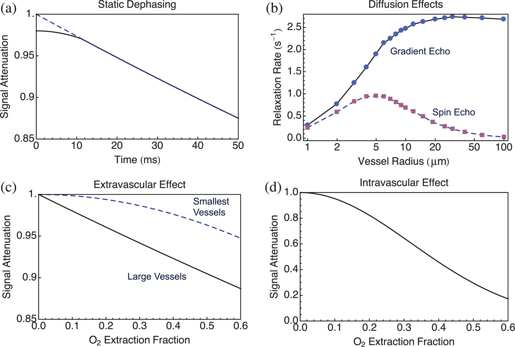
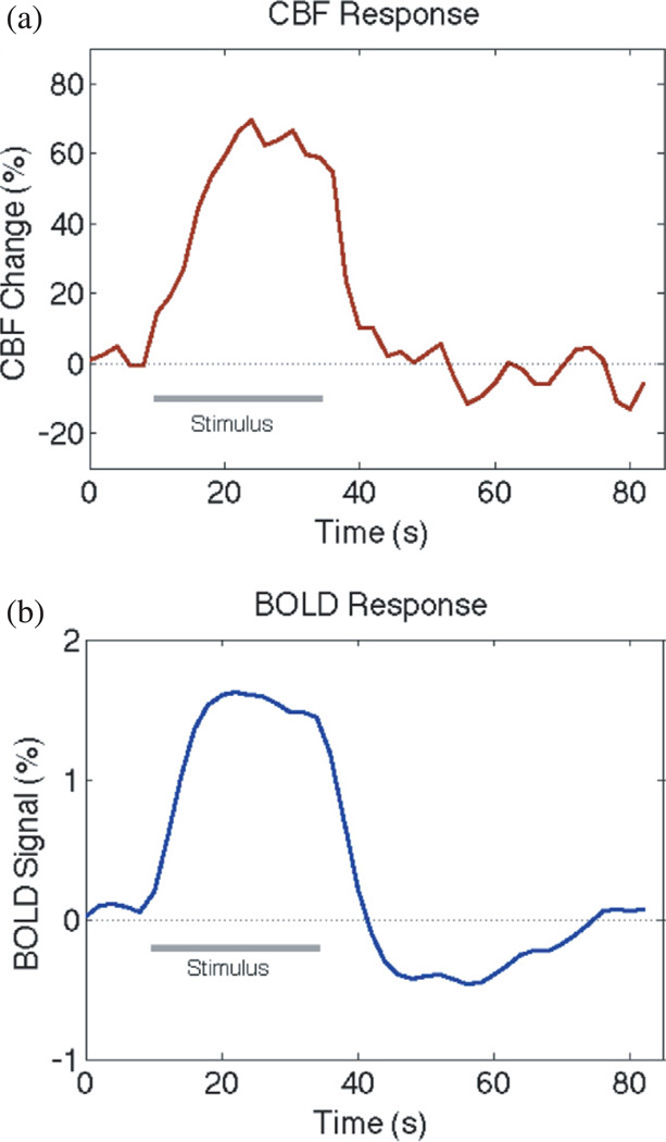
Functional magnetic resonance imaging (fMRI) is a methodology for detecting dynamic patterns of activity in the working human brain. Although the initial discoveries that led to fMRI are only about 20 years old, this new field has revolutionized the study of brain function. The ability to detect changes in brain activity has a biophysical basis in the magnetic properties of deoxyhemoglobin, and a physiological basis in the way blood flow increases more than oxygen metabolism when local neural activity increases. These effects translate to a subtle increase in the local magnetic resonance signal, the blood oxygenation level dependent (BOLD) effect, when neural activity increases. With current techniques, this pattern of activation can be measured with resolution approaching 1 mm(3) spatially and 1 s temporally. This review focuses on the physical basis of the BOLD effect, the imaging methods used to measure it, the possible origins of the physiological effects that produce a mismatch of blood flow and oxygen metabolism during neural activation, and the mathematical models that have been developed to understand the measured signals. An overarching theme is the growing field of quantitative fMRI, in which other MRI methods are combined with BOLD methods and analyzed within a theoretical modeling framework to derive quantitative estimates of oxygen metabolism and other physiological variables. That goal is the current challenge for fMRI: to move fMRI from a mapping tool to a quantitative probe of brain physiology.


Figures

Figure 1
Figure 1
CBF and BOLD responses in human primary motor cortex to 2 s of finger tapping. (a) The brief stimulus evokes a strong change in CBF measured with an ASL method. (b) The CBF change is accompanied by an increase in venous blood oxygenation, giving rise to the BOLD response measured with fMRI. Figure reproduced from [19] based on data from [228].
Figure 2
Figure 2
NMR signal decay. (a) After an initial RF pulse creates transverse magnetization, the primary signal measured in fMRI is the FID, called a GE signal in MRI terminology. The signal decays approximately with a time constant T2* due to both intrinsic T2 decay plus dephasing due to magnetic field inhomogeneities within an image voxel. (b) Subsequent RF pulses create spin echoes of the original signal, reversing the effects of the magnetic field inhomogeneities. The signal at each SE decays with time constant T2 (>T2*) on subsequent echoes. The primary origin of the BOLD effect in fMRI is that blood oxygenation affects T2*. Figure adapted from [77] with permission of the author.
Figure 3
Figure 3
Basic magnetic field gradient pulses for EPI. Applying a pattern of pulsed linear field gradients along different spatial axes ((a) shows gradient amplitude as a function of time) produces sinusoidal spatial modulations of the local signals such that the net signal from the slice traces out a trajectory in the FT space (k-space) of the image (b). The image reconstruction is then a 2D FT of the acquired data. For example, the spatial contribution to the image (c) of a single point in the measured data ((d) with the point indicated by the circle) is a single Fourier component (here emphasized by scaling up the value of that point). Current techniques of image acquisition are considerably more sophisticated, but are based on the ideas illustrated here. Adapted from [77] with permission of the author.
Figure 4
Figure 4
Basic data acquisition and analysis for fMRI. (a) Dynamic T2*-weighted images are acquired with a single-shot technique (typically EPI) while a subject performs a task, here illustrated with a simple block design alternating 20 s of finger tapping with 20 s of rest. (b) The time course of the signal for each voxel, illustrated here with a 3 × 3 display of the voxels at the intersection of the lines in (a), are correlated with the stimulus pattern (shown as the block pattern in the central voxel). (c) Voxels with a statistically significant correlation with the stimulus are classed as activated by the stimulus and displayed in color overlay on an anatomical image. As with the image acquisition methods, current fMRI data analysis methods have become considerably more sophisticated, but the idea of correlation as a measure of association remains. In resting state methods there is no external stimulus, and instead the fluctuations in the BOLD time course for different voxels are correlated with each other to identify covarying RSNs.
Figure 5
Figure 5
Diffusion-sensitive imaging. The MR signal can be sensitized to the local random diffusion of water molecules with a bipolar gradient pulse (a) that attenuates the measured signal (b) by an exponential in bD, where b depends on the gradient strength and timing parameters, and D is the local diffusion constant. This approach is sensitive to displacements of water molecules due to diffusion that are on the order of 10 µm, far smaller than the voxel resolution of the images. (c) Images are shown without the bipolar gradient pulse, with diffusion weighting, and the calculated ADC. The direction of the applied gradient pulse is arbitrary, and from measurements of multiple directions the local diffusion pattern can be determined (in the simplest case, the diffusion tensor). Diffusion in white matter is highly anisotropic due to the microscopic fiber architecture, and this has led to sophisticated techniques for mapping white matter fiber tracts to provide measures of anatomical connectivity between different brain regions. Adapted from [77] with permission of the author.
Figure 6
Figure 6
ASL to measure blood flow. (a) Magnetization of arterial blood is alternately manipulated by applying an RF inversion pulse (tag image) or leaving it relaxed (control image). (b) After a sufficient delay to allow the labeled blood to be delivered to a slice of interest, the signal difference (control–tag) subtracts out the static signal from the slice leaving a signal proportional to the volume of arterial blood delivered to each voxel during the delay time, providing a quantitative measurement of CBF.
Figure 7
Figure 7
Possible physiological origin of the mismatch of blood flow and oxygen metabolism changes as a mechanism to maintain tissue pO2. (a) Physical and physiological variations in the partial pressure of oxygen (pO2), with the O2 saturation curve of hemoglobin as an inset. To maintain constant tissue pO2 with increased oxygen metabolism (CMRO2), the capillary pO2 must increase to increase the diffusion gradient, and this means that the O2 extraction fraction (E) must decrease. The reduction in E with brain activation is the origin of the BOLD effect. (Adapted from [77] with permission of the author.) (b) Observed fractional changes in blood flow (CBF) and CMRO2 from a number of activation studies, with lines of constant ratio of fractional changes in CMRO2 to CBF (λ, with λ < 1 indicating a decrease in E). The solid line is a modeling prediction of the CBF/CMRO2 coupling ratio needed to preserve tissue pO2 (adapted from [19]).
Figure 8
Figure 8
The energy cost of neural activity. (a) Estimates of the ATP consumed for different aspects of neural activity from [80]. Primates have more synapses per neuron, resulting in the dominant energy cost being recovery from synaptic signaling. (b) The primary excitatory synaptic signaling involving pre-synaptic Ca2+ influx, release of neurotransmitter (glutamate, Glu), opening of post-synaptic Na+ channels, and inward Na+ currents are all thermodynamically downhill events, and the recovery from these events requires energy metabolism: clearing neurotransmitter through the astrocytes (1), conversion to glutamine (Gln) (2), release of glutamine, uptake by the pre-synaptic terminal, conversion back to glutamate and repackaging the neurotransmitter in vesicles (3), pumping out Ca2+ (4) and pumping out Na+. The last event consumes the most ATP, consistent with post-synaptic Na+ influx acting like an amplifier of the initial signaling. (Adapted from [77] with permission of the author.)
Figure 9
Figure 9
Magnetic field distortions around a magnetized cylinder. This is the basic physical model for the extravascular effects of a blood vessel containing deoxyhemoglobin, with the dipole pattern of distortions on the left and the geometry of equation (4) illustrated in the other panels.
Figure 10
Figure 10
Model curves of signal decay effects due to magnetized venous blood vessels. (a) For static dephasing (no diffusion) the extravascular signal initially decays slowly but then settles to an exponential decay with R2 given by equation (7). Projecting this portion back to t = 0, the difference with the actual curve is the blood volume fraction (0.02 in this example). (b) When effects of diffusion are included, the change in relaxation rate depends on the vessel size, with motional averaging reducing the net effect for the smallest vessels (data from Monte Carlo simulations reported in [131] with TE = 30 ms for the GE curve and TE = 60 ms for the SE curve). (c) Extravascular signal attenuation as a function of the O2 extraction fraction for a population of large vessels with radius > 10 µm and a population of the smallest vessels with radius = 2.5 µm, each with the same total volume of deoxyhemoglobin, calculated from equations (7) and (8). (d) Intravascular signal as a function of O2 extraction fraction based on experimental curves measured at a field strength of 3 T [145]. Curves in (a), (c) and (d) were calculated to be consistent with the GE curve in (b) with B0 = 3T, TE = 30 ms, OEF = 0.4, V = 0.02, and δω0 = 200 rad s−1 (ν = 32 s−1).
Figure 11
Figure 11
The BOLD post-stimulus undershoot in human visual cortex. The CBF response (a) and the BOLD response (b) to a 24s visual stimulus show a strong positive response to the stimulus plus a prominent post-stimulus undershoot of the BOLD signal. Data are from a study comparing luminance and color stimuli which found no difference in the responses [229], and the data for the two types of stimulus were combined for these curves. The origin of the BOLD undershoot is still debated, and it could potentially be due to vascular or metabolic effects.

References

    1. Ogawa S, et al. Brain magnetic resonance imaging with contrast dependent on blood oxygenation. Proc. Natl Acad. Sci. USA. 1990;87:9868–9872. - PMC - PubMed
    1. Ogawa S, et al. Intrinsic signal changes accompanying sensory stimulation: functional brain mapping with magnetic resonance imaging. Proc. Natl Acad. Sci. USA. 1992;89:5951–5955. - PMC - PubMed
    1. Kwong KK, et al. Dynamic magnetic resonance imaging of human brain activity during primary sensory stimulation. Proc. Natl Acad. Sci. USA. 1992;89:5675–5679. - PMC - PubMed
    1. Bandettini PA. Twenty years of functional MRI: the science and the stories. Neuroimage. 2012;62:575–588. - PubMed
    1. Bandettini PA. Functional MRI: a confluence of fortunate circumstances. Neuroimage. 2012;61:A3–A11. - PMC - PubMed

Publication types