. 2013 Nov;87(22):12166-75.
doi: 10.1128/JVI.01965-13. Epub 2013 Sep 4.

Atomic structure of Cucumber necrosis virus and the role of the capsid in vector transmission

Affiliations

Atomic structure of Cucumber necrosis virus and the role of the capsid in vector transmission

Ming Li et al. J Virol. 2013 Nov.

Abstract

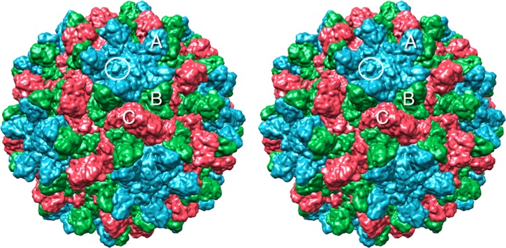
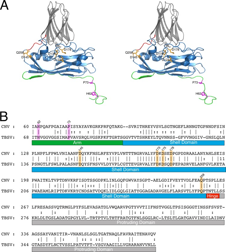
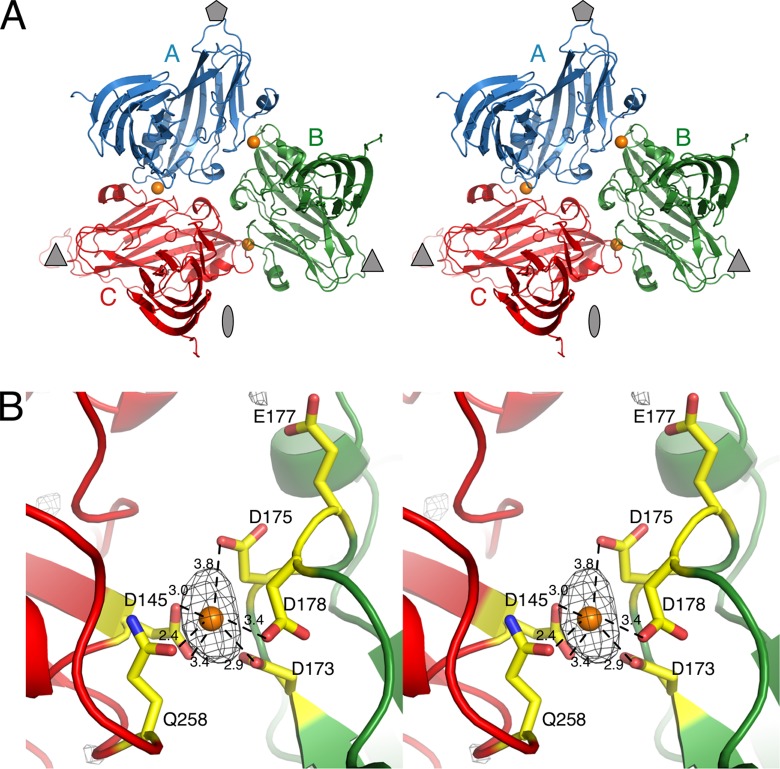
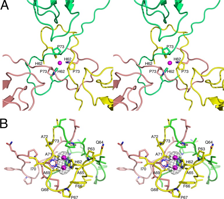
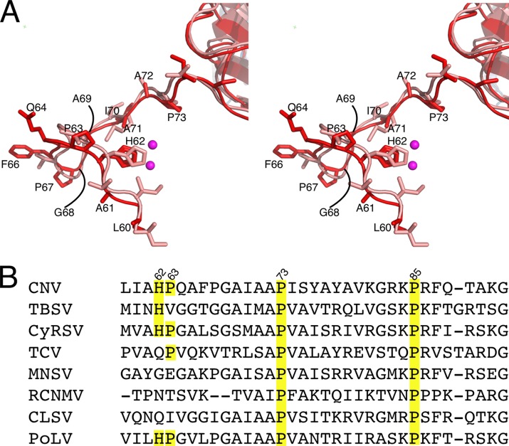
Cucumber Necrosis Virus (CNV) is a member of the genus Tombusvirus and has a monopartite positive-sense RNA genome packaged in a T=3 icosahedral particle. CNV is transmitted in nature via zoospores of the fungus Olpidium bornovanus. CNV undergoes a conformational change upon binding to the zoospore that is required for transmission, and specific polysaccharides on the zoospore surface have been implicated in binding. To better understand this transmission process, we have determined the atomic structure of CNV. As expected, being a member of the Tombusvirus genus, the core structure of CNV is highly similar to that of Tomato bushy stunt virus (TBSV), with major differences lying on the exposed loops. Also, as was seen with TBSV, CNV appears to have a calcium binding site between the subunits around the quasi-3-fold axes. However, unlike TBSV, there appears to be a novel zinc binding site within the β annulus formed by the N termini of the three C subunits at the icosahedral 3-fold axes. Two of the mutations causing defective transmission map immediately around this zinc binding site. The other mutations causing defective transmission and particle formation are mapped onto the CNV structure, and it is likely that a number of the mutations affect zoospore transmission by affecting conformational transitions rather than directly affecting receptor binding.


Figures

Fig 1
Fig 1
Surface rendering of CNV. The A, B, and C subunits are shown in blue, green, and red, respectively. White circle, location of the knob discussed in the text where the N109D mutation causing a transmission defect resides.
Fig 2
Fig 2
Structure-based sequence alignment of CNV and TBSV using the alignment tools on the PDB server (28). (A) Stereo ribbon diagram of CNV color coded as per the various regions of the capsid protein. The arm, shell, hinge, and protruding domains are shown in green, blue, orange, and gray, respectively. (B) The primary sequences of CNV and TBSV aligned as per the atomic structure. In both panels, the putative calcium and zinc binding residues are highlighted in orange and mauve, respectively.
Fig 3
Fig 3
Location of putative calcium ions in CNV. (A) Stereo ribbon diagram of the structure of a CNV icosahedral unit. The A, B, and C subunits are displayed in blue, green, and red, respectively. The approximate location of the 5-, 3-, and 2-fold axes are denoted as a gray pentagon, triangle, and ellipse, respectively. The locations of calcium ions are denoted by orange spheres. (B) Details of the interactions for the bound ion. The black mesh represents the peak from the Fo-Fc electron density contoured at 4σ. This particular ion is at the B (green) and C (red) subunit interface.
Fig 4
Fig 4
Location and binding environment of putative zinc ion. (A) The putative zinc ion (mauve sphere) binding site is located at the icosahedral 3-fold axes. The three icosahedrally related subunits are colored yellow, pink, and green. The view is from the exterior of the capsid looking into the 3-fold axis. The ligating residue H62 and the location of the P73 mutation for defective transmission are also noted. (B) Side-on view of the putative zinc binding site. The black mesh represents the Fo-Fc map contoured at 4σ. The two partially occupied zinc atoms are shown along with distance between one of the two bound zinc atoms and the closest ε2 nitrogen atom of the two alternative histidine side chain conformations.
Fig 5
Fig 5
Conservation of the zinc binding structure at the β annulus. (A) The N termini of the C subunit around the putative zinc binding site for CNV (red) and TBSV (pink). The mauve spheres represent the zinc ions. Not only is the position of H62 highly conserved, but the site that affects transmission, P73, is also conserved. (B) Sequence alignment of this region in Cymbidium ring spot virus (CyRSV; a tombusvirus), Turnip crinkle virus (TCV; a carmovirus), Melon necrotic spot virus (MNSV; a carmovirus), Red clover necrotic mosaic virus (RCNMV; a dianthovirus), Cucumber leaf spot virus (CLSV; an aureusvirus), and Pathos latent virus (PoLV; an aureusvirus).
Fig 6
Fig 6
(A) Locations of mutations causing defective transmission in CNV isolated via mechanical passaging. Shown here is the C subunit of CNV, with the mutation sites denoted by spheres. The orientation here is with the RNA at the bottom and the icosahedral 3-fold axis running vertically on the right side. The various mutations and their effects are shown in Table 2. (B) Site-directed mutagenesis of CNV around the locations of the naturally occurring mutants. The various mutations and their effects are summarized in Table 3. Red asterisks, the three sites where mutations affected transmission, but not particle formation.

References

    1. Rochon DM, Tremaine JH. 1989. Complete nucleotide sequence of the cucumber necrosis virus genome. Virology 169:251–259 - PubMed
    1. Rochon D, Kakani K, Robbins M, Reade R. 2004. Molecular aspects of plant virus transmission by Olpidium and Plasmodiophorid vectors. Annu. Rev. Phytopathol. 42:211–241 - PubMed
    1. Campbell RN. 1996. Fungal transmission of plant viruses. Annu. Rev. Phytopathol. 34:87–108 - PubMed
    1. Dias HF. 1970. The relationship between cucumber necrosis virus by Olpidium cucurbitacaerum. Virology 40:828–839 - PubMed
    1. Harrison SC, Olson AJ, Schutt CE, Winkler FK, Bricogne G. 1978. Tomato bushy stunt virus at 2.9Å resolution. Nature 276:368–373 - PubMed

Publication types

MeSH terms

LinkOut - more resources