Review
. 2013 Sep;77(3):357-79.
doi: 10.1128/MMBR.00061-12.

Functions, compositions, and evolution of the two types of carboxysomes: polyhedral microcompartments that facilitate CO2 fixation in cyanobacteria and some proteobacteria

Affiliations
Review

Functions, compositions, and evolution of the two types of carboxysomes: polyhedral microcompartments that facilitate CO2 fixation in cyanobacteria and some proteobacteria

Benjamin D Rae et al. Microbiol Mol Biol Rev. 2013 Sep.

Abstract

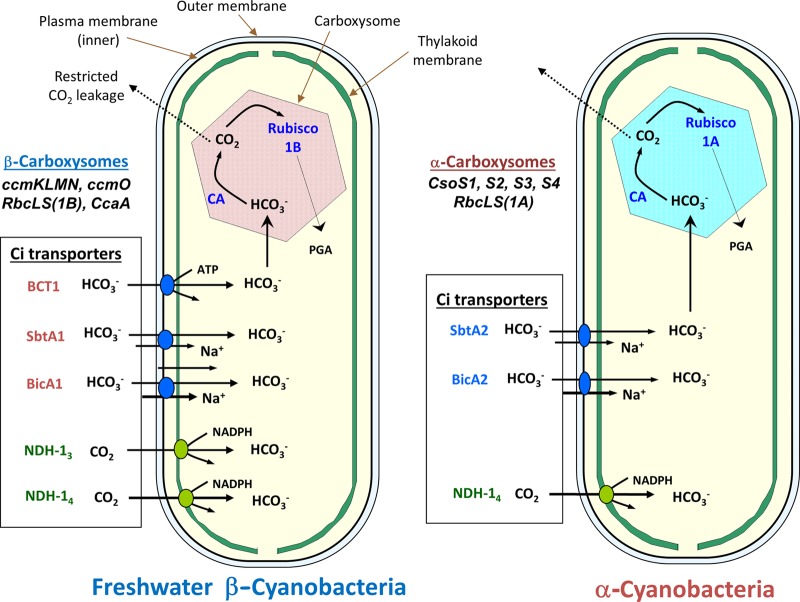
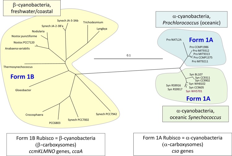
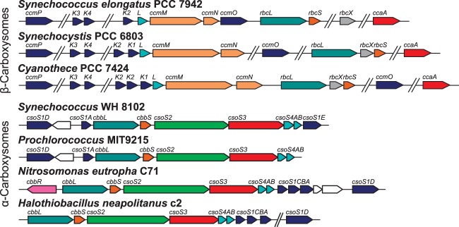
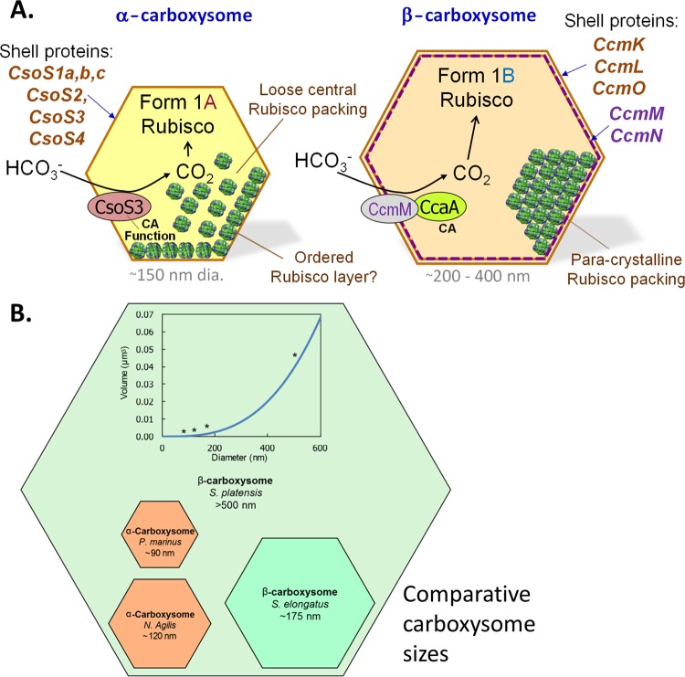
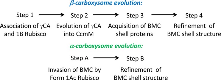
Cyanobacteria are the globally dominant photoautotrophic lineage. Their success is dependent on a set of adaptations collectively termed the CO2-concentrating mechanism (CCM). The purpose of the CCM is to support effective CO2 fixation by enhancing the chemical conditions in the vicinity of the primary CO2-fixing enzyme, D-ribulose 1,5-bisphosphate carboxylase/oxygenase (RubisCO), to promote the carboxylase reaction and suppress the oxygenase reaction. In cyanobacteria and some proteobacteria, this is achieved by encapsulation of RubisCO within carboxysomes, which are examples of a group of proteinaceous bodies called bacterial microcompartments. Carboxysomes encapsulate the CO2-fixing enzyme within the selectively permeable protein shell and simultaneously encapsulate a carbonic anhydrase enzyme for CO2 supply from a cytoplasmic bicarbonate pool. These bodies appear to have arisen twice and undergone a process of convergent evolution. While the gross structures of all known carboxysomes are ostensibly very similar, with shared gross features such as a selectively permeable shell layer, each type of carboxysome encapsulates a phyletically distinct form of RubisCO enzyme. Furthermore, the specific proteins forming structures such as the protein shell or the inner RubisCO matrix are not identical between carboxysome types. Each type has evolutionarily distinct forms of the same proteins, as well as proteins that are entirely unrelated to one another. In light of recent developments in the study of carboxysome structure and function, we present this review to summarize the knowledge of the structure and function of both types of carboxysome. We also endeavor to cast light on differing evolutionary trajectories which may have led to the differences observed in extant carboxysomes.


Figures

Fig 1
Fig 1
Overview of the general CCM characteristics of α- and β-cyanobacteria, showing the types of Ci transporters typically present in each cyanobacterial type. Typical β-cyanobacteria possess up to two types of CO2 pumps (green) and up to three types of HCO3 transporters (orange) and make use of β-carboxysomes (form 1B RubisCO plus ccm gene products), while typical α-cyanobacteria possess only two or three identifiable Ci transporters and make use of α-carboxysomes (form 1A RubisCO plus cso gene products). In the case of Prochlorococcus species (oceanic α-cyanobacteria), the NDH-1-based CO2 pump genes are entirely missing, and the only candidates for HCO3 transport are unproven (blue; BicA2 and SbtA2).
Fig 2
Fig 2
Carboxysomes and their subcellular context (arrowheads in panels A and B indicate the positions of carboxysomes). (A) β-Carboxysomes present in Synechococcus elongatus PCC 7942. (B) α-Carboxysomes present in Cyanobium PCC 7001. (Courtesy of Lynne Whitehead.) (C) Close-up of a β-carboxysome from S. elongatus PCC 7942. (D) Close-up of a β-carboxysome from Anabaena variabilis M3. Note the size differences between the different types of β-carboxysomes in panels C and D.
Fig 3
Fig 3
Phylogenies of representative sets of cyanobacteria, based on RubisCO large-subunit proteins (RbcL and CbbL proteins). Phylogenies were constructed as detailed previously (40). Note that oceanic cyanobacteria, i.e., α-cyanobacteria (Prochlorococcus and Synechococcus species), have form 1A RubisCO enzymes, fall into two minor groups, and are readily distinguished from the more diverse β-cyanobacterial group (form 1B RubisCO).
Fig 4
Fig 4
Cyanobacterial species phylogeny. α- (blue) and β-cyanobacterial (green) clades are highlighted, and marine species are shown in orange. The phylogeny shows Bayesian posterior probability values of <1.0, and AMPHORA (232), MrBayes 3.1.2 (233, 234), SeaView 4.2 (235), and GBlocks (236) were used to generate the phylogeny.
Fig 5
Fig 5
Genomic organization of representative β-carboxysomal ccm operons (top) and α-carboxysomal cso operons (bottom). Genes with structurally and/or functionally similar products are the same color. Data were adapted from the MicrobesOnline database (237).
Fig 6
Fig 6
(A) Comparison of the components and possible RubisCO packing within α- and β-carboxysomes. Note that the latter structures are always bigger than the former (see Fig. 4B). It is likely that the β-carboxysome has an outer layer composed of CcmK, CcmO, and CcmL, while the inner, less-dense, RubisCO-attached layer is composed of CcmM, CcaA, and CcmN; the interior appears to be paracrystalline and possibly organized by the shorter form of CcmM. For comparison, the smaller α-carboxysomes may feature a shell that is composed mostly of CsoS1 and CsoS4 forms, with less organization of internal RubisCO. (B) Comparison of the diameters and volumes of extreme carboxysomes of both types (112, 159, 238, 239). The inset shows the relationship between internal volume and maximum cross-sectional diameter, assuming that this measurement is the same as the diameter of a sphere circumscribing a perfectly icosahedral carboxysome. The volume of each carboxysome type is shown and is indicated by an asterisk on the curve.
Fig 7
Fig 7
Model for the outer shell structure of β-carboxysomes from Synechococcus elongatus PCC 7942. CcmK2, -K3, and -K4 produce flattened hexamers, and CcmK2 is by far the most abundant form. CcmO is postulated to form flattened trimers that could potentially interface with the triangular facets. The rarer CcmL pentamers would close the 5-fold vertices. The carboxysome model is not drawn to scale. Protein structure images were generated using Jmol (240), based on structures of the following proteins: CcmK (Protein Data Bank [PDB] entry 2A1B) (147), CcmO (represented by the structure of CsoS1D [PDB entry 3F56]) (120), and CcmL (PDB entry 2QW7) (147).
Fig 8
Fig 8
Current models of the interactions of proteins within β-carboxysomes. The CcmM-58 and CcmM-35 protein isoforms have independent roles, with the larger isoform (red) occupying the inner shell bicarbonate dehydration/RubisCO-organizing layer (inset) and recruiting the outer shell BMC layer via CcmN (blue), as well as recruiting the carboxysomal carbonic anhydrase CcaA (pink). Stoichiometric models suggest that the CcmM-35 isoform is probably localized predominantly to the interior RubisCO layers and interlinks adjacent RubisCO enzymes (green and tan) in three dimensions. Protein structure images were generated in Jmol (240), and protein threading was performed in Swiss-MODEL (–243), using the following protein structures: CcmM-58 (PDB entry 3KWC) (138), CcmN (generated by threading the CcmN protein sequence onto PDB entry 3KWD chain A) (138), Rubisco (PDB entry 1RBL) (244), and CcaA (threaded onto PDB entry 1EKJG chain A) (245).
Fig 9
Fig 9
Putative evolutionary transitions from naive cells to those containing α- and β-carboxysomes. The schemes are described in the text.

Similar articles

Cited by

References

    1. Badger MR, Hanson D, Price GD. 2002. Evolution and diversity of CO2 concentrating mechanisms in cyanobacteria. Funct. Plant Biol. 29:161–173 - PubMed
    1. Badger MR, Price GD. 2003. CO2 concentrating mechanisms in cyanobacteria: molecular components, their diversity and evolution. J. Exp. Bot. 54:609–622 - PubMed
    1. Raven JA. 2003. Inorganic carbon concentrating mechanisms in relation to the biology of algae. Photosynth. Res. 77:155–171 - PubMed
    1. Badger MR, Andrews TJ, Whitney SM, Ludwig M, Yellowlees DC, Leggat W, Price GD. 1998. The diversity and coevolution of Rubisco, plastids, pyrenoids, and chloroplast-based CO2-concentrating mechanisms in algae. Can. J. Bot. 76:1052–1071
    1. Cleland WW, Andrews TJ, Gutteridge S, Hartman FC, Lorimer GH. 1998. Mechanism of RUBISCO—the carbamate as general base. Chem. Rev. 98:549–561 - PubMed

Publication types

MeSH terms

Substances

LinkOut - more resources