Review
. 2013 Sep;77(3):440-75.
doi: 10.1128/MMBR.00018-13.

The TetR family of regulators

Affiliations
Review

The TetR family of regulators

Leslie Cuthbertson et al. Microbiol Mol Biol Rev. 2013 Sep.

Abstract

The most common prokaryotic signal transduction mechanisms are the one-component systems in which a single polypeptide contains both a sensory domain and a DNA-binding domain. Among the >20 classes of one-component systems, the TetR family of regulators (TFRs) are widely associated with antibiotic resistance and the regulation of genes encoding small-molecule exporters. However, TFRs play a much broader role, controlling genes involved in metabolism, antibiotic production, quorum sensing, and many other aspects of prokaryotic physiology. There are several well-established model systems for understanding these important proteins, and structural studies have begun to unveil the mechanisms by which they bind DNA and recognize small-molecule ligands. The sequences for more than 200,000 TFRs are available in the public databases, and genomics studies are identifying their target genes. Three-dimensional structures have been solved for close to 200 TFRs. Comparison of these structures reveals a common overall architecture of nine conserved α helices. The most important open question concerning TFR biology is the nature and diversity of their ligands and how these relate to the biochemical processes under their control.


Figures
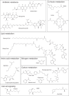
Fig 1
Fig 1
TFRs are known to interact with an exceptionally diverse set of small molecules, including antibiotics, metabolites, and cell-cell signaling molecules.
Fig 2
Fig 2
TetR regulates the expression of the tetracycline resistance determinant encoded by tetA. (A) In the absence of tetracycline, a pair of TetR dimers bind to repeated palindromic sequences in the intergenic region between tetR and tetA. (B) When present, tetracycline is bound by TetR, causing a conformational change such that TetR can no longer bind DNA. This allows for expression of the tetracycline efflux pump encoded by tetA.
Fig 3
Fig 3
Distribution of TFRs in sequenced genomes. Large genomes with a low number of TFRs are highlighted with a yellow box.
Fig 4
Fig 4
Classification of TFRs based on the orientation and proximity of adjacent genes. (A) Type I TFRs are transcribed divergently from an adjacent gene. A regulatory relationship is predicted when this intergenic region is less than 200 bp. (B) Type II TFRs are predicted to be cotranscribed with and to regulate an adjacent gene based on a distance of less than 35 bp between genes. (C) Type III TFRs show neither of the above-described relationships with adjacent genes, and a regulatory relationship with the adjacent genes cannot be predicted.
Fig 5
Fig 5
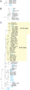
Phylogenomics can be used to predict small-molecule ligands for TFRs of unknown function. (A) The TFR of unknown function SSQG_00958 is predicted to bind a polyether ionophore based on grouping with MonRII and SchR3. (B) TFRs encoded in the biosynthesis clusters for macrolactam antibiotics cluster together, leading to the prediction that all of the TFRs in this group interact with macrolactam antibiotics. (C) AefR may recognize a phytosterol based on clustering with BreR. (Adapted from reference .)
Fig 6
Fig 6
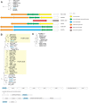
Combining information from genomic context with phylogenomics can also lead to ligand predictions for TFRs. (A and C) All of the TFRs in the group shown (A) (data are from reference 25) are type I TFRs predicted to regulate genes involved in streptogramin resistance (C). (B) Structure of the streptogramin antibiotic pristinamycin.
Fig 7
Fig 7
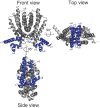
TFRs share nine conserved α helices. In the front view, the DNA-binding domain is made up of helices 1 to 3. In the side view, helices 5 to 7 in the ligand-binding domain form a central triangle. In the top view, helices 8 and 9 from each monomer form a four-helical bundle that makes up the dimer interface. The structure of Rha06780 (PDB ID 2NX4) is shown, as it shows a structure typical of TFRs (24).
Fig 8
Fig 8
TFRs display different ligand entry points. Based on current TFR-ligand structures, the ligand-binding cavity may be accessible from the side (e.g., ActR), front (e.g., SimR), or top (e.g., DesT) of the TFR. In some structures (e.g., RolR), the ligand is not accessible to the external environment and the entry point cannot be determined. SlmA interacts with a protein rather than a small-molecule ligand. Residues involved in protein-protein interactions are colored in red.
Fig 9
Fig 9
Grouping of TFRs involved in antibiotic resistance. (A and B) KijA8 and KijR (A) and VarR and Pip (B) group together in phylogenomics analysis, indicating that KijR and Pip may have been horizontally acquired from an antibiotic-producing organism. (C) Many TFRs controlling the expression of multidrug efflux pumps cluster together in phylogenomics analysis. (Adapted from reference .)
Fig 10
Fig 10
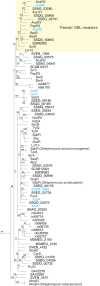
All known TFRs involved in gamma-butyrolactone (GBL) signaling form a single group (data are from reference 25). Within the GBL group, a subclade of TFRs known as the “pseudo”-GBL receptors are highlighted with a yellow box.
Fig 11
Fig 11
TFRs involved in nitrogen metabolism. (A) Homologs of AmtR, a global regulator of nitrogen metabolism in Corynebacterium, may act as local regulators in related organisms. (B) RutR and PydR homologs from separate clades within a larger group of TFRs predicted to be involved in nucleotide metabolism. (C) Homologs of XdhR may be involved in purine metabolism. (Adapted from reference .)
Fig 12
Fig 12
TFRs involved in lipid metabolism. TFRs involved in lipid metabolism are found in many groups. (A to C) TFRs involved in fatty acid biosynthesis and degradation. (D) TFRs regulating fatty acid saturation. (E) TFRs involved in the synthesis and degradation of storage polymers. (F and G) TFRs involved in terpene utilization. (Adapted from reference .)
None
None

Similar articles

Cited by

References

    1. Laub MT, Goulian M. 2007. Specificity in two-component signal transduction pathways. Annu. Rev. Genet. 41:121–145 - PubMed
    1. Ulrich LE, Koonin EV, Zhulin IB. 2005. One-component systems dominate signal transduction in prokaryotes. Trends Microbiol. 13:52–56 - PMC - PubMed
    1. Schreiter ER, Drennan CL. 2007. Ribbon-helix-helix transcription factors: variations on a theme. Nat. Rev. Microbiol. 5:710–720 - PubMed
    1. Pérez-Rueda E, Collado-Vides J. 2000. The repertoire of DNA-binding transcriptional regulators in Escherichia coli K-12. Nucleic Acids Res. 28:1838–1847 - PMC - PubMed
    1. Pérez-Rueda E, Collado-Vides J. 2001. Common history at the origin of the position-function correlation in transcriptional regulators in archaea and bacteria. J. Mol. Evol. 53:172–179 - PubMed

Publication types

MeSH terms

Substances

LinkOut - more resources