Review
. 2013 Sep;77(3):497-526.
doi: 10.1128/MMBR.00021-13.

Ecology, diversity, and evolution of magnetotactic bacteria

Affiliations
Review

Ecology, diversity, and evolution of magnetotactic bacteria

Christopher T Lefèvre et al. Microbiol Mol Biol Rev. 2013 Sep.

Abstract

Magnetotactic bacteria (MTB) are widespread, motile, diverse prokaryotes that biomineralize a unique organelle called the magnetosome. Magnetosomes consist of a nano-sized crystal of a magnetic iron mineral that is enveloped by a lipid bilayer membrane. In cells of almost all MTB, magnetosomes are organized as a well-ordered chain. The magnetosome chain causes the cell to behave like a motile, miniature compass needle where the cell aligns and swims parallel to magnetic field lines. MTB are found in almost all types of aquatic environments, where they can account for an important part of the bacterial biomass. The genes responsible for magnetosome biomineralization are organized as clusters in the genomes of MTB, in some as a magnetosome genomic island. The functions of a number of magnetosome genes and their associated proteins in magnetosome synthesis and construction of the magnetosome chain have now been elucidated. The origin of magnetotaxis appears to be monophyletic; that is, it developed in a common ancestor to all MTB, although horizontal gene transfer of magnetosome genes also appears to play a role in their distribution. The purpose of this review, based on recent progress in this field, is focused on the diversity and the ecology of the MTB and also the evolution and transfer of the molecular determinants involved in magnetosome formation.


Figures

Fig 1
Fig 1
Sampling for magnetotactic bacteria using different strategies: from the shore of the Salton Sea with a scooper (A), underwater in the Mediterranean Sea by free diving (B), and with a bottom sampler in Lake Chiemsee, Bavaria (C). (Panel C courtesy of S. Kolinko and D. Schüler, reproduced with permission.)
Fig 2
Fig 2
Transmission electron microscope (TEM) images of magnetosomes and the magnetosome membrane. (A) TEM micrograph of a cell of Magnetospirillum magneticum strain AMB-1 deposited onto a Formvar-coated electron microscope grid showing a chain of cuboctahedral magnetosomes. (B) TEM micrograph of an ultrathin section of a cell of “Ca. Magnetoovum mohavensis” showing the magnetosome membrane (arrow) surrounding bullet-shaped magnetite crystals. (C) TEM micrograph of an extracted and purified magnetosome chain from a Magnetococcus marinus MC-1 cell showing prismatic magnetite crystals surrounded by the magnetosome membrane (arrow).
Fig 3
Fig 3
Phylogenetic distribution of cultured and uncultured magnetotactic bacteria in the Alpha-, Gamma-, and Deltaproteobacteria classes of the Proteobacteria phylum, the Nitrospirae phylum, and the candidate division OP3. Magnetotactic bacteria are in boldface type. The tree is based on neighbor-joining analyses. The bar represents 2% sequence divergence.
Fig 4
Fig 4
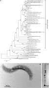
(A) Neighbor-joining phylogenetic tree, based on 16S rRNA gene sequences, showing the phylogenetic position of MTB closely related to the genus Magnetospirillum (in boldface type) in the family Rhodospirillaceae of the Alphaproteobacteria class. GenBank accession numbers are in parentheses. (B) TEM image of a cell of the cultured vibrioid strain LM-1 isolated from Lake Mead, NV, whose phylogenetic position is basal to the Magnetospirillum. (C) TEM image of a chain of cuboctahedral magnetite magnetosomes within a cell of the cultured strain CB-1 that belongs to the genus Magnetospirillum.
Fig 5
Fig 5
(A) Neighbor-joining phylogenetic tree, based on 16S rRNA gene sequences, showing the phylogenetic positions of MTB of the Magnetococcales order (in boldface type) in the phylum Proteobacteria. Bootstrap values (higher than 50) at nodes are percentages of 1,000 replicates. The bar represents 2% sequence divergence. GenBank accession numbers are in parentheses. (B to E) TEM images of different types of uncultured MTB of the order Magnetococcales. (B) Cell of a magnetotactic coccus that biomineralizes a single magnetite magnetosome chain. (C) Cell of a magnetotactic coccus that biomineralizes two magnetite magnetosome chains. (D) Cell of a magnetotactic coccus that biomineralizes a clump of magnetite magnetosomes rather than a chain. (E) Rod-shaped cell that biomineralizes a single chain of magnetite. (Panel E courtesy of E. Katzmann, S. Kolinko, and D. Schüler, reproduced with permission.)
Fig 6
Fig 6
(A) Neighbor-joining phylogenetic tree, based on 16S rRNA gene sequences, showing the phylogenetic positions of the magnetotactic marine spirilla Magnetospira thiophila and strain QH-2 and the magnetotactic marine vibrio Magnetovibrio blakemorei in the family Rhodospirillaceae (in boldface type). Bootstrap values (higher than 50) at nodes are percentages of 1,000 replicates. The bar represents 2% sequence divergence. GenBank accession numbers are in parentheses. (B to D) TEM images of a cell of Magnetovibrio blakemorei (B), a cell of Magnetospira thiophila (C), and a thin-sectioned cell of Magnetospira thiophila showing the magnetosome chain consisting of elongated octahedral crystals of magnetite surrounded by the magnetosome membrane (arrow) (D).
Fig 7
Fig 7
(A) Neighbor-joining phylogenetic tree, based on 16S rRNA gene sequences, showing the phylogenetic positions of the magnetotactic Deltaproteobacteria (in boldface type) of the orders Desulfovibrionales and Desulfobacterales. Bootstrap values (higher than 50) at nodes are percentages of 1,000 replicates. The bar represents 2% sequence divergence. GenBank accession numbers are in parentheses. (B to E) TEM images of a cell of Desulfovibrio magneticus (B); a cell of strain AV-1, an obligately alkaliphilic magnetotactic bacterium isolated from a brackish spring in Armagosa Valley, CA (C); a cell of a greigite-producing, large rod-shaped bacterium collected from a spring at ambient temperature in the Great Boiling Springs geothermal field in Gerlach, NV (D); and a greigite-producing, magnetotactic multicellular prokaryote (MMP) collected from the Salton Sea, CA (E).
Fig 8
Fig 8
(A) Neighbor-joining phylogenetic tree, based on 16S rRNA gene sequences, showing phylogenetic positions of the magnetotactic Gammaproteobacteria (in boldface type) of the orders Chromatiales and Thiotrichales. Bootstrap values (higher than 50) at nodes are percentages of 1,000 replicates. The bar represents 1% sequence divergence. GenBank accession numbers are in parentheses. (B and C) TEM images of a cell of strain SS-5 (B) and a cell of strain BW-2 (C). Cells of strains SS-5 and BW-2 biomineralize prismatic elongated and cuboctahedral magnetite magnetosomes, respectively.
Fig 9
Fig 9
(A) Neighbor-joining phylogenetic tree, based on 16S rRNA gene sequences, showing phylogenetic positions of the magnetotactic Nitrospirae and strain SKK-01, the only known magnetotactic bacterium belonging to the OP3 division of the PVC (Planctomycetes-Verrucomicrobia-Chlamydiae) superphylum. Bootstrap values (higher than 50) at nodes are percentages of 1,000 replicates. The bar represents 2% sequence divergence. GenBank accession numbers are in parentheses. (B) TEM image of a cell of “Candidatus Magnetoovum mohavensis.” (C) False-color scanning TEM image of a cell of “Ca. Magnetobacterium bavaricum.” Magnetosome crystals consisting of magnetite are visualized through back-scattered electrons (magenta) by material contrast. (D) TEM image of a cell of the uncultured “Ca. Thermomagnetovibrio paiutensis.” All magnetotactic Nitrospirae biomineralize bullet-shaped crystals of magnetite in their magnetosomes. (Panel C courtesy of G. Wanner and D. Schüler, reproduced with permission.)
Fig 10
Fig 10
(A and B) Congruency of the phylogenetic trees based on 16S rRNA gene sequences that reflect the evolution of MTB (A) and on concatenated magnetosome protein sequences (MamABIKQ) that reflect the evolution of magnetotaxis (B). (C) Gene synteny (organization) of the conserved magnetosome genes of Magnetospirillum magnetotacticum (MS-1), Ms. magneticum (AMB-1), Ms. gryphiswaldense (MSR-1), Magnetovibrio blakemorei (MV-1), Magnetospira sp. strain QH-2, Magnetococcus marinus (MC-1), strain SS-5, the magnetotactic multicellular prokaryote “Candidatus Magnetoglobus multicellularis” (MMP), “Ca. Desulfamplus magnetomortis” (BW-1), Desulfovibrio magneticus (RS-1), strain ML-1, and “Ca. Magnetobacterium bavaricum.”
Fig 11
Fig 11
Schematic representation of the evolution of magnetotactic bacteria (MTB) from a common ancestor that transferred the genes involved in magnetosome formation by descent to the groups known to display magnetotaxis. Species that do not have this ability but appear to have an ancestor that had it have likely lost the genes involved in magnetosome formation.
None
None

Similar articles

Cited by

References

    1. Bazylinski DA, Frankel RB. 2004. Magnetosome formation in prokaryotes. Nat. Rev. Microbiol. 2:217–230 - PubMed
    1. Blakemore R. 1975. Magnetotactic bacteria. Science 190:377–379 - PubMed
    1. Gorby YA, Beveridge TJ, Blakemore RP. 1988. Characterization of the bacterial magnetosome membrane. J. Bacteriol. 170:834–841 - PMC - PubMed
    1. Grünberg K, Müller E-C, Otto A, Reszka R, Linder D, Kube M, Reinhardt R, Schüler D. 2004. Biochemical and proteomic analysis of the magnetosome membrane in Magnetospirillum gryphiswaldense. Appl. Environ. Microbiol. 70:1040–1050 - PMC - PubMed
    1. Frankel RB, Bazylinski DA, Johnson MS, Taylor BL. 1997. Magneto-aerotaxis in marine coccoid bacteria. Biophys. J. 73:994–1000 - PMC - PubMed

Publication types

MeSH terms

Substances

LinkOut - more resources