Review
. 2013 Sep;77(3):527-39.
doi: 10.1128/MMBR.00013-13.

What a difference a Dalton makes: bacterial virulence factors modulate eukaryotic host cell signaling systems via deamidation

Affiliations
Review

What a difference a Dalton makes: bacterial virulence factors modulate eukaryotic host cell signaling systems via deamidation

Erica J Washington et al. Microbiol Mol Biol Rev. 2013 Sep.

Abstract

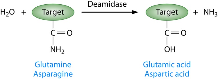
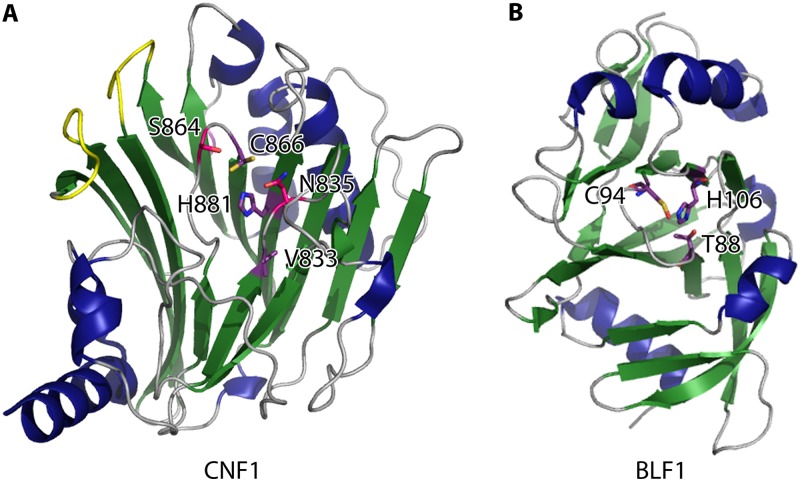
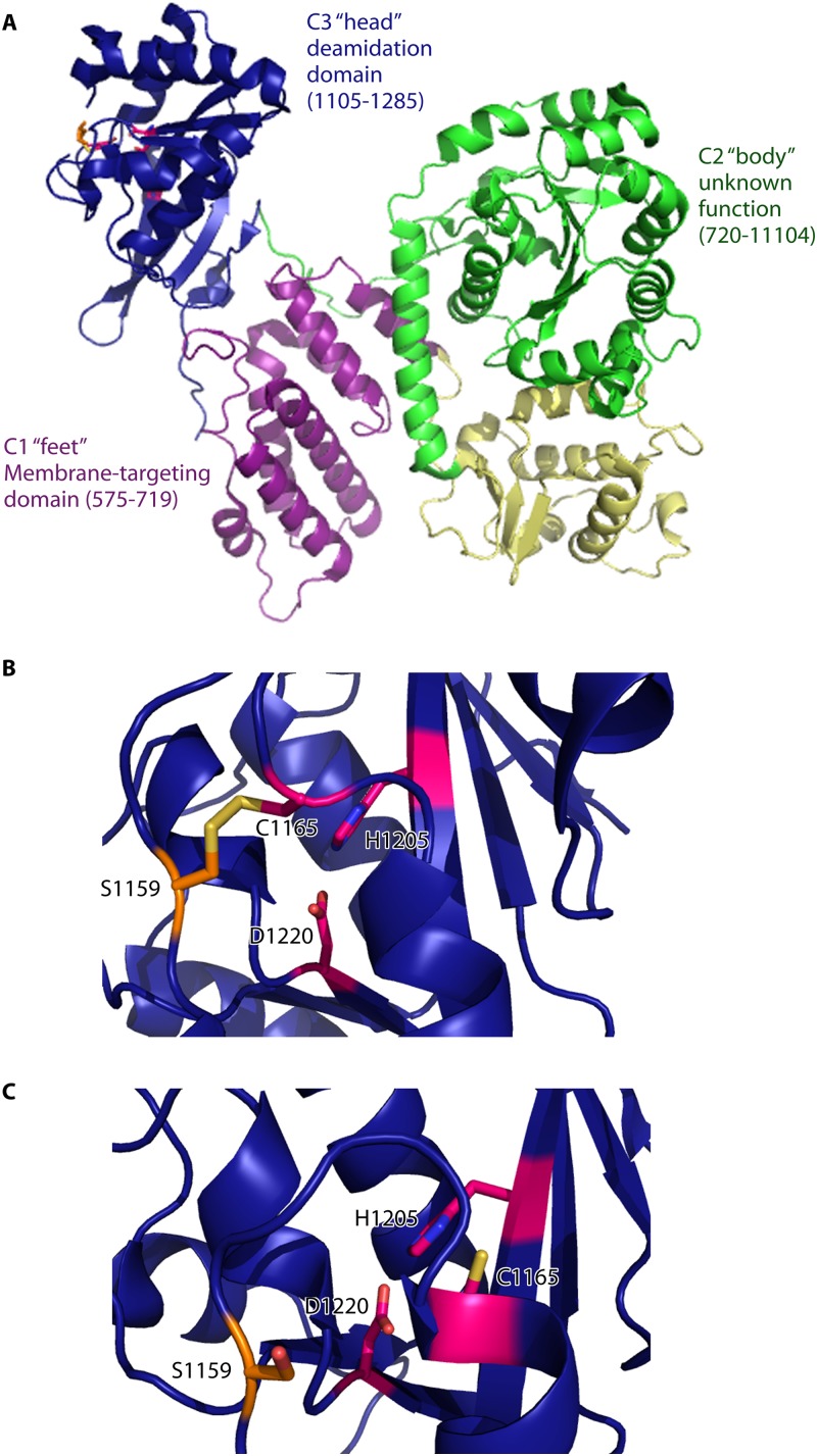
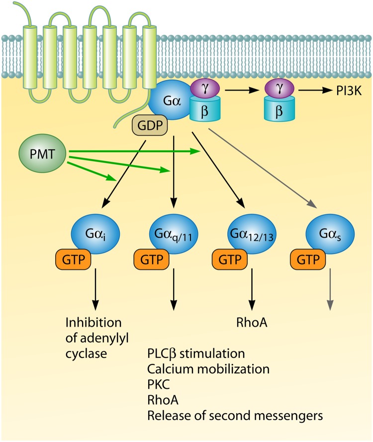
Pathogenic bacteria commonly deploy enzymes to promote virulence. These enzymes can modulate the functions of host cell targets. While the actions of some enzymes can be very obvious (e.g., digesting plant cell walls), others have more subtle activities. Depending on the lifestyle of the bacteria, these subtle modifications can be crucially important for pathogenesis. In particular, if bacteria rely on a living host, subtle mechanisms to alter host cellular function are likely to dominate. Several bacterial virulence factors have evolved to use enzymatic deamidation as a subtle posttranslational mechanism to modify the functions of host protein targets. Deamidation is the irreversible conversion of the amino acids glutamine and asparagine to glutamic acid and aspartic acid, respectively. Interestingly, all currently characterized bacterial deamidases affect the function of the target protein by modifying a single glutamine residue in the sequence. Deamidation of target host proteins can disrupt host signaling and downstream processes by either activating or inactivating the target. Despite the subtlety of this modification, it has been shown to cause dramatic, context-dependent effects on host cells. Several crystal structures of bacterial deamidases have been solved. All are members of the papain-like superfamily and display a cysteine-based catalytic triad. However, these proteins form distinct structural subfamilies and feature combinations of modular domains of various functions. Based on the diverse pathogens that use deamidation as a mechanism to promote virulence and the recent identification of multiple deamidases, it is clear that this enzymatic activity is emerging as an important and widespread feature in bacterial pathogenesis.


Figures

Fig 1
Fig 1
Schematic representation of enzymatic deamidation in proteins. Deamidases act on specific residues in the target protein. For all currently studied bacterial virulence factors, the targets of deamidation are glutamine side chains, which are converted to glutamic acids. However, it is also possible for the amide of the side chains of asparagines to be converted to aspartic acid. Deamidation results in an increase in the negative charge of the target protein, an increase of approximately 1 Da in the mass of the target protein, and the release of ammonia. Each of these outputs can be measured experimentally to characterize the activity of deamidases.
Fig 2
Fig 2
Catalytic domains of diverse deamidases. (A) Catalytic domain of CNF1 from E. coli (Protein Data Bank [PDB] entry 1HQ0), with invariant catalytic residues Cys866 and His881 shown in purple. The main chain carbonyl of Val833 (also in purple) is involved in coordinating the imidazole ring of His881. Residues in pink are conserved residues important for CNF1 function, while loops 8 and 9 (likely important for substrate recognition) are shown in yellow. (B) BLF1 (PDB entry 3TU8) is a 23-kDa toxin and deamidase that forms a compact catalytic domain structurally related to those of CNF1 from E. coli and other papain-like superfamily enzymes. The residues that compose the catalytic triad (Cys94, His106, and Thr88) are shown in purple. α-Helices are shown in blue, β-sheets in green, and loops in gray. All structure figures were prepared with PyMol.
Fig 3
Fig 3
Crystal structure of the C-terminal region of Pasteurella multocida toxin. (A) The crystal structure of the C-terminal region of PMT has been solved (residues 575 to 1285; PDB entry 2EBF). The catalytic region consists of several domains. The helical C1 domain (purple) forms the “feet” of the Trojan horse-like structure and contains membrane-targeting properties. The C2 “body” domain contains 2 α/β subdomains (shown in green and yellow) of unknown function. The domain responsible for the deamidase activity of PMT is the C3 “head” C-terminal domain (blue). (B) The crystal structure of the PMT catalytic domain reveals a disulfide bond between the catalytic cysteine, Cys1165, and Cys1159 in the binding pocket. Reduction of this disulfide bond is required for interaction of the catalytic triad and catalytic activity. His1205 and Asp1220 form the other residues of the triad. (C) The crystal structure of the catalytic domain of PMT(Cys1159Ser) (PDB entry 2EC5) demonstrates the position of the catalytic triad when the disulfide bond between Cys1159 and Cys1165 is reduced.
Fig 4
Fig 4
PMT deamidates and activates heterotrimeric Gα proteins. The activation of diverse G protein-coupled receptor signal transduction pathways by PMT leads to several phenotypes that are disruptive to the host. PMT activates Gαi, Gαq/11, and Gα12/13 but not Gαs. PMT also triggers the release of Gβγ from the heterotrimeric complex, which leads to activation of the phosphoinositide-3-kinase (PI3K) pathway.
Fig 5
Fig 5
Model of Cif inactivation of CRL activity and ubiquitination. (A) In uninfected cells, the COP9 signalosome (CSN) deneddylates CRLs. NEDD8 (yellow) conjugation and removal represent an important mechanism by which CRL activity is regulated. When CRLs are neddylated with wild-type (WT) NEDD8 (green), they are maintained in an open conformation. This allows substrates (blue) to bind the CRLs, become polyubiquitinated, and then be degraded. (B) In cells infected with bacteria that express Cif, deamidation of NEDD8 (yellow) may prevent CRL (green) from forming an open conformation. This prevents CRLs from associating with the substrate-binding module that allows downstream ubiquitination and degradation of substrate proteins (blue) to occur. This may lead to a reduction in the rate of deneddylation and a decrease in unmodified CRLs (red).
Fig 6
Fig 6
Crystal structures of cell cycle-inhibiting factors. (A) Crystal structures of Cif deamidases from E. coli (PDB entry 3EFY), B. pseudomallei (PDB entry 3GQM), P. luminescens (PDB entry 3GQJ), and Y. pseudotuberculosis (PDB entry 4F8C). Shown in blue are the α-helices of the core globular domain. In teal are the α-helices of the tail helical extension domain. The antiparallel β-sheet is shown in green, and the loops are colored gray, except for the occluding loop, which is colored black. The conserved catalytic residues histidine, cysteine, and glutamine are shown in purple. (B) Amino acids in the catalytic domain of CHBP are shown as purple sticks.
Fig 7
Fig 7
Crystal structure of the CifYp deamidase-NEDD8 complex. (A) The CHYP (α-helices are shown in blue, β-sheets in green, and loops in gray)-NEDD8 (red) complex consists of an extensive interaction surface (PDB entry 4FBJ). The catalytic Cys117 position is modeled based on the CHYP(Cys117Ala)-NEDD8 complex to demonstrate the proposed molecular mechanism of deamidation. (B) Close-up of the catalytic domain of CHYP showing that the deamidation target, Gln40 of NEDD8, is positioned near the Cif catalytic residues (shown in purple). (C) The proposed molecular mechanism of deamidation involves deprotonation of the cysteine by the imidazolium group of histidine (step 1) followed by a nucleophilic attack by the thiol group of the catalytic cysteine on the δ-carbon of glutamine (step 2).
Fig 8
Fig 8
Crystal structures of unbound OspI and the OspI-Ubc13 complex. (A) OspI (α-helices are shown in blue, β-sheets in green, and loops in gray) (PDB entry 3B21) forms a compact catalytic domain structurally similar to those of papain-like superfamily enzymes. The residues in the catalytic triad (Cys62, His145, and Asp160) are shown in purple. The catalytic pocket is blocked by Asn61 in free OspI. (B) The OspI(Cys62Ala)-Ubc13 (red) complex includes an extensive interaction surface involving Ubc13 α-helix 1 and loops L1 and L2 (PDB entry 4IP3). (C) Close-up of the OspI(Cys62Ala)-Ubc13 complex showing Gln100 of Ubc13 positioned adjacent to the OspI catalytic center (purple; OspI residue 62 was modeled as a cysteine to demonstrate the nucleophilic attack [yellow dashes] by the thiol group of Cys62 on Gln100 in Ubc13).
None
None
None

References

    1. Cui J, Shao F. 2011. Biochemistry and cell signaling taught by bacterial effectors. Trends Biochem. Sci. 36:532–540 - PubMed
    1. Ribet D, Cossart P. 2010. Pathogen-mediated posttranslational modifications: a re-emerging field. Cell 143:694–702 - PMC - PubMed
    1. Yarbrough ML, Orth K. 2009. AMPylation is a new post-translational modiFICation. Nat. Chem. Biol. 5:378–379 - PubMed
    1. Robinson AB, Rudd CJ. 1974. Deamidation of glutaminyl and asparaginyl residues in peptides and proteins. Curr. Top. Cell. Regul. 8:247–295 - PubMed
    1. Chao X, Muff TJ, Park SY, Zhang S, Pollard AM, Ordal GW, Bilwes AM, Crane BR. 2006. A receptor-modifying deamidase in complex with a signaling phosphatase reveals reciprocal regulation. Cell 124:561–571 - PubMed

Publication types

LinkOut - more resources