. 2013 Nov 1;208(9):1529-37.
doi: 10.1093/infdis/jit335. Epub 2013 Sep 4.

Profiling of Candida albicans gene expression during intra-abdominal candidiasis identifies biologic processes involved in pathogenesis

Affiliations

Profiling of Candida albicans gene expression during intra-abdominal candidiasis identifies biologic processes involved in pathogenesis

Shaoji Cheng et al. J Infect Dis. .

Abstract

Background: The pathogenesis of intra-abdominal candidiasis is poorly understood.

Methods: Mice were intraperitoneally infected with Candida albicans (1 × 10(6) colony-forming units) and sterile stool. nanoString assays were used to quantitate messenger RNA for 145 C. albicans genes within the peritoneal cavity at 48 hours.

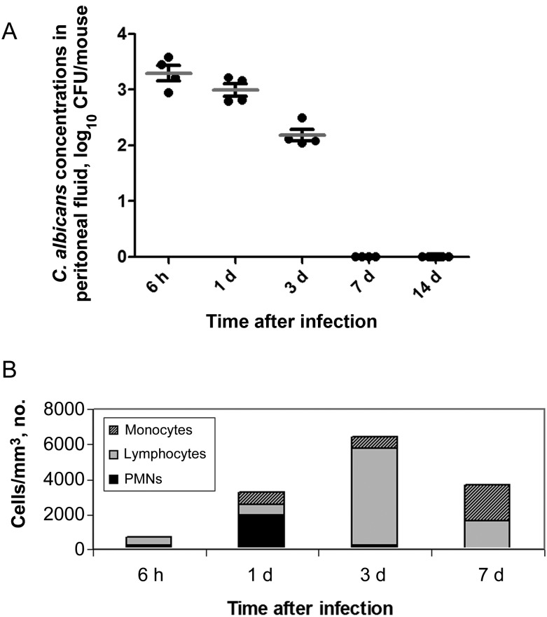
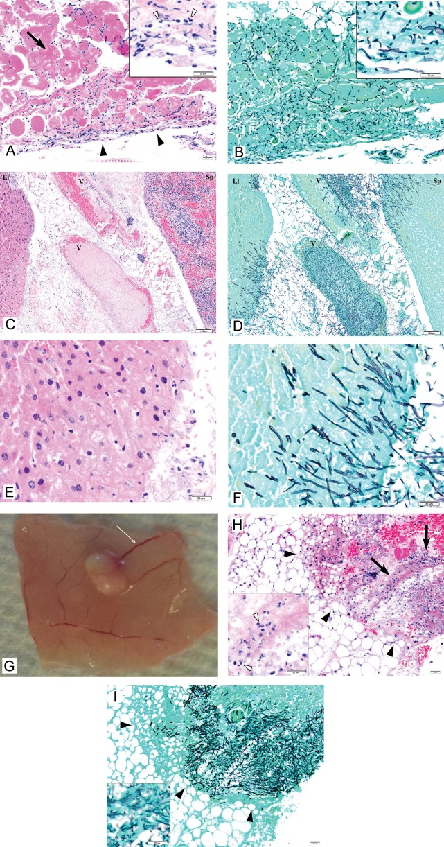
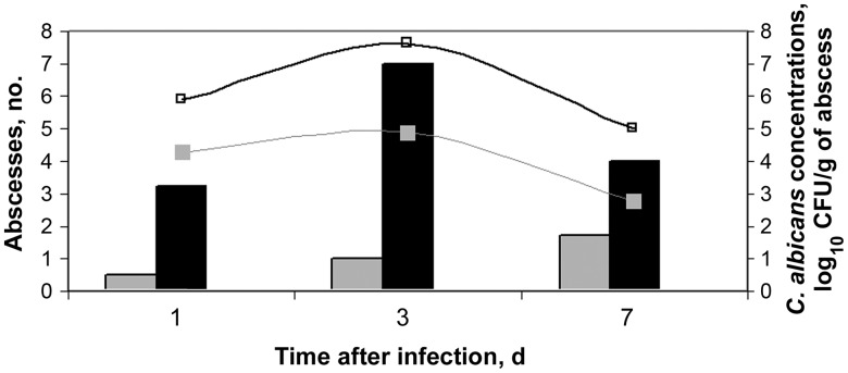
Results: Within 6 hours after infection, mice developed peritonitis, characterized by high yeast burdens, neutrophil influx, and a pH of 7.9 within peritoneal fluid. Organ invasion by hyphae and early abscess formation were evident 6 and 24 hours after infection, respectively; abscesses resolved by day 14. nanoString assays revealed adhesion and responses to alkaline pH, osmolarity, and stress as biologic processes activated in the peritoneal cavity. Disruption of the highly-expressed gene RIM101, which encodes an alkaline-regulated transcription factor, did not impact cellular morphology but reduced both C. albicans burden during early peritonitis and C. albicans persistence within abscesses. RIM101 influenced expression of 49 genes during intra-abdominal candidiasis, including previously unidentified Rim101 targets. Overexpression of the RIM101-dependent gene SAP5, which encodes a secreted protease, restored the ability of a rim101 mutant to persist within abscesses.

Conclusions: A mouse model of intra-abdominal candidiasis is valuable for studying pathogenesis and C. albicans gene expression. RIM101 contributes to persistence within intra-abdominal abscesses, at least in part through activation of SAP5.

Keywords: Candida albicans; RIM101; SAP5; in vivo gene expression; intra-abdominal candidiasis; nanoString.


Figures

Figure 1.
Figure 1.
Tissue burdens (A) and white blood cell (WBC) counts (B) in peritoneal fluid of mice infected with C. albicans intraperitoneally. Abbreviations: CFU, colony-forming units; PMN, polymorphonuclear cell.
Figure 2.
Figure 2.
Necropsy findings and histopathology of tissues of mice infected with C. albicans intraperitoneally. 2A and 2B. Peritoneum on day 1 of infection. 2A: Acute inflammation of the peritoneum is apparent (solid arrowheads). The inflammatory response is strongest on the surface, consistent with an inflammatory stimulus arising in the peritoneal cavity. The inflammatory response penetrates the serosa, resulting in necrosis of underlying diaphragmatic muscle (arrow). The inflammatory response comprises predominantly neutrophils (inset, empty arrowheads) with a smaller number of admixed lymphocytes and macrophages. 2B: C. albicans within the peritoneum are a mixture of hyphae and yeasts (inset). The density of organisms is greatest in the serosa, a finding in keeping with the gradient of the inflammatory response (Hematoxylin Eosin (A) and Grocott Methenamine Silver (B), original x 100, insets original x 600). Bars at low magnification: 50 μm. Bars at high magnification: 20 μm. 2C-2F. Liver and spleen on day 1 of infection. 2C, 2D: C. albicans infiltrating liver (Li, left), veins (V, center), and spleen (Sp, right). 2E, 2F: Higher magnification of liver showing inflammation (predominantly neutrophils) and mats of hyphae. Hematoxylin Eosin (2C, 2E) and Grocott Methenamine Silver (2D, 2F), original x 100 (2F, 2H), original x 600 (2G, 2I). Bars at low magnification: 50 μm. Bars at high magnification: 20 μm. 2G. Necropsy on day 3 of infection shows an abscess on the peritoneal membrane. Note the engorged blood vessels (thin arrow) consistent with tissue inflammation. 2H and 2I. Mesenteric abscess on day 3 of infection. 2H: Mesenteric fat showing a large area of fat necrosis (solid arrowheads) with fibrin exudates and acute inflammation (arrows). The mixed inflammatory infiltrate comprises predominantly neutrophils (inset, empty arrowheads) and few admixed lymphocytes. 2I: Grocott stain of the same area highlights numerous hyphae and yeasts in the area of necrosis and inflammation (Hematoxylin Eosin (2H) and Grocott Methenamine Silver (2I), original x 100, insets original x 600). Bars at low magnification: 50 μm. Bars at high magnification: 20 μm.
Figure 3.
Figure 3.
Impact of addition of sterile stool on number of abscesses and burdens within abscess in mice infected with C. albicans intraperitoneally. Mice were infected with 1x10⁶ CFU. Bar graphs represent the mean number of abscesses for 8 mice infected with C. albicans either alone (gray bar) or with sterile stool (black bar). Curved lines represent the mean number of log10 CFU for mice infected with C. albicans alone (gray line) or with sterile stool (gray bar). All abscesses resolved by day 14 (data not shown). Abbreviation: CFU, colony-forming units.

Similar articles

Cited by

References

    1. Pappas PG. Invasive candidiasis. Infect Dis Clin North Am. 2006;20:485–506. - PubMed
    1. Blot SI, Vandewoude KH, De Waele JJ. Candida peritonitis. Curr Opin Crit Care. 2007;13:195–9. - PubMed
    1. Carneiro HA, Mavrakis A, Mylonakis E. Candida peritonitis: an update on the latest research and treatments. World J Surg. 2011;35:2650–9. - PubMed
    1. Party BSfACW. Management of deep Candida infection in surgical and intensive care unit patients. Intensive Care Med. 1994;20:522–8. - PubMed
    1. Blot S, De Waele JJ. Critical issues in the clinical management of complicated intra-abdominal infections. Drugs. 2005;65:1611–20. - PubMed

Substances