. 2013 Nov;24(21):3449-59.
doi: 10.1091/mbc.E12-10-0776. Epub 2013 Sep 4.

Targeted inactivation of β1 integrin induces β3 integrin switching, which drives breast cancer metastasis by TGF-β

Affiliations

Targeted inactivation of β1 integrin induces β3 integrin switching, which drives breast cancer metastasis by TGF-β

Jenny G Parvani et al. Mol Biol Cell. 2013 Nov.

Erratum in

  • Mol Biol Cell. 2014 Feb;25(4):548

Abstract

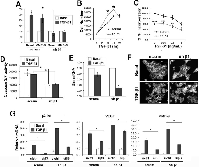
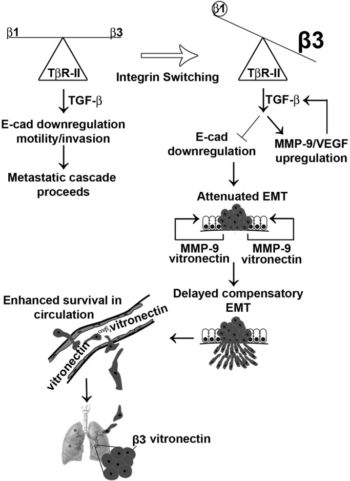
Mammary tumorigenesis and epithelial-mesenchymal transition (EMT) programs cooperate in converting transforming growth factor-β (TGF-β) from a suppressor to a promoter of breast cancer metastasis. Although previous reports associated β1 and β3 integrins with TGF-β stimulation of EMT and metastasis, the functional interplay and plasticity exhibited by these adhesion molecules in shaping the oncogenic activities of TGF-β remain unknown. We demonstrate that inactivation of β1 integrin impairs TGF-β from stimulating the motility of normal and malignant mammary epithelial cells (MECs) and elicits robust compensatory expression of β3 integrin solely in malignant MECs, but not in their normal counterparts. Compensatory β3 integrin expression also 1) enhances the growth of malignant MECs in rigid and compliant three-dimensional organotypic cultures and 2) restores the induction of the EMT phenotypes by TGF-β. Of importance, compensatory expression of β3 integrin rescues the growth and pulmonary metastasis of β1 integrin-deficient 4T1 tumors in mice, a process that is prevented by genetic depletion or functional inactivation of β3 integrin. Collectively our findings demonstrate that inactivation of β1 integrin elicits metastatic progression via a β3 integrin-specific mechanism, indicating that dual β1 and β3 integrin targeting is necessary to alleviate metastatic disease in breast cancer patients.


Figures

FIGURE 1:
FIGURE 1:
Functional disruption of β1 integrin attenuates TGF-β–mediated motility in normal NMuMG cells. (A) Confluent NMuMG cell monolayers were wounded and allowed to heal for 24 h in the absence (unstim) or presence of TGF-β1 (5 ng/ml), neutralizing β1 integrin antibodies (β1 N.A.; 5 μg/ml), or the p38 MAPK inhibitor SB203580 (p38 Inh; 10 μM) as indicated. Representative photomicrographs from a single experiment performed three times in triplicate. (B) Quantification of wounded NMuMG cultures at 24 h was conducted using ImageJ (v1.34S; National Institutes of Health, Bethesda, MD). Data are mean (±SE) percentage wound closure of three independent experiments completed in triplicate. (C, D) NMuMG cells were stimulated for 24 h with TGF-β1 (5 ng/ml) in the absence (diluent) or presence of either neutralizing β1 integrin antibodies (β1 N.A.; 5 μg/ml) or p38 MAPK inhibitor SB203580 (p38 Inh; 10 μM) as indicated. Afterward, total RNA was isolated to monitor changes in the expression of PAI-1 (C) or Cox-2 (D) by semiquantitative real-time PCR. Data are mean (±SE) of three independent experiments completed in triplicate. In B–D, *,#p < 0.05.
FIGURE 2:
FIGURE 2:
Inactivation of β1 integrin elicits compensatory expression of β3 integrin in metastatic 4T1 cells. Parental (scram) and β1 integrin–deficient (shβ1) 4T1 cells were stimulated with TGF-β1 (5 ng/ml) for 24 h before monitoring alterations in integrin expression by immunoblotting or semiquantitative real-time PCR. (A) Monitoring the extent of β1 integrin deficiency. Data are representative of at least three independent analyses. (B–D) Inactivating β1 integrin expression (B, C) or activity by administration of neutralizing β1 integrin antibodies (β1; D) elicited compensatory up-regulation of β3 integrin transcripts (B) and protein (C, D). Shown in B is the mean (±SE) of three independent experiments completed in triplicate (*,#p < 0.0005), and results in C and D are representative of three independent experiments. (E) Parental 4T1 cells were stimulated for 24 h with TGF-β1 (5 ng/ml) in the absence or presence of neutralizing β1 integrin antibodies (β1; 5 μg/ml) or the p38 MAPK inhibitor SB203580 (p38i; 10 μM). Data are representative of three independent experiments. β1, neutralizing β1 integrin antibody; IgM, nonspecific control antibody; N.A., neutralizing antibody.
FIGURE 3:
FIGURE 3:
Heterogeneous invasive and EMT phenotypes elicited by β1 integrin deficiency in 4T1 cells. (A) Parental (scram) and β1 integrin–deficient 4T1 cells were allowed to invade reconstituted basement membranes in the absence or presence of either TGF-β1 (5 ng/ml) or the MMP-9 inhibitor (MMP-9i; 10 μM) as indicated. Data are mean (±SE) of three independent experiments completed in triplicate (#p < 0.0008). (B) Accumulation of TGF-β–stimulated (5 ng/ml) parental (scram) and β1 integrin–deficient 4T1 cells measured longitudinally by trypan blue exclusion. Data are mean (±SE) of four independent experiments. (C) Alterations in DNA synthesis of parental (scram) and β1 integrin–deficient 4T1 cells in response to increasing concentrations of TGF-β1 as determined by [3H]thymidine incorporation assays. Data are mean (±SE) of three independent experiments completed in triplicate (****p < 0.0001, ***p < 0.005, **p < 0.007, and *p < 0.02). (D, E) Parental (scram) and β1 integrin–deficient 4T1 cells were stimulated with TGF-β1 (5 ng/ml) for 48 h before monitoring caspase 3/7 activity by Caspase-Glo 3/7 assays (D) or Bim transcript expression by semiquantitative real-time PCR (E). Data are mean (±SE) of three (D) or two (E) independent experiments completed in triplicate. *p < 0.005. (F) Alterations in the actin cytoskeletons of parental (scram) and β1 integrin–deficient 4T1 cells determined by phalloidin immunofluorescence as indicated. Data are representative images (200×) of four independent experiments. (G) Parental (scram) and β1 integrin–deficient 4T1 cells were transfected with a control or β3 integrin–specific siRNA and subsequently stimulated with TGF-β1 (5 ng/ml) for 48 h before monitoring β3 integrin, VEGF, and MMP-9 transcript expression by semiquantitative real-time PCR. Data are mean (±SE) of three independent experiments completed in triplicate (*p < 0.02).
FIGURE 4:
FIGURE 4:
Compensatory β3 integrin expression enhances TGF-β signaling in β1 integrin–deficient 4T1 cells. (A) Quiescent parental (scram) and β1 integrin-deficient 4T1 cells were stimulated with TGF-β1 (5 ng/ml) for 0–120 min as indicated, at which point the phosphorylation status of Smad2 and Smad3 was analyzed by immunoblotting. Data are representative of three independent analyses. (B) Smad2/3 immunofluorescence (200×) depicts the subcellular localization of Smad2/3 in basal and TGF-β1 (5 ng/ml; 30 min)–stimulated parental (scram) and β1 integrin–deficient 4T1 cells. Data are representative of three independent experiments. (C) Parental (scram) and β1 integrin–deficient 4T1 cells were transiently transfected with pCMV-β-gal and pSBE-luciferase reporter genes and subsequently stimulated with TGF-β1 (5 ng/ml) for 24 h. Data are mean (±SE) of four independent experiments completed in triplicate. (D) Parental (scram) and β1 integrin–deficient 4T1 cells were stimulated with TGF-β1 (5 ng/ml) for 48 h, at which point alterations in PAI-1 mRNA were analyzed by semiquantitative real-time PCR. Data are mean (±SE) of three independent experiments completed in triplicate. (E, F) Parental (scram) and β1 integrin–deficient 4T1 cells were stimulated for 0–120 min (E) or 24 h (F) with TGF-β1 (5 ng/ml) before monitoring the phosphorylation status and expression levels of p38 MAPK. Data are representative of three (E) or two (F) independent analyses.
FIGURE 5:
FIGURE 5:
Inactivation of β1 integrin elicits compensatory expression of β3 integrin in human MDA-MB-231 cells. Parental (scram) and β1 integrin–deficient (shβ1) MDA-MB-231 cells were stimulated with TGF-β1 (5 ng/ml) for 4 d before monitoring alterations in the extent of β1 integrin deficiency, as well as β3 integrin compensation and the expression of vimentin, ZO-2, and β-actin by immunoblotting (A), and to assess changes in their morphologies by light microscopy (100×; B). Data are representative of at least three independent analyses. (C) Parental (scram) and β1 integrin–deficient MDA-MB-231 cells were propagated in compliant 3D-organotypic cultures for 4 d. The growth of these organoids was monitored by longitudinal bioluminescence. Data are representational (±SE) of three independent experiments completed in triplicate (*p < 0.025).
FIGURE 6:
FIGURE 6:
Compensatory β3 integrin expression is essential in enhancing acinar growth of β1 integrin–deficient 4T1 cells. (A, B) Parental (scram) and β1 integrin–deficient 4T1 cells were propagated in rigid 3D-organotypic cultures (3 mg/ml type I collagen) for 4 d, at which point differences in organoid growth and morphology were monitored by phase contrast microscopy (50×; A) and longitudinal bioluminescence (B). Data are mean (±SE) of three independent experiments completed in triplicate. (C, D) Parental (scram) and β1 integrin–deficient 4T1 cells were propagated in compliant 3D-organotypic cultures for 12 d in the absence or presence of the neutralizing αvβ3 integrin antibody LM609 (15 μg/ml). The growth and morphology of the resulting organoids were monitored by phase contrast microscopy (50×; C) and longitudinal bioluminescence (D). Data are mean (±SE) of three independent experiments completed in triplicate (*p < 0.035).
FIGURE 7:
FIGURE 7:
Compensatory β3 integrin expression is essential for the growth and metastasis of β1 integrin-–deficient 4T1 tumors in mice. (A) Parental (scram) and β1 integrin–deficient 4T1 cells (12,000 cells/mouse) were engrafted into the fat pads of female BALB/c mice. Tumor growth was monitored using digital calipers on the indicated days postengraftment. Data are mean (±SE; n = 5) tumor volumes. (B) Primary tumors from A were excised and weighed at the time of killing. Data are mean tumor weights (±SE; n = 5). (C) Bioluminescence imaging of pulmonary metastasis from parental (scram) and β1 integrin–deficient 4T1 tumors from A at weeks 1 and 4 postengraftment. Inset, representative bioluminescence images of parental (scram) and β1 integrin–deficient 4T1 lung metastases. Data are mean (±SE) pulmonary area flux units detected at the indicated time points. (D) Parental (scram) and dual β1/β3 integrin (shβ1/shβ3 Int)–deficient 4T1 cells (10,000 cells/mouse) were engrafted into the fat pads of female BALB/c mice. Tumor growth was monitored using digital calipers on the indicated days postengraftment. Data are mean tumor volumes (±SE; n = 5). (E) Primary tumors from D were excised and weighed at the time of killing. Data are mean tumor weights (±SE; n = 5). (F) Bioluminescence imaging of pulmonary metastasis from parental (scram) and shβ1/shβ3 integrin–deficient 4T1 tumors from D at weeks 1 and 4 postengraftment. Inset, representative bioluminescence images of parental (scram) and β1 integrin–deficient 4T1 lung metastases. Data are mean (±SE) pulmonary area flux units detected at the indicated time points. *p < 0.05, **p < 0.0005, ***p < 0.00005.
FIGURE 8:
FIGURE 8:
Model of the dichotomous roles of β1 and β3 integrins in mediating breast cancer metastasis. Integrin switching between β1 and β3 integrins in metastatic 4T1 cells uncouples TGF-β from down-regulating E-cadherin expression, thereby attenuating the acquisition of EMT and migratory phenotypes. Elevated expression of MMP-9 and VEGF is associated with this integrin switching event and contributes to autocrine TGF-β signaling and activation of compensatory EMT programs. The physiological distribution of vitronectin expression may selectively mediate the pulmonary outgrowth of cells that underwent β1 → β3 integrin switching.

Similar articles

Cited by

References

    1. Barkan D, et al. Inhibition of metastatic outgrowth from single dormant tumor cells by targeting the cytoskeleton. Cancer Res. 2008;68:6241–6250. - PMC - PubMed
    1. Barkan D, et al. Metastatic growth from dormant cells induced by a col-I-enriched fibrotic environment. Cancer Res. 2010;70:5706–5716. - PMC - PubMed
    1. Bei L, Lu Y, Bellis SL, Zhou W, Horvath E, Eklund EA. Identification of a HoxA10 activation domain necessary for transcription of the gene encoding b3 integrin during myeloid differentiation. J Biol Chem. 2007;282:16846–16859. - PubMed
    1. Ben-Ze'ev A. The dual role of cytoskeletal anchor proteins in cell adhesion and signal transduction. Ann NY Acad Sci. 1999;886:37–47. - PubMed
    1. Bhowmick NA, Zent R, Ghiassi M, McDonnell M, Moses HL. Integrin b1 signaling is necessary for transforming growth factor-b activation of p38MAPK and epithelial plasticity. J Biol Chem. 2001;276:46707–46713. - PubMed

Publication types

MeSH terms