. 2014 Feb;95(2):215-24.
doi: 10.1189/jlb.0313180. Epub 2013 Sep 4.

Lung-resident memory CD8 T cells (TRM) are indispensable for optimal cross-protection against pulmonary virus infection

Affiliations

Lung-resident memory CD8 T cells (TRM) are indispensable for optimal cross-protection against pulmonary virus infection

Tao Wu et al. J Leukoc Biol. 2014 Feb.

Abstract

Previous studies have shown that some respiratory virus infections leave local populations of tissue TRM cells in the lungs which disappear as heterosubtypic immunity declines. The location of these TRM cells and their contribution to the protective CTL response have not been clearly defined. Here, fluorescence microscopy is used to show that some CD103(+) TRM cells remain embedded in the walls of the large airways long after pulmonary immunization but are absent from systemically primed mice. Viral clearance from the lungs of the locally immunized mice precedes the development of a robust Teff response in the lungs. Whereas large numbers of virus-specific CTLs collect around the bronchial tree during viral clearance, there is little involvement of the remaining lung tissue. Much larger numbers of TEM cells enter the lungs of the systemically immunized animals but do not prevent extensive viral replication or damage to the alveoli. Together, these experiments show that virus-specific antibodies and TRM cells are both required for optimal heterosubtypic immunity, whereas circulating memory CD8 T cells do not substantially alter the course of disease.

Keywords: cellular immunity; fluorescence microscopy; immunization; influenza virus.


Figures

Figure 1.
Figure 1.. Cellular immunity requires pulmonary immunization and declines with time.
B6 mice were primed with WSN-OVAI by pulmonary inoculation (IN-1 and IN-7) or i.p. injection (IP-1). Viral titers in the lungs were measured by plaque assay, 3 dpi (A) and 5 dpi (B; n=5/group; *P<0.05; **P<0.01).
Figure 2.
Figure 2.. Virus-specific CD8 T cells and antibodies participate in optimal heterosubtypic immunity.
(A) B6 mice received 103 naive OTI cells, 48 h before i.n. or i.p. infection with WSN-OVAI. Either 1 (IN-1 and IP-1) or 7 (IN-7) months after priming, the mice were reinfected with X31-OVA, and body weights were recorded daily (mean±sd; n=16–45/group). Data are pooled from five independent experiments. UP mice that lost 25% body weight were given 300 μl saline daily by s.c. injections. (B) μMT and B6 mice were primed with WSN-OVAI by pulmonary inoculation (IN-1) or i.p. injection (IP-1). Fifty percent of the UP μMT mice died within 5 dpi. (C) Body weights were recorded daily (mean±sd; n=3–5/group).
Figure 3.
Figure 3.. Pulmonary inoculation is required to induce PD-1 and CD103 expression on TRM OTI cells in the lungs.
B6 mice received 103 naive OTI cells 48 h before i.n. or i.p. infection with WSN-OVAI. Either 1 (IN-1 and IP-1) or 7 (IN-7) months after priming, the lungs were harvested for quantitative and phenotypic analysis of the OTI cells. (A) Total numbers of OTI cells in the lungs. (B) CD62L and CD103 expression was used to gate subsets of TRM, TEM, and TCM OTI cells in the lungs. (C) Overlaid histograms show CD62L expression on OTI in the lungs (mean±sd; n=3–6 animals). (D) Total numbers of CD62L+ and CD62L− OTI cells in the lungs. (E) Subsets of OTI cells in the lungs of IN-1 mice were analyzed for CCR7, CD69, PD-1, and CD44 expression. Shaded histograms are naive CD8 T cells in the spleen. (F) Percentages of OTI cells in the lungs of IN-1, IP-1, and IN-7 mice expressing PD-1, CD103, or both. Labels show percentages of cells in each quadrant (mean±sd; n=4–9 animals). (G) Total numbers of OTI cells expressing PD-1, CD103, or both (DP). Data are pooled from three independent experiments (*P<0.05; **P<0.01; ***P<0.001).
Figure 4.
Figure 4.. OVA-specific TRM cells line the walls of the large airways after pulmonary inoculation.
B6 mice received 103 naive OTI cells, 48 h before i.n. or i.p. infection with WSN-OVAI. The lungs were harvested 1 or 7 months after priming to visualize OTI cells in situ. (A) Fixed lung tissue was analyzed with anti-CD45.1 (green) to identify transferred OTI cells, anti-EpCAM (red) for epithelial cells, and anti-CD31 (blue) for blood vessels (BV). Z-Stack images were acquired at normal magnification, ×20. Representative images show sections across large airways (AW; upper) and areas of lung parenchyma (P; lower) from groups of four to 10 animals. (B) OTI cells (green) in the lungs of IN-1 mice were analyzed for CD103 expression (blue). Cross-sections of large airways from groups of three animals. b, Imaris software was used to identify overlapping blue and green staining shown in pink.
Figure 5.
Figure 5.. The protective CTL response is focused around the large airways early after reinfection.
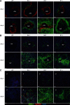
B6 mice received 103 naive OTI cells, 48 h before i.n. or i.p. infection with WSN-OVAI. One (IN-1 and IP-1) or 7 (IN-7) months later, the mice were challenged with X31-OVA. Fragments of fixed lung tissue were stained 3 and 6 dpi using antibodies to EpCAM (red), CD45.1 (green), and CD31 (blue). Z-Stack images were taken at normal magnification, ×20. Representative fields from groups of three to six animals are shown. (A) Cross-sections of large airways. (B) Cross-sections of large blood vessels. (C) Lung parenchyma.
Figure 6.
Figure 6.. Large accumulations of OTI cells appear in the lungs ∼5 dpi after reinfection.
B6 mice received 103 naive OTI cells, 48 h before i.n. or i.p. infection with WSN-OVAI. One (IN-1 and IP-1) or 7 (IN-7) months later, the mice were challenged with X31-OVA. (A) Total numbers of OTI cells in the lungs. (B) Frequencies of OTI cells with a TRM phenotype. (C) Total numbers of TRM cells in the lungs. Graphs show means ± sd from three to 12 animals/group. Data were pooled from four independent experiments. Symbols indicate significance among different groups (*P<0.05, **P<0.01, ***P<0.001 IN-1 vs. IN-7; §P<0.05, §§P<0.01 IN-1 vs. IP-1; †P<0.05, ††P<0.01, †††P<0.001 IP-1 vs. IN-7; ‡P<0.05 IN-1 Day 0 vs. IN-1 Day 3).
Figure 7.
Figure 7.. Nascent Teff cells proliferate in the MLNs and lungs after reinfection.
B6 mice received 103 naive OTI cells, 48 h before i.n. or i.p. infection with WSN-OVAI. One (IN-1 and IP-1) or 7 (IN-7) months later, the mice were challenged with X31-OVA. BrdU was given by i.p. injection, 3 or 4 dpi, and OTI cells were analyzed 2 h later. The data are means ± sd from four animals/group (*P<0.05; **P<0.01). A) Histograms show gated OTI cells with percentages BrdU+ cells in the MLN. B) Total numbers BrdU+ OTI cells in the MLN. C) Histograms show gated OTI cells with percentages BrdU+ cells in the lungs. D) Total numbers BrdU+ OTI cells in the lungs.
Figure 8.
Figure 8.. Nascent Teff cells do not participate in protection.
B6mice were primed with WSN-OVAI and challenged 1 (IN-1 and IP-1) or 7 (IN-7) months later with X31-OVA. Mice were treated four times (48-h intervals) with FTY720 or vehicle control, beginning 24 h before X31-OVA infection. (A) Percentages of CD8 T cells in the blood, 8 dpi. (B) Numbers of NP-specific CD8 T cells in the MLN, 5 dpi (IN-1) or 8 dpi (IP-1). (C) Numbers of NP-specific CD8 T cells in the lungs, 5 dpi (IN-1) or 8 dpi (IP-1). (D) Mean body weights (±sd) from four to five mice/group (*P<0.05; **P<0.01; ***P<0.001).

Comment in

Similar articles

Cited by

References

    1. Topham D. J., Tripp R. A., Doherty P. C. (1997) CD8+ T cells clear influenza virus by perforin or Fas-dependent processes. J. Immunol. 159, 5197–5200 - PubMed
    1. Belz G. T., Xie W., Altman J. D., Doherty P. C. (2000) A previously unrecognized H-2D(b)-restricted peptide prominent in the primary influenza A virus-specific CD8(+) T-cell response is much less apparent following secondary challenge. J. Virol. 74, 3486–3493 - PMC - PubMed
    1. Liang S., Mozdzanowska K., Palladino G., Gerhard W. (1994) Heterosubtypic immunity to influenza type A virus in mice. Effector mechanisms and their longevity. J. Immunol. 152, 1653–1661 - PubMed
    1. Nguyen H. H., Moldoveanu Z., Novak M. J., van Ginkel F. W., Ban E., Kiyono H., McGhee J. R., Mestecky J. (1999) Heterosubtypic immunity to lethal influenza A virus infection is associated with virus-specific CD8(+) cytotoxic T lymphocyte responses induced in mucosa-associated tissues. Virology 254, 50–60 - PubMed
    1. Kreijtz J. H., Bodewes R., van Amerongen G., Kuiken T., Fouchier R. A., Osterhaus A. D., Rimmelzwaan G. F. (2007) Primary influenza A virus infection induces cross-protective immunity against a lethal infection with a heterosubtypic virus strain in mice. Vaccine 25, 612–620 - PubMed

Publication types

MeSH terms