. 2014 Jan;95(1):191-5.
doi: 10.1189/jlb.0513272. Epub 2013 Sep 4.

Novel ex vivo culture method for human monocytes uses shear flow to prevent total loss of transendothelial diapedesis function

Affiliations

Novel ex vivo culture method for human monocytes uses shear flow to prevent total loss of transendothelial diapedesis function

Yoshiaki Tsubota et al. J Leukoc Biol. 2014 Jan.

Abstract

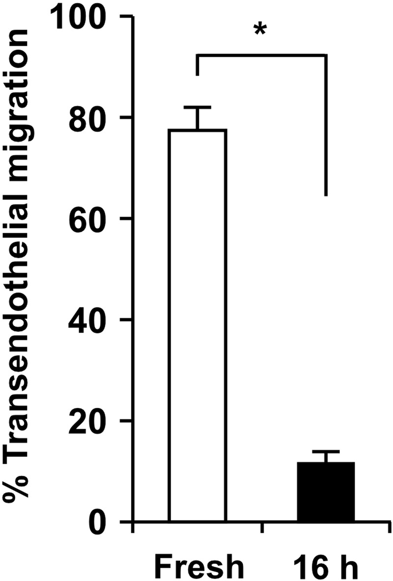
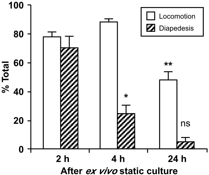
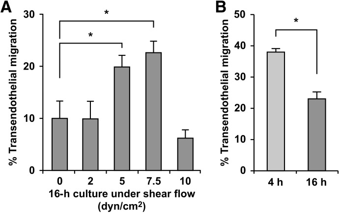
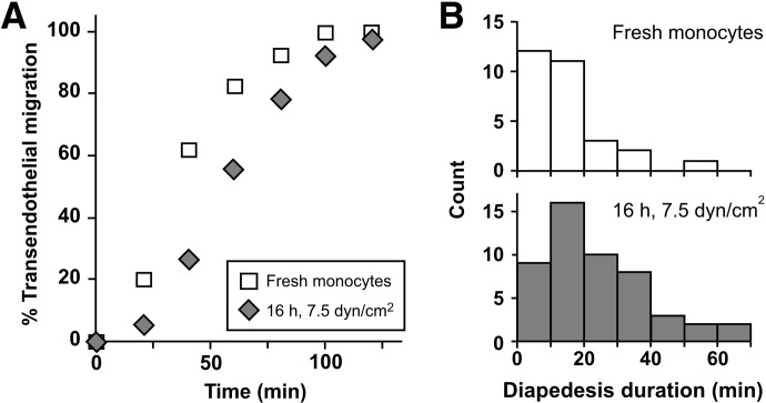
Monocyte recruitment to inflammatory sites and their transendothelial migration into tissues are critical to homeostasis and pathogenesis of chronic inflammatory diseases. However, even short-term suspension culture of primary human monocytes leads to phenotypic changes. In this study, we characterize the functional effects of ex vivo monocyte culture on the steps involved in monocyte transendothelial migration. Our data demonstrate that monocyte diapedesis is impaired by as little as 4 h culture, and the locomotion step is subsequently compromised. After 16 h in culture, monocyte diapedesis is irreversibly reduced by ∼90%. However, maintenance of monocytes under conditions mimicking physiological flow (5-7.5 dyn/cm²) is sufficient to reduce diapedesis impairment significantly. Thus, through the application of shear during ex vivo culture of monocytes, our study establishes a novel protocol, allowing functional analyses of monocytes not currently possible under static culture conditions. These data further suggest that monocyte-based therapeutic applications may be measurably improved by alteration of ex vivo conditions before their use in patients.

Keywords: macrophages; migration; transfusion medicine; vascular biology.


Figures

Figure 1.
Figure 1.. Primary monocytes lose their ability to complete transendothelial migration after 16 h ex vivo incubation at 37°C under static conditions.
Freshly isolated human monocytes (left column; Supplemental Video 1) or monocytes incubated for 16 h at 37°C in a polypropylene tube (right column; Supplemental Video 2) were added to TNF-α-activated HUVEC monolayers, and monocyte transendothelial migration under static conditions was monitored for 2 h by time-lapse video microscopy. The ex vivo incubation time was selected to allow optimal knockdown of proteins of interest in primary monocytes using siRNA constructs [24]. Individual monocytes were tracked on the apical surfaces up to the last frame or completion of diapedesis, which was defined as the frame where the monocyte cell bodies, retracting between endothelial junctions, disappeared from the apical surface of HUVEC monolayers. A monocyte that underwent diapedesis was evaluated as one that completed transendothelial migration, and the data are expressed as percent of total monocytes in the microscopic field over a 2-h observation period. Means ± sem from independent experiments using three different monocyte donors are shown; *P < 0.01 (paired t-test).
Figure 2.
Figure 2.. Static incubation of primary monocytes first impairs monocyte diapedesis and subsequently their locomotion.
Human monocytes were incubated ex vivo for 2 h (Supplemental Video 3), 4 h (Supplemental Video 4), and 24 h and then subjected to the transendothelial migration assay, as described in Fig. 1. The numbers of monocytes that undergo locomotion and/or diapedesis are expressed as percent of total monocytes in the microscopic field (>25). Means ± sem of three fields/incubation time are shown; *P < 0.02 relative to diapedesis of monocytes after 2 h incubation; **P < 0.002 relative to locomotion of monocytes after 4 h incubation (unpaired t-test); ns, not significant for comparison with diapedesis after 4 h incubation. Representative data from two independent experiments using monocytes from two different donors are shown.
Figure 3.
Figure 3.. Application of shear flow during ex vivo incubation leads to less impairment of monocyte transendothelial migration than static incubation.
To maintain monocytes under conditions during ex vivo incubation that more closely mimic those in the circulation, shear flow was introduced by shaking the freshly prepared monocytes in polypropylene tubes with a rotary shaker. (A) Monocytes were shaken for 16 h at various speeds to attain fluid flow conditions that are estimated as shear stresses of 2, 5, 7.5, and 10 dyn/cm2, and their frequencies of diapedesis on TNF-activated HUVEC monolayers were evaluated by 2-h time-lapse microscopy, as described in Fig. 1. Means ± sem from experiments using four different monocyte donors for 5 dyn/cm2, and three for other conditions are shown; *P < 0.05 (unpaired t-test). (B) Monocytes were isolated from individual donors and shaken separately at 7.5 dyn/cm2 for 4 and 16 h (fourth column of Fig. 3A), and the frequencies of diapedesis were evaluated immediately after the incubation and then compared. Means ± sem from independent experiments using three different monocyte donors are shown; *P < 0.05 (paired t-test).
Figure 4.
Figure 4.. Analysis of diapedesis further establishes that monocytes cultured under shear flow conditions retain their ability to undergo diapedesis.
Monocytes freshly prepared or incubated under conditions of 7.5 dyn/cm2 for 16 h were analyzed by time-lapse microscopy of transendothelial migration. Individual monocytes that successfully underwent diapedesis were analyzed. Representative data from one of at least four experiments with different monocyte donors are shown (n=29 and 54 monocytes for monocytes freshly isolated and incubated with shear flow, respectively). (A) Elapsed times for individual monocytes to complete their transendothelial migration were defined as in Fig. 1. Means ± sem are 29.9 ± 3.8 and 50.7 ± 4.3 min, respectively; P < 0.005 (unpaired t-test). (B) Duration times for diapedesis were calculated as an interval between the frames where monocytes arrived at the place of diapedesis and where monocytes completed diapedesis as defined in Fig. 1. Means ± sem are 14.4 ± 2.0 and 25.0 ± 2.6 min, respectively; P < 0.01 (unpaired t-test). Note that although diapedesis is delayed following 16 h incubation under flow conditions, the ability of monocytes to undergo diapedesis is not lost, as observed under static incubation conditions.

Similar articles

Cited by

References

    1. Ley K., Laudanna C., Cybulsky M. I., Nourshargh S. (2007) Getting to the site of inflammation: the leukocyte adhesion cascade updated. Nat. Rev. Immunol. 7, 678–689 - PubMed
    1. Thomas-Ecker S., Lindecke A., Hatzmann W., Kaltschmidt C., Zanker K. S., Dittmar T. (2007) Alteration in the gene expression pattern of primary monocytes after adhesion to endothelial cells. Proc. Natl. Acad. Sci. USA 104, 5539–5544 - PMC - PubMed
    1. Williams M. R., Sakurai Y., Zughaier S. M., Eskin S. G., McIntire L. V. (2009) Transmigration across activated endothelium induces transcriptional changes, inhibits apoptosis, and decreases antimicrobial protein expression in human monocytes. J. Leukoc. Biol. 86, 1331–1343 - PMC - PubMed
    1. Randolph G. J., Beaulieu S., Lebecque S., Steinman R. M., Muller W. A. (1998) Differentiation of monocytes into dendritic cells in a model of transendothelial trafficking. Science 282, 480–483 - PubMed
    1. Muller W. A., Randolph G. J. (1999) Migration of leukocytes across endothelium and beyond: molecules involved in the transmigration and fate of monocytes. J. Leukoc. Biol. 66, 698–704 - PubMed

Publication types

MeSH terms

LinkOut - more resources