. 2013 Sep 4;7(1):21.
doi: 10.1186/1754-1611-7-21.

Contribution of cytoskeletal elements to the axonal mechanical properties

Affiliations

Contribution of cytoskeletal elements to the axonal mechanical properties

Hui Ouyang et al. J Biol Eng. .

Abstract

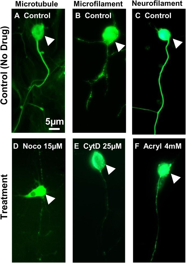
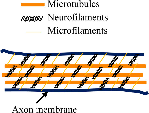
Background: Microtubules, microfilaments, and neurofilaments are cytoskeletal elements that affect cell morphology, cellular processes, and mechanical structures in neural cells. The objective of the current study was to investigate the contribution of each type of cytoskeletal element to the mechanical properties of axons of dorsal root and sympathetic ganglia cells in chick embryos.

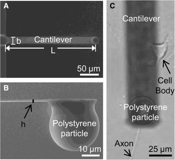
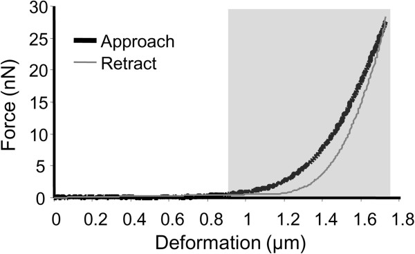
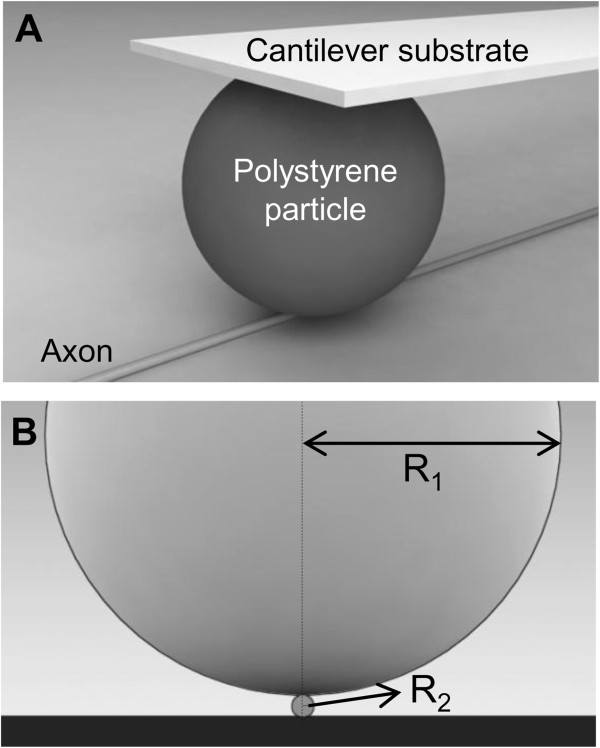
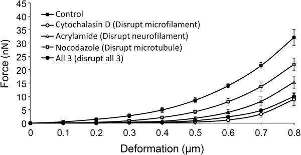
Results: Microtubules, microfilaments, and neurofilaments in axons were disrupted by nocodazole, cytochalasin D, and acrylamide, respectively, or a combination of the three. An atomic force microscope (AFM) was then used to compress the treated axons, and the resulting corresponding force-deformation information was analyzed to estimate the mechanical properties of axons that were partially or fully disrupted.

Conclusion: We have found that the mechanical stiffness was most reduced in microtubules-disrupted-axons, followed by neurofilaments-disrupted- and microfilaments-disrupted-axons. This suggests that microtubules contribute the most of the mechanical stiffness to axons.


Figures

Figure 1
Figure 1
Diagrams illustrating dimension of cantilever and axon being compressed by cantilever in atomic force microscopy (AFM). (A) Top view of a cantilever obtained by scanning electric microscopy (SEM). (B) Side view of a cantilever tip obtained by SEM. (C) Top view of the particle of a cantilever compressing the axon of a dorsal root ganglia (DRG) cell. Image was taken by a digital camera from the eyepiece of the optical part of the AFM.
Figure 2
Figure 2
Sample raw data of the force-deformation responses obtained from atomic force microscopy (AFM). Only the approach curve was analyzed as it is the initial measurement of force responses in the compression test. The deformation at which the force amplitude was at least 2% of the maximum force was chosen to be the initial contact point between the cantilever and the axon (approximately 0.9 μm, gray area).
Figure 3
Figure 3
Illustration of contact and dimensional ratio between the polystyrene particle and the axon in 3-dimension (A) and 2-dimension (B). R1 is the radius of a polystyrene particle (12.5 μm), while R2 is the radius of the axon under compression (0.5 μm).
Figure 4
Figure 4
Immunocytochemistry of dorsal root and sympathetic ganglia cells with disrupted cytoskeletal elements. (A-C) Normal axons showed significant staining in microtubules, microfilaments, and neurofilaments. (D-F) Axons treated with 15 μM Nocodazole, 25 μM Cytochalasin D, and 4 mM Acrylamide showed significantly less staining in microtubules, microfilaments, and neurofilaments in axons, respectively. White arrows indicate cell bodies. Scale bar = 5 μm.
Figure 5
Figure 5
Average force responses at each increment of deformation as the control and treated axons were compressed from 0 (no compression) to 0.8 μm. Untreated control axons had the highest force response in each increment of deformation (n = 6), followed by cytochalasin D-treated (n = 6, microfilament disruption) and acrylamide treated axons (n = 6, neurofilament disruption). Nocodazole-treated axons (n = 6, microtubule disruption) and axons treated with all three drugs had similar force response in each increment of deformation.
Figure 6
Figure 6
Proposed distribution of cyctoskeletal elements in an axon.

References

    1. Fletcher DA, Mullins RD. Cell mechanics and the cytoskeleton. Nature. 2010;463:485–492. doi: 10.1038/nature08908. - DOI - PMC - PubMed
    1. Baas PW, Ahmad FJ. Force generation by cytoskeletal motor proteins as a regulator of axonal elongation and retraction. Trends Cell Biol. 2001;11:244–249. doi: 10.1016/S0962-8924(01)02005-0. - DOI - PubMed
    1. Ahmad FJ, Hughey J, Wittmann T, Hyman A, Greaser M, Baas PW. Motor proteins regulate force interactions between microtubules and microfilaments in the axon. Nat Cell Biol. 2000;2:276–280. doi: 10.1038/35010544. - DOI - PubMed
    1. Kolodney MS, Elson EL. Contraction due to microtubule disruption is associated with increased phosphorylation of myosin regulatory light chain. Proc Natl Acad Sci U S A. 1995;92:10252–10256. doi: 10.1073/pnas.92.22.10252. - DOI - PMC - PubMed
    1. Hirose M, Ishizaki T, Watanabe N, Uehata M, Kranenburg O, Moolenaar WH, Matsumura F, Maekawa M, Bito H, Narumiya S. Molecular dissection of the Rho-associated protein kinase (p160ROCK)-regulated neurite remodeling in neuroblastoma N1E-115 cells. J Cell Biol. 1998;141:1625–1636. doi: 10.1083/jcb.141.7.1625. - DOI - PMC - PubMed

LinkOut - more resources