. 2014 Jan 1;23(1):5-15.
doi: 10.1089/scd.2013.0309. Epub 2013 Oct 8.

Detailed expression analysis of regulatory genes in the early developing human neural tube

Affiliations

Detailed expression analysis of regulatory genes in the early developing human neural tube

Ulrika Marklund et al. Stem Cells Dev. .

Abstract

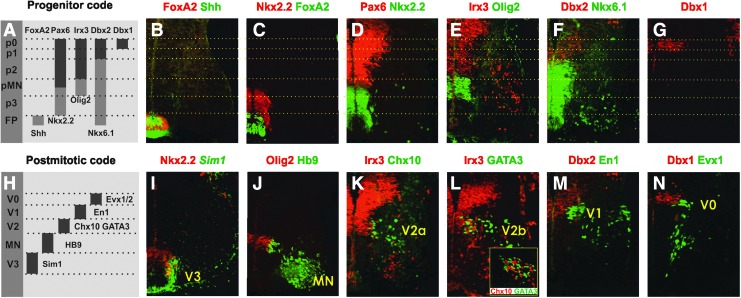
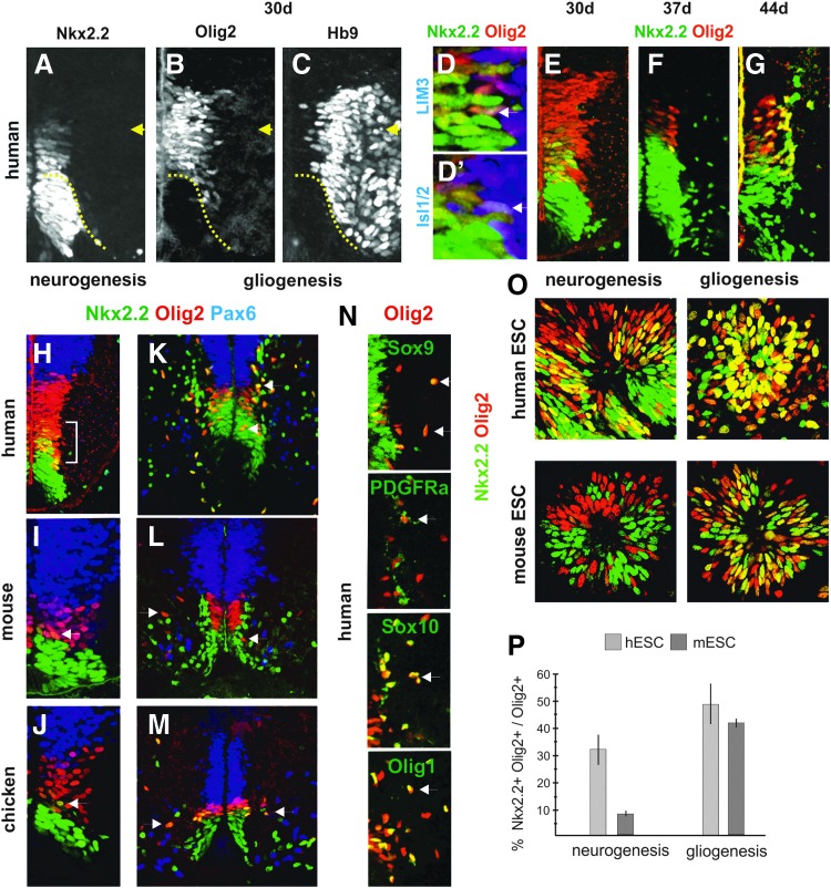
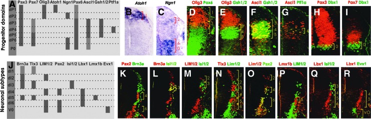
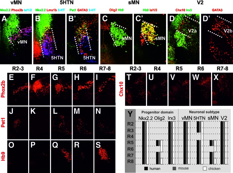
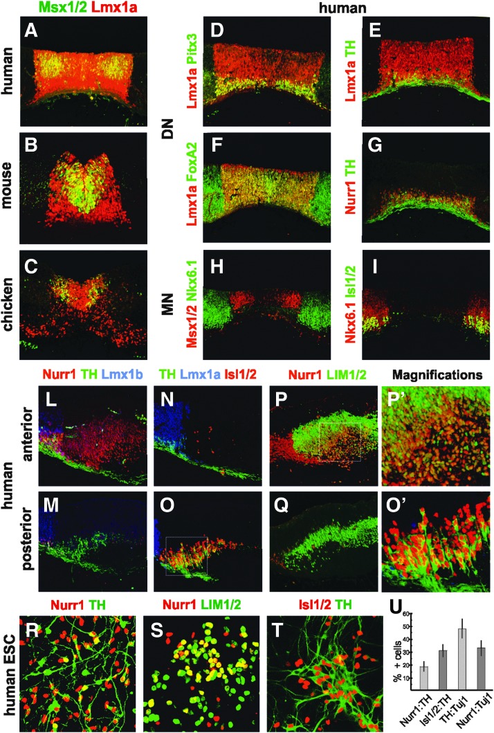
Studies in model organisms constitute the basis of our understanding of the principal molecular mechanisms of cell fate determination in the developing central nervous system. Considering the emergent applications in stem cell-based regenerative medicine, it is important to demonstrate conservation of subtype specific gene expression programs in human as compared to model vertebrates. We have examined the expression patterns of key regulatory genes in neural progenitor cells and their neuronal and glial descendants in the developing human spinal cord, hindbrain, and midbrain, and compared these with developing mouse and chicken embryos. As anticipated, gene expression patterns are highly conserved between these vertebrate species, but there are also features that appear unique to human development. In particular, we find that neither tyrosine hydroxylase nor Nurr1 are specific markers for mesencephalic dopamine neurons, as these genes also are expressed in other neuronal subtypes in the human ventral midbrain and in human embryonic stem cell cultures directed to differentiate towards a ventral mesencephalic identity. Moreover, somatic motor neurons in the ventral spinal cord appear to be produced by two molecularly distinct ventral progenitor populations in the human, raising the possibility that the acquisition of unique ventral progenitor identities may have contributed to the emergence of neural subtypes in higher vertebrates.


Figures

<b>FIG. 1.</b>
FIG. 1.
Gene expression in progenitor domains and neuronal subtypes in the human ventral spinal cord. (A) Schematic drawing of the domain specific expression profile of ventral progenitor cells. (B–G) Expression of regulatory genes defining the major progenitor domains (p0–p3, pMN) and the FP (5.5 weeks). Note the mutually exclusive expression of repressor pairs in (D), (E), and (F). (H) Schematic drawing of marker gene expression in ventral neuronal subtypes. (I–N) Correlation between the generation of neuronal subtypes (V0–V3, MN) and progenitor domains, each marked by representative marker genes (5–5.5 weeks). Note that Chx10 and GATA3 were expressed in distinct cell populations (Fig. 1L infold). Dotted lines indicate borders between the progenitor domains. Pictures of two consecutive immunohistochemical stained sections were merged in (K). MN, motor neuron.
<b>FIG. 2.</b>
FIG. 2.
Sequential generation of MNs and OLPs from ventral spinal cord progenitor cells and ESCs. (A–C) The Nkx2.2+Olig2+ domain (between arrow and dotted line) correlates to Hb9 positive cells in the human neural tube at the D-V level (30 days). Single Nkx2.2+Olig2+ cells coexpressed LIM3 (D) or Isl1/2 (D’) (arrows) at the border between the progenitor and postmitotic zones. See Supplementary Fig. S1A for separate pictures of each staining. (E–G) Dynamic temporal expression of Nkx2.2 relative Olig2 in human embryos (30–44d). (H–J) Expression of Nkx2.2, Olig2, and Pax6 in human (4.5 weeks), mouse (E10.5), and chicken (day 4) at neurogenic stages. Bracket and arrows indicate Nkx2.2+Olig2+ cells. (K–M) Expression of progenitor and OLP markers in human (9 weeks), mouse (E12.5), and chicken (day 7.5) at gliogenic stages. Many early migratory OLPs expressed both Nkx2.2 and Olig2 in chicken and human (Fig. 2K, M, arrows) but only Olig2 in mouse (L, arrows). (N) Expression of OLP markers (arrows) in human (9 weeks). (O) Coexpression of Nkx2.2 and Olig2 in differentiating hESCs and mESCs at neurogenic stages (20 DDC) and 5 DDC, respectively) and gliogenic stages (45 DDC and 8 DDC, respectively). (P) Diagram showing the percentage of Olig2+ cells that coexpress Nkx2.2 in O. n=3, mean±SD. Immunohistochemistry was used in all pictures. D-V, dorsoventral; mESC, mouse embryonic stem cell; hESC, human embryonic stem cell; OLP, oligodendrocyte precursor cell; DDC, days of differentiation culture.
<b>FIG. 3.</b>
FIG. 3.
Gene expression in progenitor domains and neuronal subtypes in the human dorsal spinal cord. (A) Schematic drawing of the domain specific gene expression profile in dorsal progenitor cells. (B–I) Nested expression of transcription factors in six dorsal progenitor domains (dP1–dP6) (5 weeks). (J) Schematic drawing of the subtype-specific expression in postmitotic neurons. (K–R) Combinatorial expression of marker genes defining six major dorsal neuronal subtypes (dI1–dI6) (5 weeks). Light gray indicates weak expression in A and J. Brackets indicate progenitor (dP) or neuronal (dI) domains. In (B) and (C) in situ hybridization was used, and in (D–I); (K–R) immunohistochemistry was applied.
<b>FIG. 4.</b>
FIG. 4.
Generation of four ventral neuronal subtypes from three progenitor domains along the A-P axis of human hindbrain. Expression of specific marker proteins for vMN (A), 5HTN (B, B’), sMN (C, C’), and V2 IN (D, D’) in relation to respective progenitor domain expression (5.5 weeks). Dotted lines indicate the borders of neuronal subtypes emerging from the characteristic progenitor domains. Generation of specific neuronal subtypes along the A-P axis using specific marker expressions for vMN (E–I), 5HTN (J–N), sMN (O–S), and V2a IN (T–X) (5.5 weeks). (Y) Schematic drawing comparing the progenitor gene expression and neuronal distribution along the A-P axis of human (black), mouse (gray), and chicken (white). Immunohistochemistry was used in all pictures. A-P, anterioposterior; IN, interneuron; vMN, visceral motor neuron; sMN, somatic motor neuron; 5HTN, serotonin neuron.
<b>FIG. 5.</b>
FIG. 5.
Gene expression in the ventral midbrain and differentiated hESCs. (A–C) Expression of mesDN progenitor marker genes in the human (5.5 weeks), mouse (E12.5) and chicken (day 5). (D–G) Lmx1a expression demarcates the mesDN progenitor domain, generating mesDNs as assessed by specific marker expression in human (5.5 weeks). (H, I) MN (Isl1/2+) generation from Nkx6.1+ progenitor cells, abutting the Msx1/2+ domain. (L, N, P) A large anterior population of Nurr1+ cells, devoid of mesDN (Lmx1a, Lmx1b, TH) or MN (Isl1/2) markers coexpressed the IN marker LIM1/2. (M, O, Q) MNs (Isl1/2+LIM1/2) coexpressed TH but not Lmx1b, Nurr1, or LIM1/2. Magnifications of Nurr1+LIM1/2+ cells (P’) indicated in box (P), and of TH+ Isl1/2+ cells (O’) indicated in box (O). (R–T) Significant populations of Nurr1+TH+, Nurr1+ LIM1/2+, and Isl1/2+TH+ neurons were found in differentiating hESCs at 40 DDC. (U) Diagram shows Nurr1+ out of TH+ (Nurr:TH), Isl1/2+ out of TH+ (Isl1/2:TH), TH+ out of Tuj1+ (TH:Tuj1), or Nurr1+ out of Tuj1+ (Nurr1:Tuj1). n=3 mean±SD. Immunohistochemistry was used in all pictures. mesDN, mesencephalic dopamine neuron; TH, tyrosine hydroxylase.

References

    1. Arenas E. (2010). Towards stem cell replacement therapies for Parkinson's disease. Biochem Biophys Res Commun 396:152–156 - PubMed
    1. Chipman PH, Toma JS. and Rafuse VF. (2012). Generation of motor neurons from pluripotent stem cells. Prog Brain Res 201:313–331 - PubMed
    1. Patani R. and Chandran S. (2012). Experimental and therapeutic opportunities for stem cells in multiple sclerosis. Int J Mol Sci 13:14470–14491 - PMC - PubMed
    1. Liu H. and Zhang SC. (2011). Specification of neuronal and glial subtypes from human pluripotent stem cells. Cell Mol Life Sci 68:3995–4008 - PMC - PubMed
    1. Panman L, Andersson E, Alekseenko Z, Hedlund E, Kee N, Mong J, Uhde CW, Deng Q, Sandberg R, et al. (2011). Transcription factor-induced lineage selection of stem-cell-derived neural progenitor cells. Cell Stem Cell 8:663–675 - PubMed

Publication types

MeSH terms

Substances