

An anti-CD154 domain antibody prolongs graft survival and induces Foxp3(+) iTreg in the absence and presence of CTLA-4 Ig
- PMID: 24007441
- PMCID: PMC4287239
- DOI: 10.1111/ajt.12417
An anti-CD154 domain antibody prolongs graft survival and induces Foxp3(+) iTreg in the absence and presence of CTLA-4 Ig
Abstract
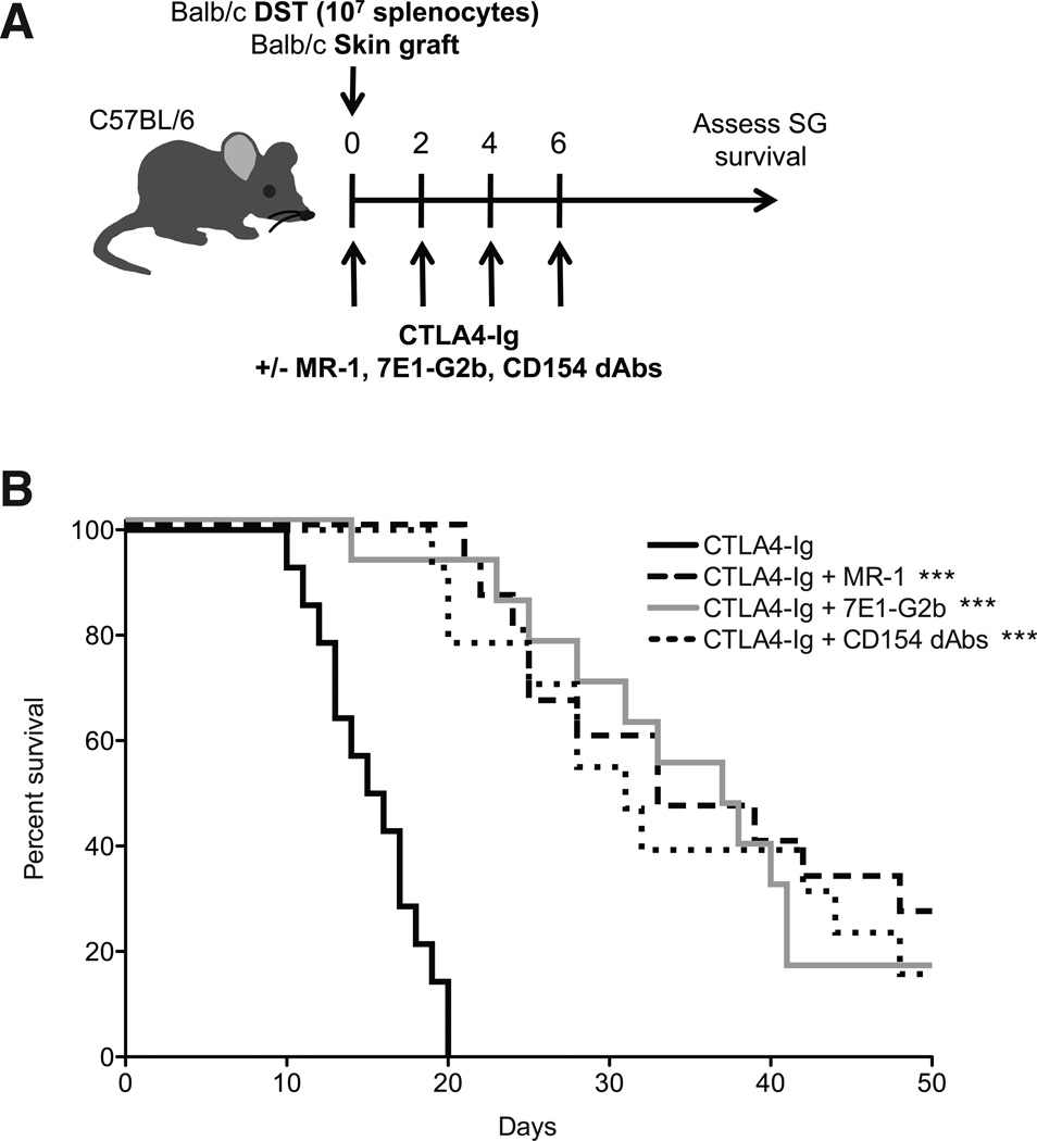
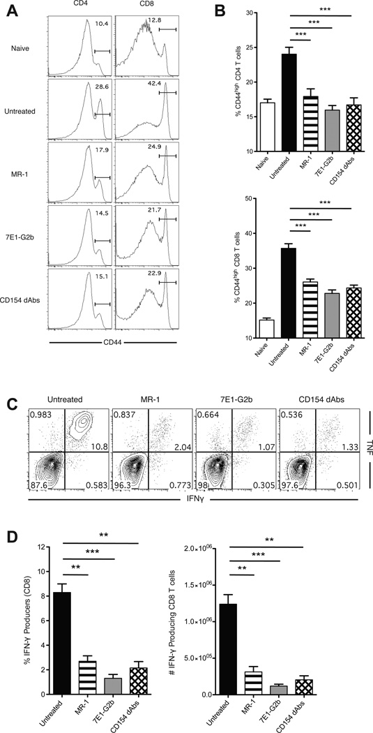
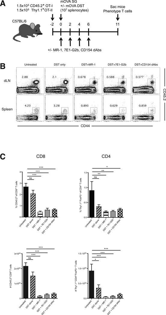
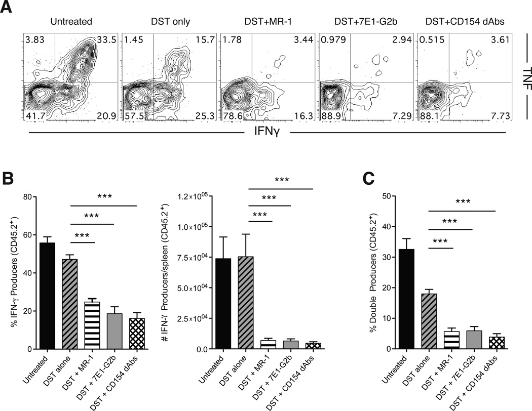
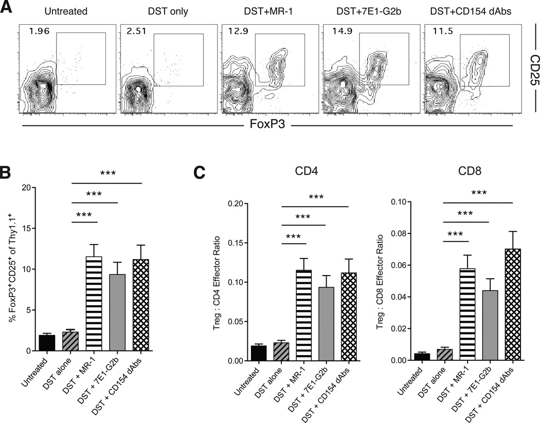
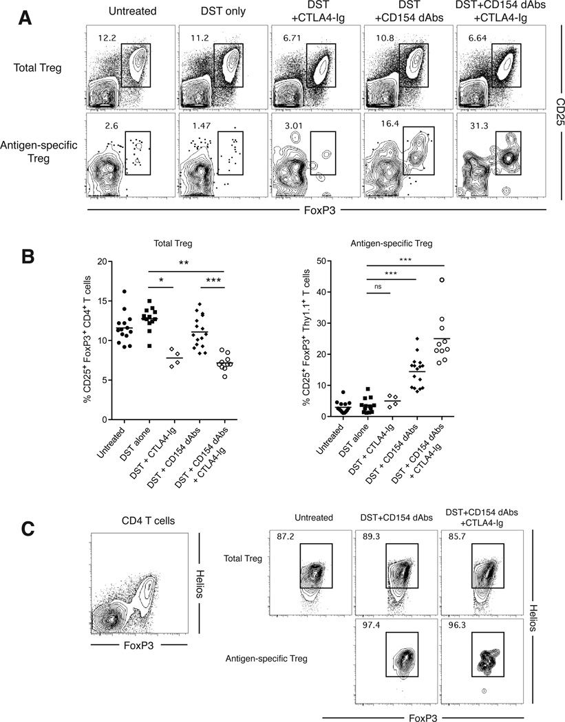
The use of monoclonal antibodies targeting the CD154 molecule remains one of the most effective means of promoting graft tolerance in animal models, but thromboembolic complications during early clinical trials have precluded their use in humans. Furthermore, the role of Fc-mediated deletion of CD154-expressing cells in the observed efficacy of these reagents remains controversial. Therefore, determining the requirements for anti-CD154-induced tolerance will instruct the development of safer but equally efficacious treatments. To investigate the mechanisms of action of anti-CD154 therapy, two alternative means of targeting the CD40-CD154 pathway were used: a nonagonistic anti-CD40 antibody and an Fc-silent anti-CD154 domain antibody. We compared these therapies to an Fc-intact anti-CD154 antibody in both a fully allogeneic model and a surrogate minor antigen model in which the fate of alloreactive cells could be tracked. Results indicated that anti-CD40 mAbs as well as Fc-silent anti-CD154 domain antibodies were equivalent to Fc-intact anti-CD154 mAbs in their ability to inhibit alloreactive T cell expansion, attenuate cytokine production of antigen-specific T cells and promote the conversion of Foxp3(+) iTreg. Importantly, iTreg conversion observed with Fc-silent anti-CD154 domain antibodies was preserved in the presence of CTLA4-Ig, suggesting that this therapy is a promising candidate for translation to clinical use.
Keywords: Alloreactivity; CD8+ T-lymphocytes; costimulation; regulatory T cells.
© Copyright 2013 The American Society of Transplantation and the American Society of Transplant Surgeons.
Conflict of interest statement
The authors of this manuscript have conflicts of interest to disclose as described by the American Journal of Transplantation.
Figures
Similar articles
-
CD40-CD40L Blockade: Update on Novel Investigational Therapeutics for Transplantation.Transplantation. 2023 Jul 1;107(7):1472-1481. doi: 10.1097/TP.0000000000004469. Epub 2023 Jun 20. Transplantation. 2023. PMID: 36584382 Free PMC article. Review.
-
Treatment of allograft recipients with donor-specific transfusion and anti-CD154 antibody leads to deletion of alloreactive CD8+ T cells and prolonged graft survival in a CTLA4-dependent manner.J Immunol. 2000 Jan 1;164(1):512-21. doi: 10.4049/jimmunol.164.1.512. J Immunol. 2000. PMID: 10605049
-
Differential induction of donor-reactive Foxp3+ regulatory T cell via blockade of CD154 vs CD40.Am J Transplant. 2024 Aug;24(8):1369-1381. doi: 10.1016/j.ajt.2024.03.033. Epub 2024 Mar 27. Am J Transplant. 2024. PMID: 38552961 Free PMC article.
-
Antigen-specific induced Foxp3+ regulatory T cells are generated following CD40/CD154 blockade.Proc Natl Acad Sci U S A. 2011 Dec 20;108(51):20701-6. doi: 10.1073/pnas.1105500108. Epub 2011 Dec 5. Proc Natl Acad Sci U S A. 2011. PMID: 22143783 Free PMC article.
-
Novel insights into anti-CD40/CD154 immunotherapy in transplant tolerance.Immunotherapy. 2015;7(4):399-410. doi: 10.2217/imt.15.1. Immunotherapy. 2015. PMID: 25917630 Free PMC article. Review.
Cited by
-
Stability-Diversity Tradeoffs Impose Fundamental Constraints on Selection of Synthetic Human VH/VL Single-Domain Antibodies from In Vitro Display Libraries.Front Immunol. 2017 Dec 12;8:1759. doi: 10.3389/fimmu.2017.01759. eCollection 2017. Front Immunol. 2017. PMID: 29375542 Free PMC article.
-
T Cell Repertoire Maturation Induced by Persistent and Latent Viral Infection Is Insufficient to Induce Costimulation Blockade Resistant Organ Allograft Rejection in Mice.Front Immunol. 2018 Jun 15;9:1371. doi: 10.3389/fimmu.2018.01371. eCollection 2018. Front Immunol. 2018. PMID: 29963060 Free PMC article.
-
T cell responsiveness to IL-10 defines the immunomodulatory effect of costimulation blockade via anti-CD154 and impacts transplant survival.bioRxiv [Preprint]. 2024 Jun 14:2024.06.12.598652. doi: 10.1101/2024.06.12.598652. bioRxiv. 2024. PMID: 38915537 Free PMC article. Preprint.
-
Distinct Graft-Specific TCR Avidity Profiles during Acute Rejection and Tolerance.Cell Rep. 2018 Aug 21;24(8):2112-2126. doi: 10.1016/j.celrep.2018.07.067. Cell Rep. 2018. PMID: 30134172 Free PMC article.
-
CD40-CD40L Blockade: Update on Novel Investigational Therapeutics for Transplantation.Transplantation. 2023 Jul 1;107(7):1472-1481. doi: 10.1097/TP.0000000000004469. Epub 2023 Jun 20. Transplantation. 2023. PMID: 36584382 Free PMC article. Review.
References
-
- Kirk AD, Burkly LC, Batty DS, Baumgartner RE, Berning JD, Buchanan K, et al. Treatment with humanized monoclonal antibody against CD154 prevents acute renal allograft rejection in nonhuman primates. Nat Med. 1999;5(6):686–693. - PubMed
-
- Larsen CP, Elwood ET, Alexander DZ, Ritchie SC, Hendrix R, Tucker-Burden C, et al. Long-term acceptance of skin and cardiac allografts after blocking CD40 and CD28 pathways. Nature. 1996;30:434–438. - PubMed
-
- Kawai T, Andrews D, Colvin RB, Sachs DH, Cosimi AB. Thromboembolic complications after treatment with monoclonal antibody against CD40 ligand. Nat Med. 2000;6:114. - PubMed
-
- Monk NJ, Hargreaves RE, Marsh JE, Farrar CA, Sacks SH, Millrain M, et al. Fc-dependent depletion of activated T cells occurs through CD40L-specific antibody rather than costimulation blockade. Nat Med. 2003;9(10):1275–1280. - PubMed
-
- Daley SR, Cobbold SP, Waldmann H. Fc-disabled anti-mouse CD40L antibodies retain efficacy in promoting transplantation tolerance. Am J Transplant. 2008;8(11):2265–2271. - PubMed
Publication types
MeSH terms
Substances
Grants and funding
LinkOut - more resources
Full Text Sources
Other Literature Sources
Research Materials
