Review
. 2013 Nov;183(5):1364-1374.
doi: 10.1016/j.ajpath.2013.06.033. Epub 2013 Sep 3.

New insights into the pathology of podocyte loss: mitotic catastrophe

Affiliations
Review

New insights into the pathology of podocyte loss: mitotic catastrophe

Helen Liapis et al. Am J Pathol. 2013 Nov.

Abstract

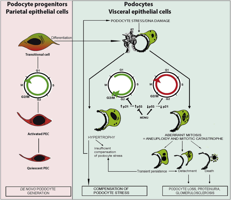
Podocytes represent an essential component of the kidney's glomerular filtration barrier. They stay attached to the glomerular basement membrane via integrin interactions that support the capillary wall to withstand the pulsating filtration pressure. Podocyte structure is maintained by a dynamic actin cytoskeleton. Terminal differentiation is coupled with permanent exit from the cell cycle and arrest in a postmitotic state. Postmitotic podocytes do not have an infinite life span; in fact, physiologic loss in the urine is documented. Proteinuria and other injuries accelerate podocyte loss or induce death. Mature podocytes are unable to replicate and maintain their actin cytoskeleton simultaneously. By the end of mitosis, cytoskeletal actin forms part of the contractile ring, rendering a round shape to podocytes. Therefore, when podocyte mitosis is attempted, it may lead to aberrant mitosis (ie, mitotic catastrophe). Mitotic catastrophe implies that mitotic podocytes eventually detach or die; this is a previously unrecognized form of podocyte loss and a compensatory mechanism for podocyte hypertrophy that relies on post-G1-phase cell cycle arrest. In contrast, local podocyte progenitors (parietal epithelial cells) exhibit a simple actin cytoskeleton structure and can easily undergo mitosis, supporting podocyte regeneration. In this review we provide an appraisal of the in situ pathology of mitotic catastrophe compared with other proposed types of podocyte death and put experimental and renal biopsy data in a unified perspective.


Figures

Figure 1
Figure 1
Working model of the role of cell cycle in proliferation of podocyte progenitor cells or podocyte hypertrophy and how aberrant podocyte proliferation may lead to podocyte loss. De novo podocyte generation starts from renal progenitors that are committed to the podocyte lineage within the parietal epithelial cell (PEC) layer. On activation, PECs enter the cell cycle (ie, proliferate and differentiate), which first creates a transitional cell that expresses both PEC and podocyte markers. Transitional cells are often found around the vascular pole of the glomerulus. Terminal differentiation into podocytes usually occurs only on the glomerular tuft, which implies loss of all parietal cell and progenitor markers so that these cells can no longer be distinguished from podocytes. Only parietal cell lineage tracing experiments are able to document that these are a progeny of former parietal epithelial cells. Once terminally differentiated (and postmitotic) podocytes are exposed to stress, such as loss of neighboring podocytes, a compensatory response becomes necessary. Functionally and structurally, the most important response to stress is podocyte hypertrophy. To undergo hypertrophy the podocyte needs to enter the S phase of the cell cycle but keep the cell cycle arrest at the G1 or G2/M restriction point. This is ensured by p53-mediated induction of cyclin kinases, such as p21. Mitogenic stimuli or DNA damage induce MDM2, which inactivates p53-mediated cell cycle arrest and forces the podocytes to complete mitosis. However, podocytes need their actin cytoskeleton to maintain their sophisticated anatomical structure; therefore, this usually leads to an incomplete formation of mitotic spindles, aberrant chromosome segregation, and/or podocyte detachment. In addition, podocytes cannot complete cytokinesis, which results in aneuploid podocytes with two or more nuclei. Such cells are susceptible to death; hence, podocyte mitosis is not a sign of podocyte regeneration but a pathologic mechanism of podocyte loss.
Figure 2
Figure 2
Mitotic podocytes in human glomerular diseases. A: IgA nephropathy from 20-year-old woman with hematuria and necrotizing IgA. Mitotic podocyte (arrow) among numerous detached counterparts (silver stain). B: Collapsing FSGS from a 54-year-old man with a protein level of 8.5 g/dL. Half mitotic spindle in detached podocyte (arrow) in a glomerulus with implosion of the capillary loops (silver stain). C: Drug-induced MCD in an 84-year-old woman with a protein level of 6 g/dL. Mitotic podocyte traveling toward the proximal tubule pole of the glomerulus (arrow) (H&E). Original magnification, ×1000 (A and B); ×400 (C).
Figure 3
Figure 3
Mitotic podocytes in human glomerular diseases. A: IgA nephropathy. Hypertrophic, binucleated podocyte with an increased number of cytoplasmic organelles apparent on a renal biopsy specimen from a 58-year-old patient with a creatinine level of 2.2 mg/dL and a protein level of 1.1 g/dL. B: Lupus membranous. Binucleated detached podocyte apparent on a renal biopsy specimen from a 31-year-old woman with a urinary protein excretion of 4.2 g per 24 hours and a normal creatinine level. C: FSGS. Trinucleated podocyte apparent on a renal biopsy specimen from a 23-year-old man with a protein level of 12.5 g/dL. D: Recurrent FSGS. Micronucleolus next to podocyte nucleus (arrow) is apparent on a transplant renal biopsy specimen from a 23-year-old man with a protein level of 12.3 g/dL.
Figure 4
Figure 4
Mitotic catastrophe in HIVAN. A: Collapsing glomerulopathy from a 34-year-old man who presented with acute renal failure; both visceral and parietal podocytes are numerous, proliferating over imploded capillary loops (silver stain). B: On high magnification, gigantic, multinucleated podocytes are apparent (arrow) (H&E). C: Electron microscopy highlights even more binucleated podocytes; podocytes also contain numerous cytoplasmic osmiophilic vacuoles characteristic of HIVAN. Podocytes are, however, still attached to foot processes, attempting to maintain functionality perhaps but apparently unable to divide. D: Podocyte death eventually ensues in binucleated (arrows) HIV-infected podocytes. Disintegrating cytoplasm and expulsion of cytoplasmic contents are shown. Original magnification, ×400 (A); ×1000 (B).
Figure 5
Figure 5
A: Podocyte hypertrophy in diabetes. Advanced diabetic glomerulosclerosis shows thick capillary loops and mesangial sclerosis. Most podocytes are hypertrophic and partially detached from the glomerular basement membrane (GBM); an occasional binucleated podocyte with decreased and condensed cytoplasm possibly due to podocyte death via MC is apparent. B: Podocyte necrosis. Exploded podocyte in the Bowman space shows ruptured plasma membrane, clear (edematous) nucleus, and dispersed cytoplasmic organelles apparent on a transplant renal biopsy specimen from a patient with severe acute renal failure secondary to antibody-mediated rejection (EM). C: Podocyte autophagy. There are variably shaped, membrane-bound organelles; some have double membrane (asterisk), and others have single membrane consistent with lysosomes or lysosomes in transition (autophagolysosomes) (thick arrows). Dilated rough endoplasmic reticulum (thin arrow), a feature of autophagy, is also present in this renal biopsy specimen from a 56-year-old man with a urinary protein excretion of 1 g per 24 hours (EM). D: Podocyte apoptosis. Detached podocytes have reduced volume and condensed and fragmented nuclei with apoptotic bodies apparent on a renal biopsy specimen from a child with congenital nephrotic syndrome (diffuse mesangial sclerosis) and a urinary protein excretion rate of 12 g per 24 hours (H&E). E: Detached viable podocyte. Detached podocyte with viable-appearing nucleus floats in the Bowman space adjacent to parietal epithelial cells apparent on a renal biopsy specimen from a 7-year-old girl after bone marrow transplantation who presented with acute renal failure (EM). Original magnification, ×1500 (B and E); ×5000 (C); ×1000 (D).

References

    1. Kriz W., Shirato I., Nagata M., Lehir M., Lemley K.V. The podocyte’s response to stress: the enigma of foot process effacement. Am J Physiol Renal Physiol. 2013;304:F333–F347. - PubMed
    1. Wharram B.L., Goyal M., Wiggins J.E., Sanden S.K., Hussain S., Filipiak W.E., Saunders T.L., Dysko R.C., Kohno K., Holzman L.B., Wiggins R.C. Podocyte depletion causes glomerulosclerosis: diphtheria toxin-induced podocyte depletion in rats expressing human diphtheria toxin receptor transgene. J Am Soc Nephrol. 2005;16:2941–2952. - PubMed
    1. Mulay S.R., Thomasova D., Ryu M., Kulkarni O.P., Migliorini A., Bruns H., Gröbmayr R., Lazzeri E., Lasagni L., Liapis H., Romagnani P., Anders A.J. Podocyte loss involves MDM2-driven mitotic catastrophe. J Pathol. 2013;230:322–335. - PubMed
    1. Migliorini A., Angelotti M.L., Mulay S.R., Kulkarni O.O., Demleitner J., Dietrich A., Shankland S., Sagrinati C., Ballerini L., Peired A., Liapis H., Romagnani P., Anders H.J. The antiviral cytokines IFN-α and IFN-β modulate podocytes and parietal epithelial cells: implications for IFN toxicity, viral glomerulonephritis and glomerular regeneration. Am J Pathol. 2013;183:431–440. - PubMed
    1. Lee H.S. Mechanisms and consequences of TGF-ss overexpression by podocytes in progressive podocyte disease. Cell Tissue Res. 2012;347:129–140. - PMC - PubMed

Publication types