

Degradation in the dentin-composite interface subjected to multi-species biofilm challenges
- PMID: 24008178
- PMCID: PMC3840069
- DOI: 10.1016/j.actbio.2013.08.034
Degradation in the dentin-composite interface subjected to multi-species biofilm challenges
Abstract
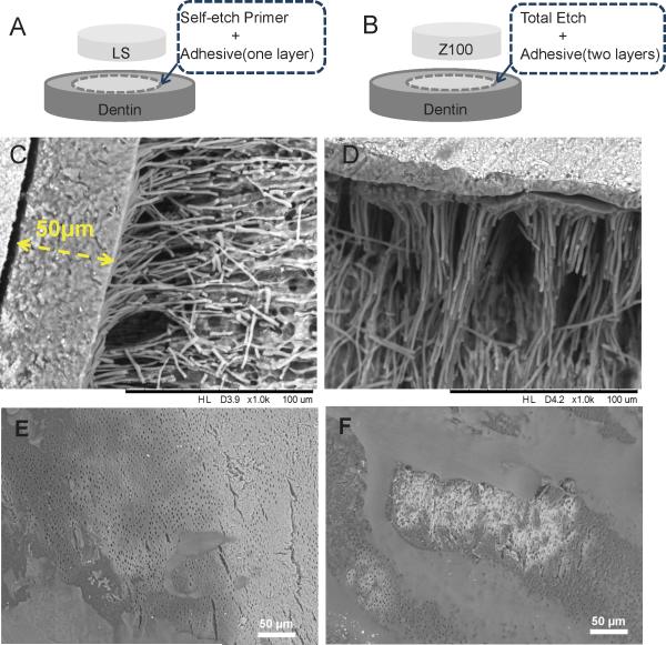
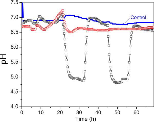
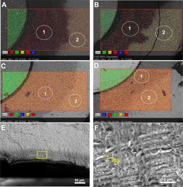
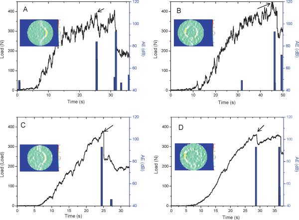
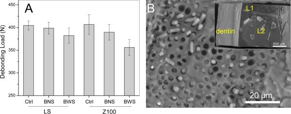
Oral biofilms can degrade the components in dental resin-based composite restorations, thus compromising marginal integrity and leading to secondary caries. This study investigates the mechanical integrity of the dentin-composite interface challenged with multi-species oral biofilms. While most studies used single-species biofilms, the present study used a more realistic, diverse biofilm model produced directly from plaques collected from donors with a history of early childhood caries. Dentin-composite disks were made using bovine incisor roots filled with Z100(TM) or Filtek(TM) LS (3M ESPE). The disks were incubated for 72 h in paired CDC biofilm reactors, using a previously published protocol. One reactor was pulsed with sucrose, and the other was not. A sterile saliva-only control group was run with sucrose pulsing. The disks were fractured under diametral compression to evaluate their interfacial bond strength. The surface deformation of the disks was mapped using digital image correlation to ascertain the fracture origin. Fracture surfaces were examined using scanning electron microscopy/energy-dispersive X-ray spectroscopy to assess demineralization and interfacial degradation. Dentin demineralization was greater under sucrose-pulsed biofilms, as the pH dropped <5.5 during pulsing, with LS and Z100 specimens suffering similar degrees of surface mineral loss. Biofilm growth with sucrose pulsing also caused preferential degradation of the composite-dentin interface, depending on the composite/adhesive system used. Specifically, Z100 specimens showed greater bond strength reduction and more frequent cohesive failure in the adhesive layer. This was attributed to the inferior dentin coverage by Z100 adhesive, which possibly led to a higher level of chemical and enzymatic degradation. The results suggested that factors other than dentin demineralization were also responsible for interfacial degradation. A clinically relevant in vitro biofilm model was therefore developed, which would effectively allow assessment of the degradation of the dentin-composite interface subjected to multi-species biofilm challenge.
Keywords: Bond strength; Dental restoration; Interfacial Degradation; Multi-species biofilm; Resin composite.
Copyright © 2013 Acta Materialia Inc. Published by Elsevier Ltd. All rights reserved.
Figures
References
-
- Bowen RL, Marjenhoff WA. Dental composites/glass ionomers: the materials. Adv Dent Res. 1992;6:44–49. - PubMed
-
- Bernardo M, Luis H, Martin MD, Leroux BG, Rue T, Leitao J, et al. Survival and reasons for failure of amalgam versus composite posterior restorations placed in a randomized clinical trial. J Am Dent Assoc. 2007;138:775–783. - PubMed
-
- Hickel R, Manhart J, Garcia-Godoy F. Clinical results and new developments of direct posterior restorations. Am J Dent. 2000;13:41D–54D. - PubMed
-
- Manhart J, Neuerer P, Scheibenbogen-Fuchsbrunner A, Hickel R. Three-year clinical evaluation of direct and indirect composite restorations in posterior teeth. J Prosthet Dent. 2000;84:289–296. - PubMed
Publication types
MeSH terms
Substances
Grants and funding
LinkOut - more resources
Full Text Sources
Other Literature Sources
Molecular Biology Databases
