

Discovery of an uncovered region in fibrin clots and its clinical significance
- PMID: 24008368
- PMCID: PMC3764439
- DOI: 10.1038/srep02604
Discovery of an uncovered region in fibrin clots and its clinical significance
Abstract
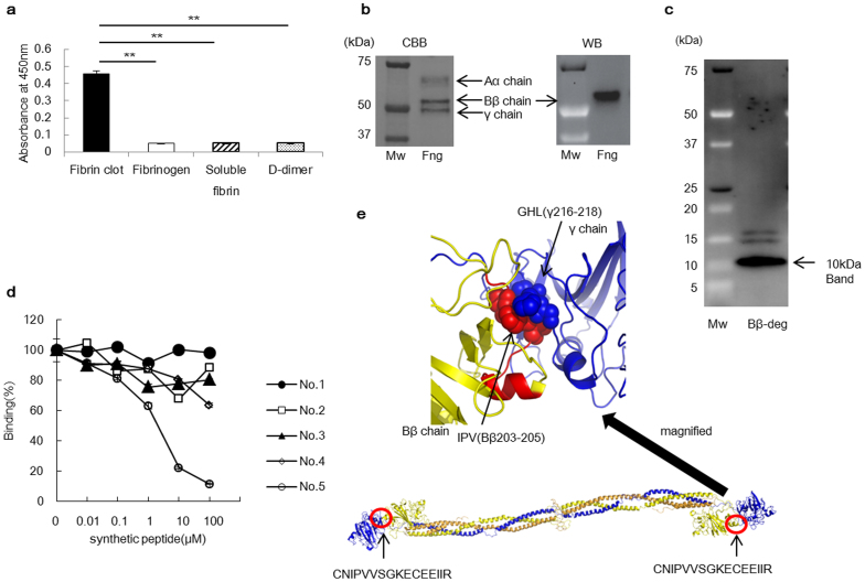
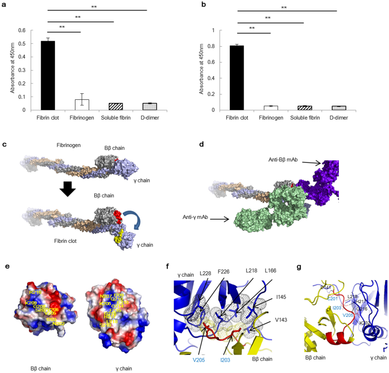
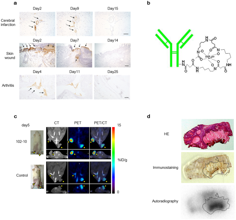
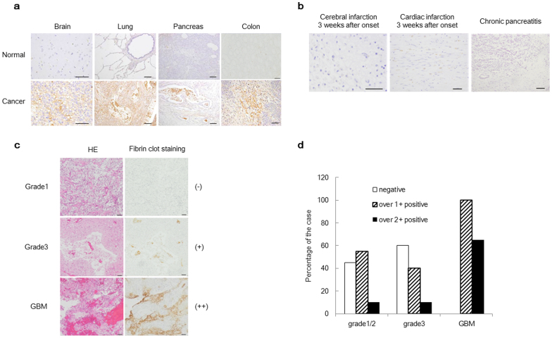
Despite the pathological importance of fibrin clot formation, little is known about the structure of these clots because X-ray and nuclear magnetic resonance (NMR) analyses are not applicable to insoluble proteins. In contrast to previously reported anti-fibrin monoclonal antibodies (mAbs), our anti-fibrin clot mAb (clone 102-10) recognises an uncovered region that is exposed only when a fibrin clot forms. The epitope of the 102-10 mAb was mapped to a hydrophobic region on the Bβ chain that interacted closely with a counterpart region on the γ chain in a soluble state. New anti-Bβ and anti-γ mAbs specific to peptides lining the discovered region appeared to bind exclusively to fibrin clots. Furthermore, the radiolabelled 102-10 mAb selectively accumulated in mouse spontaneous tumours, and immunohistochemistry using this mAb revealed greater fibrin deposition in World Health Organization (WHO) grade 4 glioma than in lower-grade gliomas. Because erosive tumours are apt to cause micro-haemorrhages, even early asymptomatic tumours detected with a radiolabelled 102-10 mAb may be aggressively malignant.
Figures
Similar articles
-
In vivo penetration of experimentally produced clots by monoclonal antibodies.Thromb Haemost. 2000 Jun;83(6):882-6. Thromb Haemost. 2000. PMID: 10896242
-
Accessibility of epitopes on fibrin clots and fibrinogen gels.Blood. 1991 Apr 1;77(7):1469-75. Blood. 1991. PMID: 1706954
-
Two monoclonal antibodies to D-dimer-specific inhibitors of fibrin polymerization.Thromb Res. 2004;113(3-4):251-9. doi: 10.1016/j.thromres.2004.03.005. Thromb Res. 2004. PMID: 15140590
-
Structural Aspects of the Allergen-Antibody Interaction.Front Immunol. 2020 Sep 2;11:2067. doi: 10.3389/fimmu.2020.02067. eCollection 2020. Front Immunol. 2020. PMID: 32983155 Free PMC article. Review.
-
Thrombin generation and fibrin clot structure.Blood Rev. 2007 May;21(3):131-42. doi: 10.1016/j.blre.2006.11.001. Epub 2007 Jan 8. Blood Rev. 2007. PMID: 17208341 Review.
Cited by
-
Enhancing the anticancer potential of metformin: fabrication of efficient nanospanlastics, in vitro cytotoxic studies on HEP-2 cells and reactome enhanced pathway analysis.Int J Pharm X. 2023 Oct 23;6:100215. doi: 10.1016/j.ijpx.2023.100215. eCollection 2023 Dec 15. Int J Pharm X. 2023. PMID: 38024451 Free PMC article.
-
Association between multiple coagulation-related factors and lymph node metastasis in patients with gastric cancer: A retrospective cohort study.Front Oncol. 2023 Feb 23;13:1099857. doi: 10.3389/fonc.2023.1099857. eCollection 2023. Front Oncol. 2023. PMID: 36910598 Free PMC article.
-
The role of molecular imaging in diagnosis of deep vein thrombosis.Am J Nucl Med Mol Imaging. 2014 Aug 15;4(5):406-25. eCollection 2014. Am J Nucl Med Mol Imaging. 2014. PMID: 25143860 Free PMC article. Review.
-
Coagulation markers as independent predictors of colorectal cancer aggressiveness.BMC Gastroenterol. 2025 Sep 2;25(1):634. doi: 10.1186/s12876-025-04249-4. BMC Gastroenterol. 2025. PMID: 40898085 Free PMC article.
-
A Novel and Potent Thrombolytic Fusion Protein Consisting of Anti-Insoluble Fibrin Antibody and Mutated Urokinase.Thromb Haemost. 2022 Jan;122(1):57-66. doi: 10.1055/a-1488-3723. Epub 2021 Jun 15. Thromb Haemost. 2022. PMID: 33882608 Free PMC article.
References
-
- Mosesson M. W. Fibrinogen and fibrin structure and functions. J Thromb Haemost 3, 1894–1904 (2005). - PubMed
-
- Pacella B. L. Jr, Hui K. Y., Haber E. & Matsueda G. R. Induction of fibrin-specific antibodies by immunization with synthetic peptides that correspond to amino termini of thrombin cleavage sites. Mol Immunol 20, 521–527 (1983). - PubMed
-
- Brass E. P., Forman W. B., Edwards R. V. & Lindan O. Fibrin formation: the role of the fibrinogen-fibrin monomer complex. Thromb Haemost 36, 37–48 (1976). - PubMed
-
- Graeff H., Hafter R. & von Hugo R. On soluble fibrinogen-fibrin complexes. Thromb Res 16, 575–576 (1979). - PubMed
-
- Drew A. F., Liu H., Davidson J. M., Daugherty C. C. & Degen J. L. Wound-healing defects in mice lacking fibrinogen. Blood 97, 3691–3698 (2001). - PubMed
Publication types
MeSH terms
Substances
LinkOut - more resources
Full Text Sources
Other Literature Sources
