

Variation in risk factors for recent small subcortical infarcts with infarct size, shape, and location
- PMID: 24008573
- PMCID: PMC3955807
- DOI: 10.1161/STROKEAHA.113.002227
Variation in risk factors for recent small subcortical infarcts with infarct size, shape, and location
Abstract
Background and purpose: Lacunar infarction is attributable to a perforating arteriolar abnormality. Possible causes include embolism, atheromatosis, or intrinsic disease. We examined whether the size, shape, or location of the lacunar infarct varied with embolic sources, systemic atheroma, or vascular risk factors.
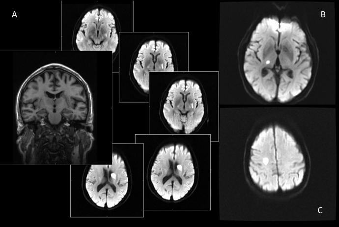
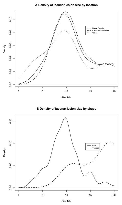
Methods: We examined data from 3 prospective studies of patients with clinical and diffusion-weighted imaging-positive symptomatic lacunar infarction who underwent full clinical assessment and investigation for stroke risk factors. Lacunar infarct sizes (maximum diameter; shape, oval/tubular; location, basal ganglia/centrum semiovale/brain stem) were coded blind to clinical details.
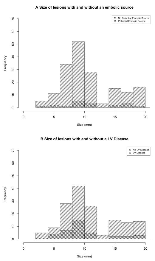
Results: Among 195 patients, 48 infarcts were tubular, 50 were 15 to 20 mm in diameter, and 97 and 74 were located in the basal ganglia and the centrum semiovale, respectively. There was no association between infarct size or shape and any of the risk factors. Centrum semiovale infarcts were less likely to have a potential relevant embolic source (4% versus 11%; odds ratio, 0.16; 95% confidence interval, 0.03-0.83) and caused a lower National Institute of Health Stroke Scale score (2 versus 3; odds ratio, 0.78; 95% confidence interval, 0.62-0.98) than basal ganglia infarcts. There were no other differences by infarct location.
Conclusions: Lacunar infarcts in the basal ganglia caused marginally severer strokes and were 3 times more likely to have a potential embolic source than those in the centrum semiovale, but the overall rate of carotid or known cardiac embolic sources (11%) was low. We found no evidence that other risk factors differed with location, size, or shape, suggesting that most lacunar infarcts share a common intrinsic arteriolar pathology.
Keywords: pathogenesis; pathology; stroke; stroke, lacunar.
Figures
References
-
- Fisher CM. Lacunar strokes and infarcts: a review. Neurology. 1982;32:871. - PubMed
-
- Caplan LR. Intracranial branch atheromatous disease: a neglected, understudied, and underused concept. Neurology. 1989 Sep;39(9):1246–50. - PubMed
-
- Ryu DW, Shon YM, Kim BS, Cho AH. Conglomerated beads shape of lacunar infarcts on diffusion-weighted MRI: what does it suggest? Neurology. 2012 May 1;78(18):1416–9. - PubMed
-
- Takase K, Murai H, Tasaki R, Miyahara S, Kaneto S, Shibata M, et al. Initial MRI findings predict progressive lacunar infarction in the territory of the lenticulostriate artery. Eur Neurol. 2011;65(6):355–60. - PubMed
-
- Del Bene A, Palumbo V, Lamassa M, Saia V, Piccardi B, Inzitari D. Progressive lacunar stroke: review of mechanisms, prognostic features, and putative treatments. Int J Stroke. 2012;7(4):321–9. - PubMed
Publication types
MeSH terms
Grants and funding
LinkOut - more resources
Full Text Sources
Other Literature Sources
Medical
