

Complications and management in Descemet's stripping endothelial keratoplasty: analysis of consecutive 430 cases
- PMID: 24008797
- PMCID: PMC4005239
- DOI: 10.4103/0301-4738.116484
Complications and management in Descemet's stripping endothelial keratoplasty: analysis of consecutive 430 cases
Abstract
Purpose: To analyze the complications and their managements in Descemet's stripping endothelial keratoplasty (DSEK) in consecutive 430 cases by single surgeon in a tertiary eye hospital.
Materials and methods: 430 eyes of 366 patients with endothelial dysfunctions scheduled for DSEK, were analyzed retrospectively. In all cases donor dissection was performed manually, and 'Taco' insertion and unfolding technique was used. Intra-operative and postoperative complications with their managements and outcomes were reviewed retrospectively. Periodic endothelial cell density was analyzed for each patient till the last visit. Follow-up period was between 3 to 60 months (mean 18.7 months).
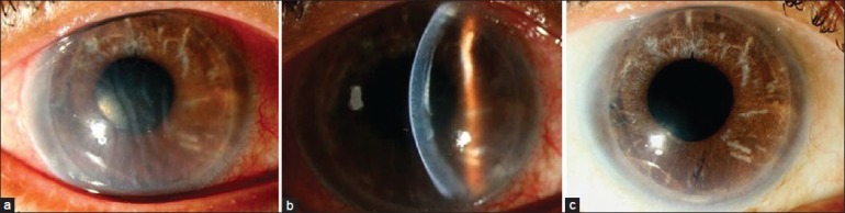
Results: 13 (3.0%) eyes had operative complications during donor dissection and 16 (3.7%) had during recipient procedure. In 7 (1.6%) eyes, donor lenticule was replaced with a new one during the surgery. In early postoperative period, 21 (4.9%) eyes had donor dislocation and 12 (2.8%) eyes had air-induced pupillary block; and they were managed immediately. 2 cases had primary graft failure and in 1 case had postoperative bacterial endophthalmitis requiring evisceration. In late postoperative period, 48 (11.3%) eyes had secondary glaucoma and 14 (3.3%) eyes had late secondary graft failure. Endothelial rejection occurred in 5 (1.2%) cases. Mean endothelial cell loss was 19.7% after 3 months and 54.2% after 5 years. Total graft failure in this series was 31 (7.2%) and in 17 cases re-DSEK was performed successfully.
Conclusions: Both operative and postoperative complications do occur in DSEK. Most of these complications can be managed by medical or appropriate surgical means. Some of the complications can be avoided and reduced with experience.
Conflict of interest statement
Figures


Similar articles
-
Outcomes of rebubbling for graft detachment after Descemet's stripping endothelial keratoplasty or Descemet's stripping automated endothelial keratoplasty.Indian J Ophthalmol. 2020 Jan;68(1):48-53. doi: 10.4103/ijo.IJO_1521_18. Indian J Ophthalmol. 2020. PMID: 31856465 Free PMC article.
-
Outcomes of Descemet's stripping endothelial keratoplasty in eyes with failed therapeutic penetrating keratoplasty.Acta Ophthalmol. 2014 Mar;92(2):167-70. doi: 10.1111/aos.12033. Epub 2013 Feb 7. Acta Ophthalmol. 2014. PMID: 23387367
-
Comparison of graft survival following penetrating keratoplasty and Descemet's stripping endothelial keratoplasty in eyes with a glaucoma drainage device.Int Ophthalmol. 2018 Feb;38(1):223-231. doi: 10.1007/s10792-017-0451-4. Epub 2017 Mar 16. Int Ophthalmol. 2018. PMID: 28303370
-
Clinical characteristics, risk factor analysis and outcomes in 61 eyes with graft rejection after descemet stripping endothelial keratoplasty, with a review of literature.Int Ophthalmol. 2024 Nov 11;44(1):423. doi: 10.1007/s10792-024-03339-8. Int Ophthalmol. 2024. PMID: 39523234 Review.
-
Descemet's stripping endothelial keratoplasty: safety and outcomes: a report by the American Academy of Ophthalmology.Ophthalmology. 2009 Sep;116(9):1818-30. doi: 10.1016/j.ophtha.2009.06.021. Epub 2009 Jul 30. Ophthalmology. 2009. PMID: 19643492 Review.
Cited by
-
Sheets glide-assisted versus Busin glide-assisted insertion techniques for descemet stripping endothelial keratoplasty (DSEK): A comparative analysis.Med J Armed Forces India. 2019 Oct;75(4):370-374. doi: 10.1016/j.mjafi.2018.02.007. Epub 2018 Apr 5. Med J Armed Forces India. 2019. PMID: 31719729 Free PMC article.
-
Post-corneal transplant Candida keratitis - Incidence and outcome.Indian J Ophthalmol. 2022 Feb;70(2):536-541. doi: 10.4103/ijo.IJO_560_21. Indian J Ophthalmol. 2022. PMID: 35086233 Free PMC article.
-
Dye-based identification of the orientation of tissue for Descemet stripping automated endothelial keratoplasty: A laboratory-based study.Indian J Ophthalmol. 2021 Jul;69(7):1741-1745. doi: 10.4103/ijo.IJO_2074_20. Indian J Ophthalmol. 2021. PMID: 34146018 Free PMC article.
-
Incidence and management of early postoperative complications in lamellar corneal transplantation.Graefes Arch Clin Exp Ophthalmol. 2023 Nov;261(11):3097-3111. doi: 10.1007/s00417-023-06073-6. Epub 2023 Apr 27. Graefes Arch Clin Exp Ophthalmol. 2023. PMID: 37103622 Free PMC article. Review.
-
Outcomes of Descemet stripping endothelial keratoplasty in cases of corneal endothelial dysfunction.Oman J Ophthalmol. 2022 Aug 3;15(3):337-341. doi: 10.4103/ojo.ojo_130_21. eCollection 2022 Sep-Dec. Oman J Ophthalmol. 2022. PMID: 36760963 Free PMC article.
References
-
- Patel SV. Keratoplasty for endothelial dysfunction. Ophthalmology. 2007;114:627–8. - PubMed
-
- Lee WB, Jacobs DS, Musch DC, Kaufman SC, Reinhart WJ, Shtein RM. Descemet's stripping endothelial keratoplasty: Safety and outcomes: A report by the American Academy of Ophthalmology. Ophthalmology. 2009;116:1818–30. - PubMed
-
- Suh LH, Yoo SH, Deobhakta A, Donaldson KE, Alfonso EC, Culbertson WW, et al. Complications of Descemet's stripping with automated endothelial keratoplasty: Survey of 118 eyes at one institute. Ophthalmology. 2008;115:1517–24. - PubMed
-
- Glasser DB. Tissue complications during endothelial keratoplasty. Cornea. 2010;29:1428–9. - PubMed
-
- Price FW, Jr, Price MO. Descemet's stripping with endothelial keratoplasty in 200 eyes: Early challenges and techniques to enhance donor adherence. J Cataract Refract Surg. 2006;32:411–8. - PubMed
Publication types
MeSH terms
LinkOut - more resources
Full Text Sources
Other Literature Sources
Medical
