

Repeat gas insufflation for successful closure of idiopathic macular hole following failed primary surgery
- PMID: 24008807
- PMCID: PMC4061686
- DOI: 10.4103/0301-4738.116452
Repeat gas insufflation for successful closure of idiopathic macular hole following failed primary surgery
Abstract
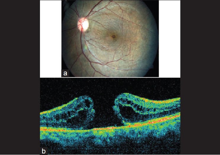
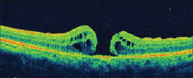
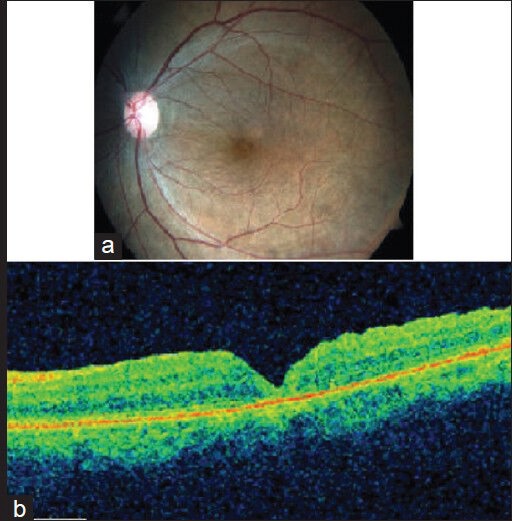
A 65-year-old lady presented with decreased vision in left eye since seven months. Vision was 6/9 in right eye and 6/36 in left. Examination revealed idiopathic, full-thickness macular hole in left eye; confirmed by optical coherence tomography (OCT). Patient underwent phacoemulsification with intraocular lens (IOL) implantation, vitrectomy, internal limiting membrane (ILM) peeling and 14% C ₃ F ₈ gas injection. OCT repeated after six weeks revealed type II closure with cuff of subretinal fluid. Four weeks later, patient underwent fluid-gas exchange with 14% C ₃ F ₈ gas and postoperative positioning. OCT was repeated after two weeks, which showed complete closure of the macular hole. OCT can help in selection of eyes for re-surgery that stand a better chance for hole closure. Macular holes with cuff of subretinal fluid are probably more likely to close on re-surgery than those without. However, larger studies with longer follow-up are required to validate this finding.
Conflict of interest statement
Figures
Comment in
-
Repeat gas insufflation for successful closure of idiopathic macular hole following failed primary surgery--our experience.Indian J Ophthalmol. 2014 Dec;62(12):1177. doi: 10.4103/0301-4738.149158. Indian J Ophthalmol. 2014. PMID: 25579370 Free PMC article. No abstract available.
-
Comment on Repeat gas insufflation for successful closure of idiopathic macular hole following failed primary surgery.Indian J Ophthalmol. 2015 Jan;63(1):79-80. doi: 10.4103/0301-4738.151494. Indian J Ophthalmol. 2015. PMID: 25686075 Free PMC article. No abstract available.
-
Author reply: To PMID 24008807.Indian J Ophthalmol. 2014 Dec;62(12):1178. doi: 10.4103/0301-4738.149159. Indian J Ophthalmol. 2014. PMID: 25723044 Free PMC article. No abstract available.
-
Author's response Comments on Repeat gas insufflation for successful closure of idiopathic macular hole following failed primary surgery.Indian J Ophthalmol. 2015 Jan;63(1):80-1. Indian J Ophthalmol. 2015. PMID: 25834855 Free PMC article. No abstract available.
-
Author response: Repeat gas insufflation for successful closure of idiopathic macular hole following failed primary surgery.Indian J Ophthalmol. 2015 Apr;63(4):364. doi: 10.4103/0301-4738.158110. Indian J Ophthalmol. 2015. PMID: 26044488 Free PMC article. No abstract available.
Similar articles
-
Value of internal limiting membrane peeling in surgery for idiopathic macular hole and the correlation between function and retinal morphology.Acta Ophthalmol. 2009 Dec;87 Thesis 2:1-23. doi: 10.1111/j.1755-3768.2009.01777.x. Acta Ophthalmol. 2009. PMID: 19912135 Clinical Trial.
-
Adjuvant methods in macular hole surgery: intraoperative plasma-thrombin mixture and postoperative fluid-gas exchange.Ophthalmic Surg Lasers. 2001 May-Jun;32(3):198-207. Ophthalmic Surg Lasers. 2001. PMID: 11371086
-
Is it necessary to cover the macular hole with the inverted internal limiting membrane flap in macular hole surgery? A case report.BMC Ophthalmol. 2015 Aug 26;15:115. doi: 10.1186/s12886-015-0104-1. BMC Ophthalmol. 2015. PMID: 26307540 Free PMC article.
-
Additional intravitreal gas injection in the early postoperative period for an unclosed macular hole treated with internal limiting membrane peeling.Retina. 2005 Feb-Mar;25(2):158-61. doi: 10.1097/00006982-200502000-00007. Retina. 2005. PMID: 15689805
-
Anatomical outcomes of surgery for idiopathic macular hole as determined by optical coherence tomography.Arch Ophthalmol. 2002 Jan;120(1):29-35. doi: 10.1001/archopht.120.1.29. Arch Ophthalmol. 2002. PMID: 11786054
Cited by
-
Author reply: To PMID 24008807.Indian J Ophthalmol. 2014 Dec;62(12):1178. doi: 10.4103/0301-4738.149159. Indian J Ophthalmol. 2014. PMID: 25723044 Free PMC article. No abstract available.
-
New Surgical Technique for Management of Recurrent Macular Hole.Middle East Afr J Ophthalmol. 2017 Jan-Mar;24(1):61-63. doi: 10.4103/meajo.MEAJO_211_15. Middle East Afr J Ophthalmol. 2017. PMID: 28546696 Free PMC article.
-
Efficacy of a simple intravitreal perfluoropropane injection in treating unclosed idiopathic macular holes following vitrectomy.BMC Ophthalmol. 2025 Feb 5;25(1):61. doi: 10.1186/s12886-024-03839-2. BMC Ophthalmol. 2025. PMID: 39910548 Free PMC article.
-
Retrospective study of changes in ocular coherence tomography characteristics after failed macular hole surgery and outcomes of fluid-gas exchange for persistent macular hole.Indian J Ophthalmol. 2018 Aug;66(8):1130-1135. doi: 10.4103/ijo.IJO_1119_17. Indian J Ophthalmol. 2018. PMID: 30038157 Free PMC article.
-
Parafoveal retinal massage combined with autologous blood cover in the management of giant, persistent or recurrent macular holes.Int J Ophthalmol. 2020 Nov 18;13(11):1773-1779. doi: 10.18240/ijo.2020.11.14. eCollection 2020. Int J Ophthalmol. 2020. PMID: 33215009 Free PMC article.
References
-
- Kelly NE, Wendel RT. Vitreous surgery for idiopathic macular holes. Results of a pilot study. Arch Ophthalmol. 1991;109:654–9. - PubMed
-
- Brooks J, Logan H. Macular hole surgery with and without internal limiting membrane peeling. Ophthalmology. 2000;107:1939–49. - PubMed
-
- Christmas NJ, Smiddy WE, Flynn HW., Jr Reopening of macular holes after initially successful repair. Ophthalmology. 1998;105:1835–8. - PubMed
-
- Sheidow TG, Blinder KJ, Holekamp N, Joseph D, Shah G, Grand MG, et al. Outcome results in macular hole surgery: An evaluation of internal limiting membrane peeling with and without indocyanine green. Ophthalmology. 2003;110:1697–701. - PubMed
-
- Tognetto D, Grandin R, Sanguinetti G, Minutola D, Di Nicola M, Di Mascio R, et al. Macular Hole Surgery Study Group. Internal limiting membrane removal during macular hole surgery: Results of A Multicenter Retrospective Study. Ophthalmology. 2006;113:1401–10. - PubMed
Publication types
MeSH terms
Substances
LinkOut - more resources
Full Text Sources
Other Literature Sources
