. 2015 Feb;54(2):134-47.
doi: 10.1002/mc.22081. Epub 2013 Sep 5.

Differential 12-O-Tetradecanoylphorbol-13-acetate-induced activation of rat mammary carcinoma susceptibility Fbxo10 variant promoters via a PKC-AP1 pathway

Affiliations

Differential 12-O-Tetradecanoylphorbol-13-acetate-induced activation of rat mammary carcinoma susceptibility Fbxo10 variant promoters via a PKC-AP1 pathway

Xin Xu et al. Mol Carcinog. 2015 Feb.

Abstract

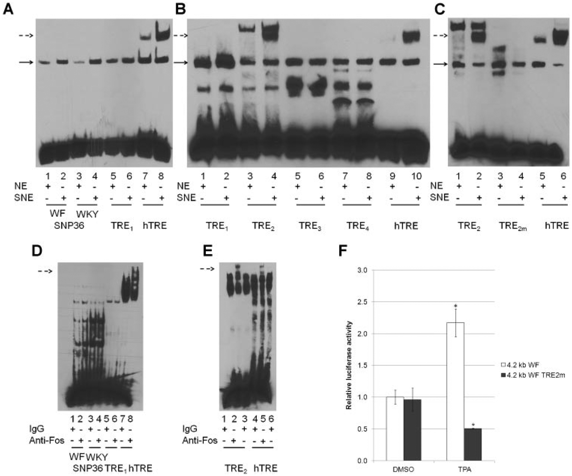
Rat mammary carcinoma susceptibility 5a1 (Mcs5a1), which is concordant to human MCS5A1 breast cancer risk locus, mediates susceptibility by a non-mammary cell-autonomous mechanism associated with T cell differential expression of F-box protein 10 (Fbxo10). Human FBXO10, an evolutionarily conserved ubiquitin ligase gene, was shown to have a potential role in regulating cell death by controlling the degradation of Bcl-2, a key protein involved in apoptosis. Breast cancer susceptibility is controlled by interactions between environmental and genetic factors; therefore, we sought to determine if breast cancer risk-associated environmental chemicals interact with Mcs5a1 variants using luciferase reporter constructs containing 4.2 kb Fbxo10 promoters based on alleles of mammary cancer susceptible Wistar Furth (WF) and resistant Wistar Kyoto (WKY) rat strains. 12-O-Tetradecanoylphorbol-13-acetate (TPA) induced activation of a 4.2 kb WF Fbxo10 promoter region, but lower levels of activation of the homologous WKY Fbxo10 promoter region. Using general and specific protein kinase inhibitors, we identified a protein kinase C (PKC) pathway that mediated TPA activation. We narrowed the possible PKCs to a member of the atypical PKC isoforms, namely PKCµ. We also determined that activator protein 1 (AP1) family member c-Fos mediated TPA activation of the 4.2 kb WF Fbxo10 promoter. TPA was shown to induce endogenous FBXO10 mRNA and FBXO10 protein in Jurkat cells, a human T cell line, with a maximal level of expression from 1.5 to 2.5 h after exposure. These results indicate that FBXO10/Fbxo10 expression is regulated by a PKC-dependent pathway acting through c-Fos, which binds AP1-specific DNA elements in Mcs5a1.

Keywords: F-box protein 10; activator protein 1; breast cancer susceptibility; mammary carcinoma susceptibility 5a1; protein kinase C.


Figures

Figure 1.
Figure 1.
A schematic of human MCS5A1 and the rat genomic region of Mcs5a1 used to make 4.2 kb Fbxo10 luciferase reporter constructs. (A) The human MCS5A1 locus is a 5.7 kb region of human chromosome 9 that contains four breast cancer risk associated polymorphisms (rs7042509, rs6476643, rs10758441, and indel138–9899, marked with black circles). (B) The rat Mcs5a1 locus is a 32 kb region of rat chromosome 5 that contains segments hypothesized to be 5′-Fbxo10 regulatory regions. Relative locations of genetic variants between mammary cancer susceptible WF and resistant WKY alleles are marked with black circles. Asterisks represent potential TRE (predicted AP1 binding sites). The lengths of all reporter constructs are shown and more detailed information regarding these variants is available in Table 1. Human and rat FBXO10/Fbxo10 conserved 5′-UTRs and an intronic segment are shown for reference. The human/rat conserved CpG islands contain FBXO10/Fbxo10 T cell preferred transcription start sites (TSS).
Figure 2.
Figure 2.
In vitro TPA activation of luciferase reporter genes containing a 5′-flanking region of either WF or WKY Fbxo10. TPA activated 4.2 kb WF and WKY Fbxo10 promoters in a time (A) and concentration (B) dependent manner. Jurkat cells were transfected with luciferase reporter genes containing either a WF (lighter bars) or WKY (darker bars) Fbxo10 segment and exposed to TPA for the time and concentrations indicated. Luciferase activities were measured in triplicate and results from triplicate samples are shown as average relative luciferase activity ± SD adjusted to the respective Renilla luciferase activity. Single or double asterisks indicate statistically significant (P ≤ 0.05) relative to DMSO control (no TPA exposure) or WF compared to WKY, respectively.
Figure 3.
Figure 3.
TPA activation of rat Fbxo10 luciferase reporter is PKC dependent and PKCμ is a potential activating isoform. (A) Jurkat cells were transfected with luciferase reporter plasmids containing either the WF (solid lines) or WKY (dashed lines) 4.2 kb Fbxo10 5′-flanking region. Twenty-four hours after transfection, cells were incubated with 1 μM TPA alone or TPA plus increasing concentrations of H-7 (squares), H-8 (triangles), or HA-1004 (circles) for 4 h. DMSO was used as a control. The results are expressed as average relative luciferase activity ± SD for three independent experiments performed in triplicate. (B) Jurkat cells transfected with WF (gray bars) or WKY (black bars) 4.2 kb Fbxo10 luciferase reporter vectors were incubated with either 1 μM TPA, 1 μM Thapsigargin (TG), TPA + TG, or 0.8 μg/mL 1,2-dioleoyl-sn-glycerol (DAG). (C) Jurkat cells transfected with WF (solid lines) or WKY (dashed lines) 4.2 kb Fbxo10 luciferase reporter were incubated with 1 μM TPA and TPA plus different concentrations of either GÖ6976 (squares) or GÖ6983 (circles) PKC inhibitors as indicated. Asterisks indicate P < 0.05 comparing inhibitor group to DMSO group.
Figure 4.
Figure 4.
c-Fos expression activated luciferase reporters containing a 5′-flanking region of either the 4.2 kb WF or WKY Fbxo10 gene in Jurkat cells. (A) Jurkat cells were transfected with 1 μg of luciferase reporter gene containing either the 5′-flanking regions of either the 4.2 kb WF (lighter black) or WKY (dark black) Fbxo10 genes and 50 ng pRL-TK plasmids plus either 1 μg c-Fos, c-Jun, NRF2, or combinations of c-Fos with either c-Jun or NRF2 as indicated. *Statistically significant (P < 0.001) as indicated. (B) The c-Jun (0.2, 0.5, and 1.0 μg) and control plasmids were transfected into Jurkat cells and TPA (1 μM) was added for 4 h. Statistically significant (*P = 0.016, **P = 0.006, and ***P = 0.019 as indicated). (C) The c-Fos (0.2, 0.5, and 1.0 μg) or control vectors were transfected into Jurkat cells; and TPA (1 μM) was added for 4 h. Statistically significant (*P < 0.02, **P = 0.002) as indicated). (D) An NRF2 (0.2, 0.5, and 1.0 μg) or empty vector was transiently transfected into Jurkat cells; TPA (1 μM) was added for 4 h. Statistically significant (*P < 0.02) as indicated). (E) The c-Maf (0.2, 0.5, or 1.0 μg) or control vector was transfected into Jurkat cells, and TPA (1 μM) was added for 4 h. Statistically significant (*P < 0.02) as indicated). (F) Luciferase reporters containing 4.2, 3.5, 1.8, or 0.7 kb WF or WKY rat strain Fbxo10 alleles were transfected into Jurkat cells with pRL-TK as a transfection control. One micromolar TPA was added for 4 h. X-Axis genotype by treatment subgroups are shown below each Fbxo10 segment group (4.2, 3.8, 1.8, and 0.7 kb). Asterisks represent P < 0.05 comparing the respective groups indicated by lines.
Figure 5.
Figure 5.
Identification of a TPA responsive element (TRE) in a 4.2 kb regulatory region of Fbxo10. (A) SNP36 WF or WKY, putative rat Mcs5a1 TRE-1 (TRE1) did not show common mobility shifts due to c-Fos antibody, as observed with the human collagenase TRE. EMSA was performed using TPA stimulated Jurkat cell nuclear extract (SNE) and control nuclear extract (NE) as described in methods. DNA–protein complexes that were increased after TPA treatment of Jurkat cells was observed for all SNP36 ds-oligonucleotides, Fbxo10 TRE1, and human collagenase TRE (solid arrow), but an additional slower moving DNA–protein complex was seen with the human TRE ds-oligo (dashed arrow). (B) Among selected rat Mcs5a1 TREs, only Mcs5a1 TRE-2 (TRE2) showed an electrophoretic mobility shift similar to that observed with the human collagenase TRE. The experiment was performed as described in (A). A dashed arrow marks the common shift between TRE2 and human TRE. (C) Oligonucleotides containing TRE2m, a mutation of the core AP1 binding TRE site, resulted in the loss of DNA–protein binding in EMSA. (D) SNP36 WF/WKY and TRE1 failed to show a supershift when anti-c-Fos was added to the reaction mixture, compared to the supershift observed with the human TRE during EMSA. A dashed arrow marks the common supershift between TRE2 and human TRE. (E) Mcs5a1 TRE-2 (TRE2) showed a supershift when anti-c-Fos is added, similar to the supershift observed with the human TRE during EMSA. Supershift was performed as described above. (F) The 4.2 kb WF Fbxo10 promoter harboring TRE2 mutation failed to respond to TPA stimulation. Jurkat cells were transfected with luciferase reporter genes containing either the 4.2 kb WF (white bars) or 4.2 kb WF with TRE2 mutation (gray bars) Fbxo10 5′-flanking region plus pRL-TK plasmids as a control. Cells were exposed to 1 μM TPA for 4 h. Luciferase activities were measured in triplicate, and results from triplicate samples are shown as average relative luciferase activity ± SD adjusted to the respective Renilla luciferase activity *P < 0.05.
Figure 6.
Figure 6.
TPA increased FBXO10 mRNA and protein levels in Jurkat cells. (A) TPA (10, 100, and 1000 nM) had no significant effect on FBXO10 mRNA levels 24 h after exposure. Jurkat cells were incubated with different concentrations of TPA indicated for 24 h. Cells were collected for RNA extraction prior to measuring relative FBXO10 mRNA levels by QPCR. (B) The effect of TPA (10, 100, and 1000 nM) on FBXO10 mRNA levels at different time points. *Statistically significant P < 0.05. (C) The effect of TPA (1.0 μM) on FBXO10 protein levels at different time points as indicated. Jurkat cells were treated with 1.0 μM TPA for times indicated and cells were collected for protein extraction and western blot analysis. Relative intensity of the FBXO10 protein was standardized relative to the intensity of GAPDH band using ImageJ.
Figure 7.
Figure 7.
A schematic of TPA-induced PKC-AP1 pathway regulation of 4.2 kb WF and WKY Fbxo10 promoters. Induction of PKCμ activity after TPA exposure results in increased c-Fos and an unidentified heterodimerization partner binding at Fbxo10-TRE (asterisk) in both WF and WKY 4.2 kb Fbxo10 promoters. Either one or both genetic variants marked by factor X? is hypothesized to be responsible for a difference in TPA-responsiveness of the WF and WKY 4.2 kb Fbxo10 promoters observed in this study. Black and white ellipses represent genetic variants between WF and WKY promoter sequences. Transcription factors c-Fos, an unidentified factor X heterodimerization partner, and possible coactivators are represented by squares, circles, and ovals, respectively. Potential differential DNA binding affinity between genetic variants is indicated by different factor X shape sizes.

References

    1. Siegel R, Naishadham D, Jemal A. Cancer statistics. CA Cancer J Clin 2012;62:10–29. - PubMed
    1. Hindorff LA, Gillanders EM, Manolio TA. Genetic architecture of cancer and other complex diseases: lessons learned and future directions. Carcinogenesis 32:945–954. - PMC - PubMed
    1. Bond GL. Hu W. Bond EE, et al. A single nucleotide polymorphism in the MDM2 promoter attenuates the p53 tumor suppressor pathway and accelerates tumor formation in humans. Cell 2004;119:591–602. - PubMed
    1. Meyer KB, Maia AT, O’Reilly M, et al. Allele-specific up-regulation of FGFR2 increases susceptibility to breast cancer. PLoS Biol 2008;6:e108. - PMC - PubMed
    1. Wu AH, Wan P, Hankin J, et al. Adolescent and adult soy intake and risk of breast cancer in Asian-Americans. Carcinogenesis 2002;23:1491–1496. - PubMed

Publication types

MeSH terms