Review
. 2013 Mar-Apr;2(2):213-27.
doi: 10.1002/wdev.71. Epub 2012 May 29.

Neural tube defects--disorders of neurulation and related embryonic processes

Affiliations
Review

Neural tube defects--disorders of neurulation and related embryonic processes

Andrew J Copp et al. Wiley Interdiscip Rev Dev Biol. 2013 Mar-Apr.

Abstract

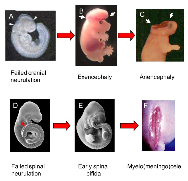
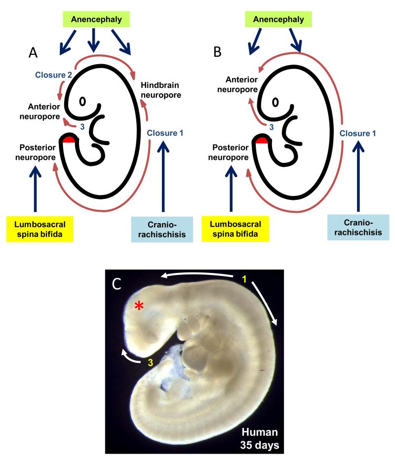
Neural tube defects (NTDs) are severe congenital malformations affecting 1 in every 1000 pregnancies. 'Open' NTDs result from failure of primary neurulation as seen in anencephaly, myelomeningocele (open spina bifida), and craniorachischisis. Degeneration of the persistently open neural tube in utero leads to loss of neurological function below the lesion level. 'Closed' NTDs are skin-covered disorders of spinal cord structure, ranging from asymptomatic spina bifida occulta to severe spinal cord tethering, and usually traceable to disruption of secondary neurulation. 'Herniation' NTDs are those in which meninges, with or without brain or spinal cord tissue, become exteriorized through a pathological opening in the skull or vertebral column (e.g., encephalocele and meningocele). NTDs have multifactorial etiology, with genes and environmental factors interacting to determine individual risk of malformation. While over 200 mutant genes cause open NTDs in mice, much less is known about the genetic causation of human NTDs. Recent evidence has implicated genes of the planar cell polarity signaling pathway in a proportion of cases. The embryonic development of NTDs is complex, with diverse cellular and molecular mechanisms operating at different levels of the body axis. Molecular regulatory events include the bone morphogenetic protein and Sonic hedgehog pathways which have been implicated in control of neural plate bending. Primary prevention of NTDs has been implemented clinically following the demonstration that folic acid (FA), when taken as a periconceptional supplement, can prevent many cases. Not all NTDs respond to FA, however, and adjunct therapies are required for prevention of this FA-resistant category.


Figures

Figure 1
Figure 1. Natural history of open cranial (A-C) and spinal (D-F) NTDs in mice.
After an initial failure of neural tube closure in either the midbrain (A) or low spine (D), the neuroepithelium continues to proliferate and undergoes neuronal differentiation, appearing to protrude from the surface of the embryo (B,E). This is termed ‘exencephaly’ in cranial lesions. With continued gestation, the exposed neuroepithelium becomes damaged by continuous exposure to the amniotic fluid. Apoptosis and necrosis intervene so that, by the time of birth, the neuroepithelium has degenerated, yielding the phenotype of anencephaly (C) or myelocele (D). Developmental stages indicated as E (embryonic day) or P (postnatal day). Figures modified from: Dunlevy et al, 2006, FEBS Letters 580, 2803-7 (A); Copp et al, 2003, Nature Reviews Genetics 4, 784–793 (B); Copp, 2005, Journal of Anatomy 207, 623-35 (C); Van Straaten & Copp, 2001, Anatomy and Embryology, 203, 225-237 (D,E); Stiefel et al, 2003, Journal of Neurosurgery (Spine) 99, 206–213 (F).
Figure 2
Figure 2. Diagrammatic representation of the main events of neural tube closure in mouse (A) and human (B) embryos.
The main types of NTD resulting from failure of closure at different levels of the body axis are indicated. The red shading on the tail bud indicates the site of secondary neurulation in both species. Disturbance of this process leads to closed spina bifida. In each species the initial de novo closure event (Closure 1) occurs at the hindbrain/cervical boundary and closure spreads bidirectionally from this site. In the mouse, a second de novo closure site (Closure 2) occurs at the forebrain/midbrain boundary with closure also spreading rostrally and caudally. Closure 2 does not appear to occur in human embryos (B). A third de novo initiation event (Closure 3) occurs in both species at the rostral extremity of the neural plate, with closure spreading caudally from here. Hence, in mice, closure is completed sequentially at the anterior neuropore, hindbrain neuropore and posterior neuropore. In humans, owing to the lack of Closure 2, there are likely to be only two neuropores: anterior and posterior. (C) Human embryo aged 35 days post-fertilisation from the Human Developmental Biology Resource (www.hdbr.org). Neurulation has recently been completed in the low spinal region. The positions of Closures 1 and 3, and the directions of closure are marked. Note the relatively small size of the midbrain (red asterisk) in this human embryo compared with the mouse embryo (Figure 1D,E) which, in evolution, may have rendered Closure 2 an unnecessary

Similar articles

Cited by

References

    1. Dolk H, Loane M, Garne E. The prevalence of congenital anomalies in Europe. Adv Exp Med Biol. 2010;686:349–364. - PubMed
    1. Malcoe LH, Shaw GM, Lammer EJ, Herman AA. The effect of congenital anomalies on mortality risk in white and black infants. Am J Public Health. 1999;89:887–892. - PMC - PubMed
    1. Mitchell LE. Epidemiology of neural tube defects. Am J Med Genet C Semin Med Genet. 2005;135:88–94. - PubMed
    1. Wood LR, Smith MT. Generation of anencephaly: 1. Aberrant neurulation and 2. Conversion of exencephaly to anencephaly. J Neuropath exp Neurol. 1984;43:620–633. - PubMed
    1. Seller MJ. Sex, neural tube defects, and multisite closure of the human neural tube. Am J Med Genet. 1995;58:332–336. - PubMed

Publication types