. 2013 Sep;4(9):1399-415.
doi: 10.18632/oncotarget.1202.

Repression of c-Kit by p53 is mediated by miR-34 and is associated with reduced chemoresistance, migration and stemness

Affiliations

Repression of c-Kit by p53 is mediated by miR-34 and is associated with reduced chemoresistance, migration and stemness

Helge Siemens et al. Oncotarget. 2013 Sep.

Abstract

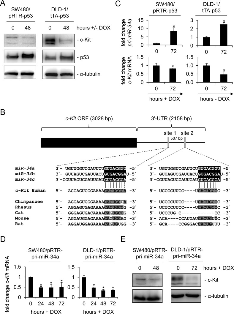
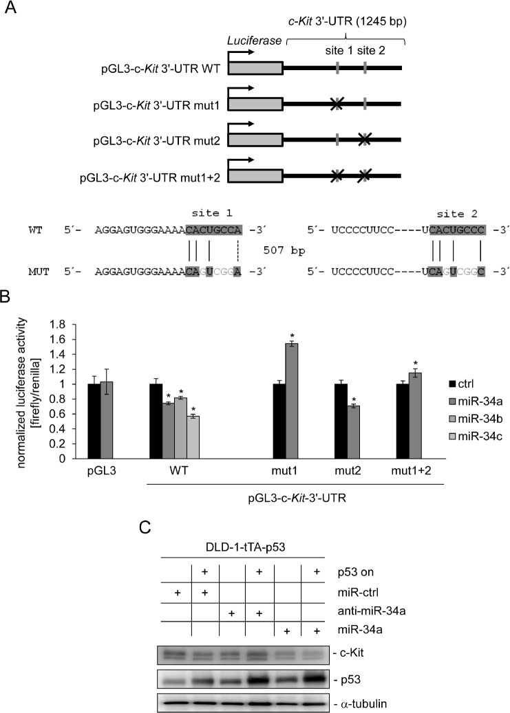
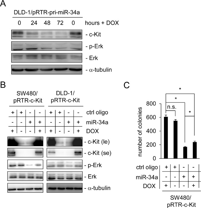
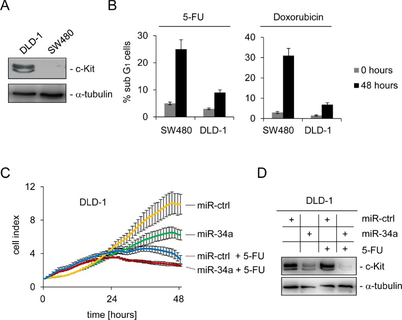
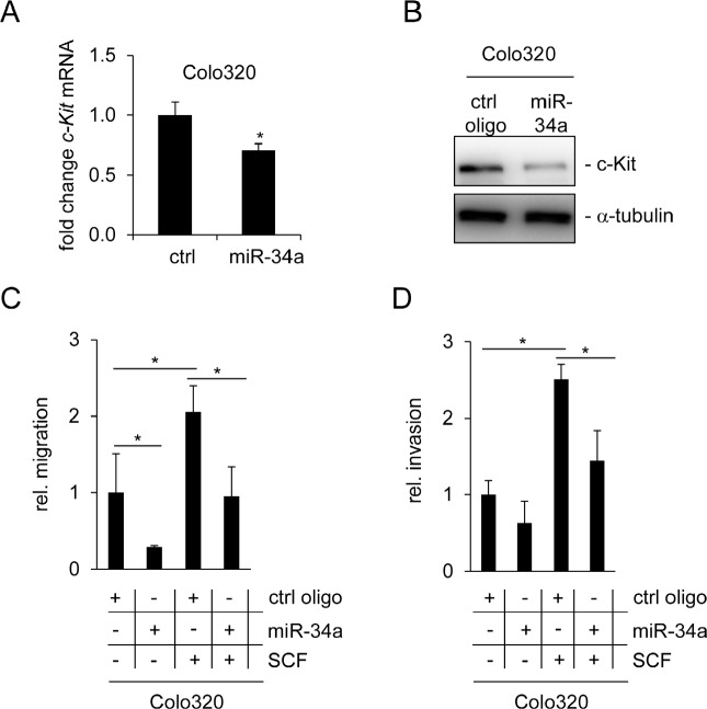
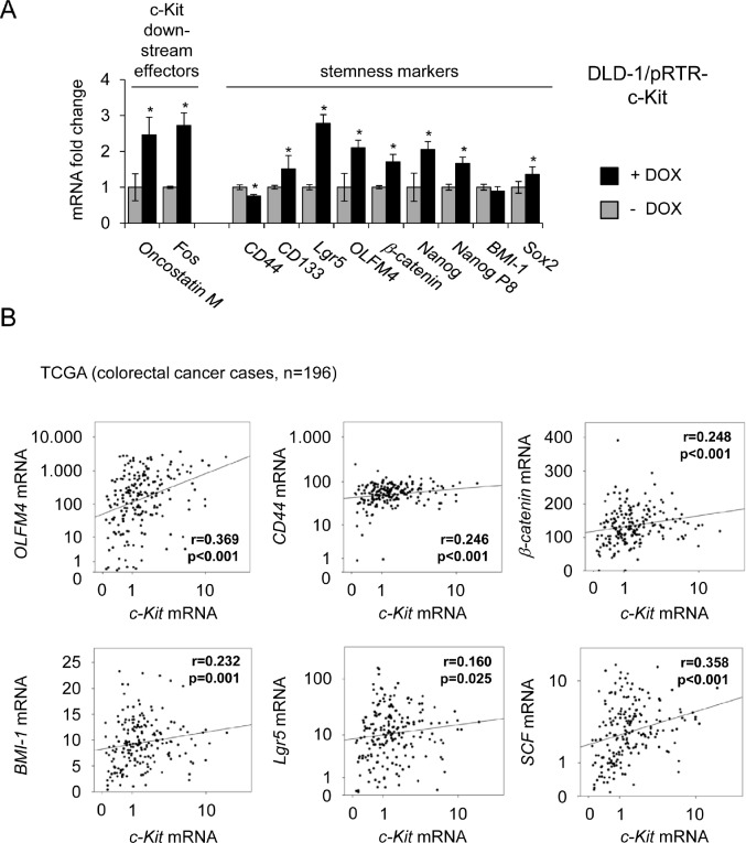
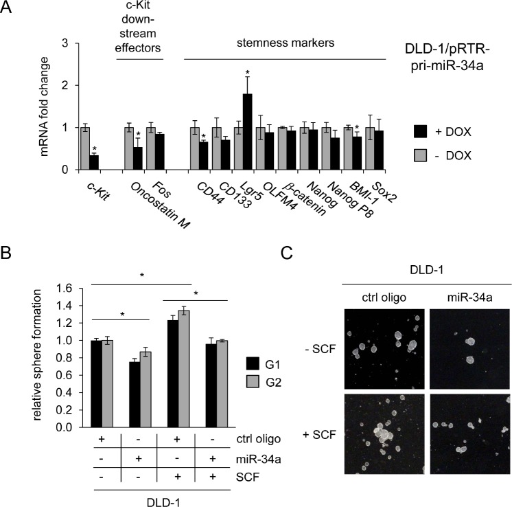
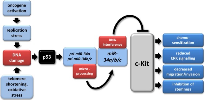
The c-Kit receptor tyrosine kinase is commonly over-expressed in different types of cancer. p53 activation is known to result in the down-regulation of c-Kit. However, the underlying mechanism has remained unknown. Here, we show that the p53-induced miR-34 microRNA family mediates repression of c-Kit by p53 via a conserved seed-matching sequence in the c-Kit 3'-UTR. Ectopic miR-34a resulted in a decrease in Erk signaling and transformation, which was dependent on the down-regulation of c-Kit expression. Furthermore, ectopic expression of c-Kit conferred resistance of colorectal cancer (CRC) cells to treatment with 5-fluorouracil (5-FU), whereas ectopic miR-34a sensitized the cells to 5-FU. After stimulation with c-Kit ligand/stem cell factor (SCF) Colo320 CRC cells displayed increased migration/invasion, whereas ectopic miR-34a inhibited SCF-induced migration/invasion. Activation of a conditional c-Kit allele induced several stemness markers in DLD-1 CRC cells. In primary CRC samples elevated c-Kit expression also showed a positive correlation with markers of stemness, such as Lgr5, CD44, OLFM4, BMI-1 and β-catenin. On the contrary, activation of a conditional miR-34a allele in DLD-1 cells diminished the expression of c-Kit and several stemness markers (CD44, Lgr5 and BMI-1) and suppressed sphere formation. MiR-34a also suppressed enhanced sphere-formation after exposure to SCF. Taken together, our data establish c-Kit as a new direct target of miR-34 and demonstrate that this regulation interferes with several c-Kit-mediated effects on cancer cells. Therefore, this regulation may be potentially relevant for future diagnostic and therapeutic approaches.


Figures

Figure 1
Figure 1. c-Kit is repressed after ectopic p53 and miR-34a expression in colorectal cancer cell lines
(A) Western blot analysis of c-Kit protein levels after induction of p53 in colorectal cancer cell lines SW480 and DLD-1. α-tubulin served as a loading control. (B) Scheme of the c-Kit mRNA and conservation of the putative miR-34 seed-matching sequences, which are represented as grey vertical bars. Detailed sequences of the two sites and phylogenetic homologies are shown below. Potential base pairing is shaded in grey. (C) qPCR analysis of pri-miR-34a and c-Kit mRNA levels in the colorectal cancer cell lines SW480 and DLD-1 after induction of p53 by addition or withdrawal of doxycycline (DOX) for 72 hours. Results were normalized to β-actin mRNA. (D) qPCR analysis of c-Kit mRNA in the colorectal cancer cell lines SW480 and DLD-1 carrying the inducible pRTR/pri-miR-34a vector after addition of doxycycline. E) Detection of c-Kit protein by Western blot analysis after induction of pri-miR-34a in the indicated cells. α-tubulin served as a loading control. C+D: results represent the mean +/−S.D. (n=3).
Figure 2
Figure 2. miR-34a directly targets c-Kit and mediates c-Kit repression by p53
(A) Scheme of constructs used for dual luciferase assays. The positions of the putative miR-34 seed-matching sequences in the c-Kit 3'-UTR are depicted as a grey vertical bars, their mutations as crosses. Sequences of the respective targeted mutations are given below. (B) Dual reporter assay in SW480 cells transfected with miR-34a/b/c mimics or control oligonucleotide and the indicated 3'-UTR-reporter constructs for the human c-Kit 3'-UTR. Data are represented as mean ± SD (n = 3). (C) DLD-1/tTA-p53 cells were either transfected with a control oligonucleotide, miR-34a or an antagomiR directed against miR-34a for 24 hours either in the presence or absence of DOX (without or with ectopic p53). Expression of the indicated proteins was detected by Western blot analysis. α-tubulin served as a loading control.
Figure 3
Figure 3. Ectopic c-Kit overrides miR-34a-dependent inhibition of Erk signaling and colony formation in soft agar
(A) DLD-1/pRTR-pri-miR-34a cells were treated with DOX for the indicated time-points. The indicated proteins were detected by Western blot analysis. α-tubulin served as a loading control. (B) Western blot analysis of the indicated SW480 and DLD-1 cells after oligonucleotide transfection for 48 hours and addition of DOX for 24 hours. α-tubulin served as a loading control. le = long exposure, se = short exposure. (C) Soft agar colony formation assay. The indicated cells were treated as described under (B). Results represent the mean +/−S.D. (n=3) and significance was calculated applying a Student's t-test.” * “: p < 0.05.
Figure 4
Figure 4. c-Kit and miR-34a modulate the apoptotic response to chemotherapeutic agents
(A) Detection of endogenous c-Kit levels by Western blot analysis of DLD-1 and SW480 colorectal cancer cells. Detection of α-tubulin served as loading control. (B) Cells were treated with either 5-FU or Doxorubicin for 48 hours and subjected to DNA content analysis by flow cytometry. Results represent the mean +/−S.D. (n=3). (C) DLD-1 cells were transfected with the indicated oligonucleotides and simultaneously treated with 5-FU or water. The cell index, which corresponds to cell proliferation, was determined by real-time impedance measurements. (D) Cells treated as described in (C) were subjected to Western blot analysis. α-tubulin served as loading control.
Figure 5
Figure 5. miR-34a inhibits basal and SCF-induced migration and invasion of Colo320 cells
(A) Colo320 CRC cells were transfected either with a control (ctrl) or a miR-34a oligonucleotide for 48 hours. qPCR analysis was used to determine c-Kit mRNA levels. (B) Detection of c-Kit protein levels by Western blot analysis 48 hours after oligonucleotide transfection. α-tubulin served as loading control. (C) Colo320 cells were transfected with the indicated oligonucleotides and simultaneously treated with SCF (or water) for 24 hours. Thereafter cells were seeded into Boyden chambers to determine migration. (D) Determination of invasion using a Boyden chamber assay. Cells were treated as in (C). (A,C,D) Results represent the mean +/−S.D. (n=3) and significance was calculated applying a Student's t-test.” * “: p < 0.05.
Figure 6
Figure 6. Ectopic c-Kit induces expression of stemness markers and correlates with their expression in primary CRC
(A) DLD-1/pRTR-c-Kit cells were treated with DOX for 48 hours. Expression of the indicated mRNAs was determined by qPCR. Results represent the mean +/−S.D. (n=3).” * “: p < 0.05. (B) mRNA expression data derived from primary CRC samples (n = 196) were obtained from the public database TCGA [54]. Correlation coefficients and p-values were calculated applying the Spearman correlation algorithm. Scatter plots show the respective correlations. Both, the c-Kit levels on the x-axes and the y-axes of the OLFM4, CD44, Lgr5 and SCF mRNA expression values are provided as log10 scale.
Figure 7
Figure 7. Role of the miR-34a/c-Kit axis in the regulation of stemness markers and sphere formation
(A) DLD-1 cells pRTR/pri-miR-34a were treated with DOX for 48 hours. Expression of the indicated mRNAs was analyzed by qPCR. (B) DLD-1 cells were transfected with the indicated oligonucleotides and treated with SCF (or water) and subjected to a sphere formation assay. Sphere numbers were determined after seven days for the first generation (G1) and seven days after seeding for G2. Treatment with oligonucleotides and SCF was repeated when cells were passaged. (C) Representative pictures of DLD-1 derived G1 spheres, magnification: 40x. (A,B) Results represent the mean +/−S.D. (n=3) and significance was calculated applying a Student's t-test.” * “: p < 0.05.
Figure 8
Figure 8. The p53/miR-34/c-Kit axis
The model depicts multiple tumor suppressive effects of miR-34a directly targeting c-Kit which were identified in this study. In tumors, p53 mutation/inactivation or CpG methylation of miR-34a/b/c promoters may abrogate this pathway [33, 91, 92]. miR-34a/b/c have multiple other targets besides c-Kit [33, 93]. The model was adapted from [94].

Similar articles

Cited by

References

    1. Besmer P, Murphy JE, George PC, Qiu FH, Bergold PJ, Lederman L, Snyder HW, Jr, Brodeur D, Zuckerman EE, Hardy WD. A new acute transforming feline retrovirus and relationship of its oncogene v-kit with the protein kinase gene family. Nature. 1986;320(6061):415–421. - PubMed
    1. Yarden Y, Kuang WJ, Yang-Feng T, Coussens L, Munemitsu S, Dull TJ, Chen E, Schlessinger J, Francke U, Ullrich A. Human proto-oncogene c-kit: a new cell surface receptor tyrosine kinase for an unidentified ligand. EMBO J. 1987;6(11):3341–3351. - PMC - PubMed
    1. Anderson DM, Lyman SD, Baird A, Wignall JM, Eisenman J, Rauch C, March CJ, Boswell HS, Gimpel SD, Cosman D, et al. Molecular cloning of mast cell growth factor, a hematopoietin that is active in both membrane bound and soluble forms. Cell. 1990;63(1):235–243. - PubMed
    1. Lennartsson J, Ronnstrand L. Stem Cell Factor Receptor/c-Kit: From Basic Science to Clinical Implications. Physiol Rev. 2012;92(4):1619–1649. - PubMed
    1. Rothschild G, Sottas CM, Kissel H, Agosti V, Manova K, Hardy MP, Besmer P. A role for kit receptor signaling in Leydig cell steroidogenesis. Biol Reprod. 2003;69(3):925–932. - PubMed

Publication types

MeSH terms