. 2013 Dec;231(4):457-65.
doi: 10.1002/path.4256.

Breast stromal fibroblasts from histologically normal surgical margins are pro-carcinogenic

Affiliations
Free PMC article

Breast stromal fibroblasts from histologically normal surgical margins are pro-carcinogenic

Maha A Al-Rakan et al. J Pathol. 2013 Dec.
Free PMC article

Abstract

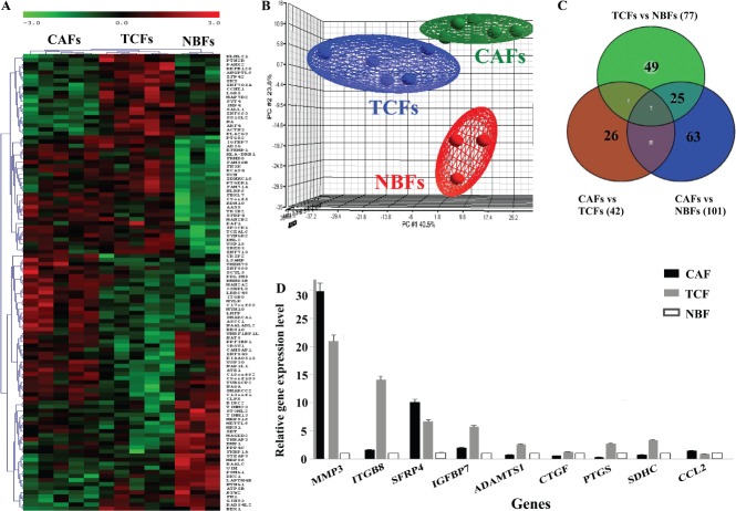
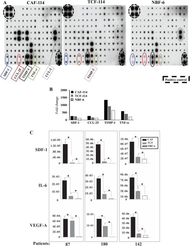
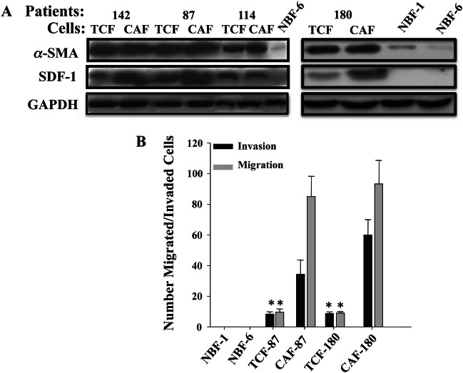
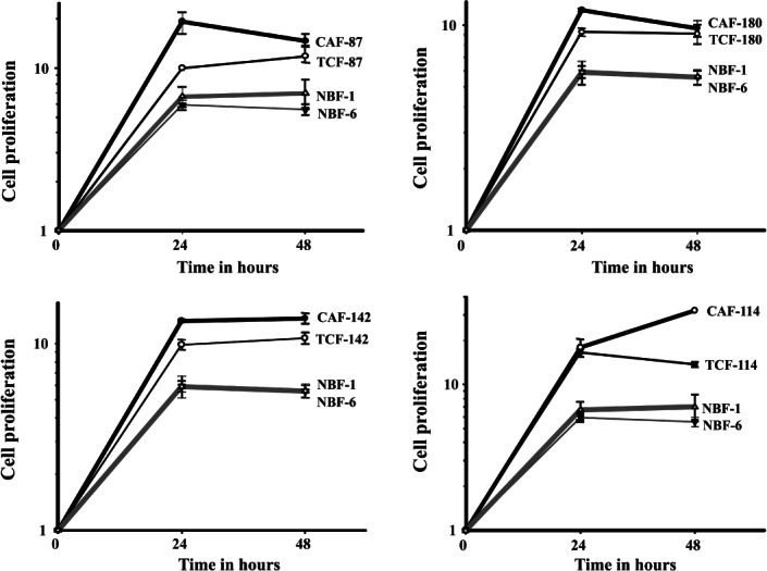
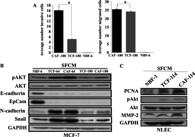
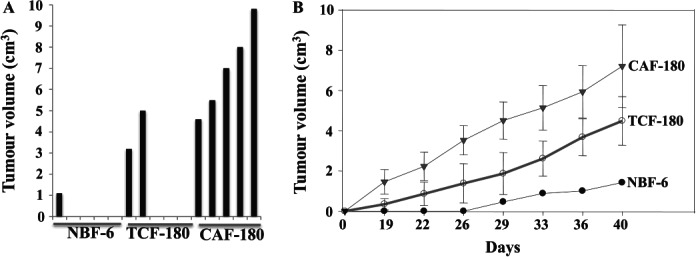
There is evidence that normal breast stromal fibroblasts (NBFs) suppress tumour growth, while cancer-associated fibroblasts (CAFs) promote tumourigenesis through functional interactions with tumour cells. Little is known about the biology and the carcinogenic potential of stromal fibroblasts present in histologically normal surgical margins (TCFs). Therefore, we first undertook gene expression analysis on five CAF/TCF pairs from breast cancer patients and three NBF samples (derived from mammoplasties). This comparative analysis revealed variation in gene expression between these three categories of cells, with a TCF-specific gene expression profile. This variability was higher in TCFs than in their paired CAFs and also NBFs. Cytokine arrays show that TCFs have a specific secretory cytokine profile. In addition, stromal fibroblasts from surgical margins expressed high levels of α-SMA and SDF-1 and exhibited higher migratory/invasiveness abilities. Indirect co-culture showed that TCF cells enhance the proliferation of non-cancerous mammary epithelial cells and the epithelial-to-mesenchymal transition of breast cancer cells. Moreover, TCF and CAF cells increased the level of PCNA, MMP-2 and the phosphorylated/activated form of Akt in normal breast luminal fibroblasts in a paracrine manner. Furthermore, TCFs were able to promote the formation and growth of humanized orthotopic breast tumours in nude mice. Interestingly, these TCF phenotypes and the extent of their effects were intermediate between those of NBFs and CAFs. Together, these results indicate that stromal fibroblasts located in non-cancerous tissues exhibit a tumour-promoting phenotype, indicating that their presence post-surgery may play important roles in cancer recurrence.

Keywords: breast cancer; epithelial-to-mesenchymal transition; gene expression; stromal fibroblasts.


Figures

Figure 1
Figure 1
Differential gene expression between CAFs, TCFs and NBFs. Total RNA from five CAFs, their corresponding TCFs as well as three NBFs was used to perform genome-wide gene expression analysis. (A) Unsupervised 2D hierarchical clustering of significantly varying genes across the three groups was performed using Pearson's correlation with average linkage clustering (only the top 112 of the most significantly dysregulated genes are shown for readability). (B) The three dominant PCA components that contained about 70% of the variance in the data matrix clearly distinguished samples as CAF, TCF and NBF. (C) Venn diagram showing differential gene expression between the groups. (D) qRT–PCR data for the indicated genes (mean ± SD)
Figure 2
Figure 2
TCF cells secrete high levels of pro-carcinogenic cytokines. SFCMs from the indicated cells were collected and applied on the human cytokine antibody array map. (A) Cytokine array maps highlighting the differentially expressed cytokines. (B) Quantification of the array data for selected cytokines. (C) SFCMs were used to assess the levels of the indicated proteins by ELISA in CAF/TCF pairs from three patients compared to NBF-6 (mean ± SD; *p < 0.005)
Figure 3
Figure 3
TCFs are active fibroblasts. (A) Immunoblotting for α-SMA and SDF-1 of the indicated cell lysates. (B) Invasion/migration assays. The numbers of invaded/migrated cells are presented in the histogram (mean ± SD; *p < 0.005 for TCFs compared to NBF-1 and NBF-6; note that CAFs are also significantly different)
Figure 4
Figure 4
TCF-conditioned medium enhances the proliferation of mammary epithelial cells. MCF10A cells were cultured in SFCMs from the indicated fibroblasts from four patients and proliferation measured at the indicated times using the WST-1 assay, compared to SFCMs from the two NBF cultures performed at the same time. Error bars represent ± SD
Figure 5
Figure 5
TCF cells enhance the migration/invasion of breast cancer cells and promote EMT. (A) SCFMs from the indicated cells were added into the lower compartments of 24-well BD BioCoat plates. MCF-7 cells (105) were seeded onto the upper compartment of the plates and incubated for 24 h. The numbers of invaded and migrated cells are presented (mean ± SD; *p < 0.005). (B, C) MCF-7 or normal luminal epithelial cells (NLEC) were treated with the indicated SFCMs for 24 h and cell lysates were used for immunoblotting with the indicated antibodies
Figure 6
Figure 6
TCFs enhance the formation and the growth of human breast tumour orthotopic xenografts in mice. Breast cancer xenografts were created by co-injecting MDA-MB-231 cells with NBF-6, TCF-180 or CAF-180 cells under the nipples of nude mice. (A) Tumour size 40 days post-injection. (B) Time-dependent tumour growth (mean ± SD)

Similar articles

Cited by

References

    1. Arendt LM, Rudnick JA, Keller PJ, et al. Stroma in breast development and disease. Semin Cell Dev Biol. 2009;21::11–18. - PMC - PubMed
    1. Polyak K, Kalluri R. The role of the microenvironment in mammarY gland development and cancer. Cold Spring Harb Perspect Biol. 2010 2010 doi: 10.1101/cshperspect.a003244. - DOI - PMC - PubMed
    1. Aboussekhra A. Role of cancer-associated fibroblasts in breast cancer development and prognosis. Int J Dev Biol. 2011;55::841–849. - PubMed
    1. Polanska UM, Orimo A. Carcinoma-associated fibroblasts: non-neoplastic tumour-promoting mesenchymal cells. J Cell Physiol. 2013;228::1651–1657. - PubMed
    1. Place AE, Jin Huh S, Polyak K. The microenvironment in breast cancer progression: biology and implications for treatment. Breast Cancer Res. 2011;13:227. - PMC - PubMed

MeSH terms