. 2014 Jul;51(4):846-57.
doi: 10.1177/0300985813501335. Epub 2013 Sep 5.

Phenotypic characterization of the KK/HlJ inbred mouse strain

Affiliations

Phenotypic characterization of the KK/HlJ inbred mouse strain

A Berndt et al. Vet Pathol. 2014 Jul.

Abstract

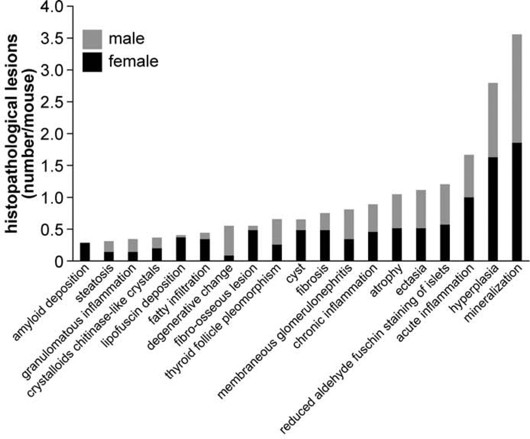
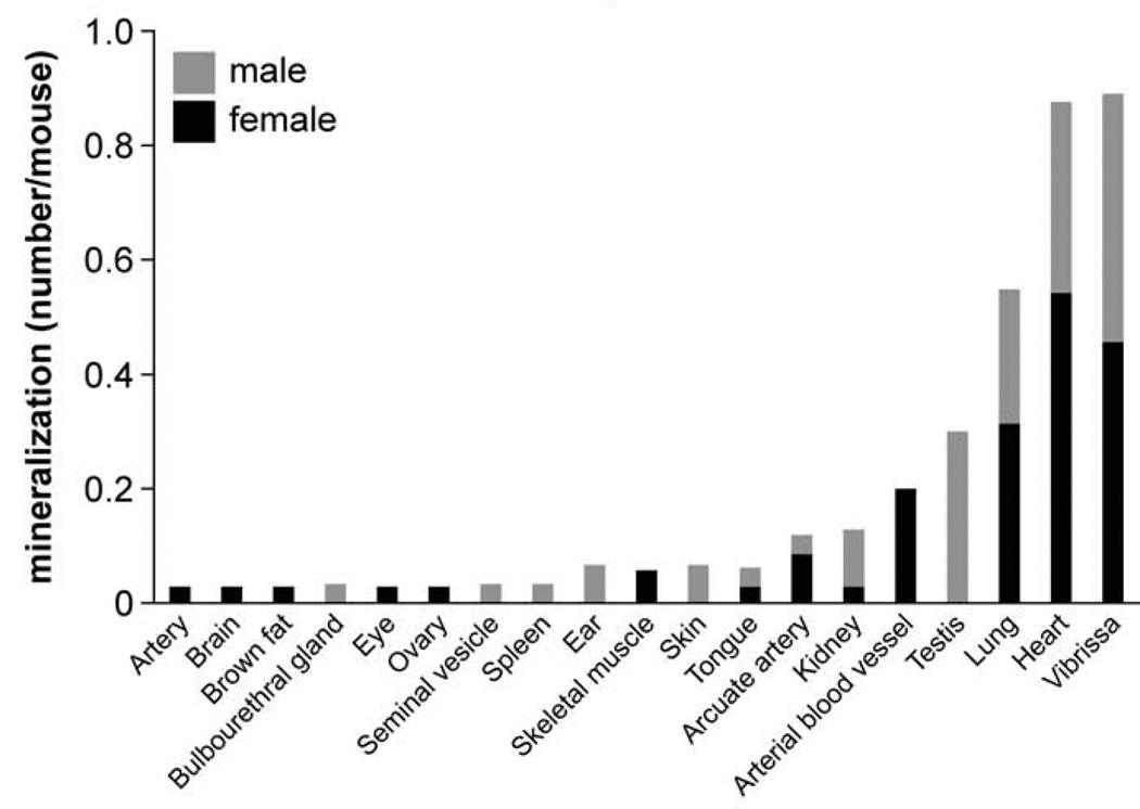
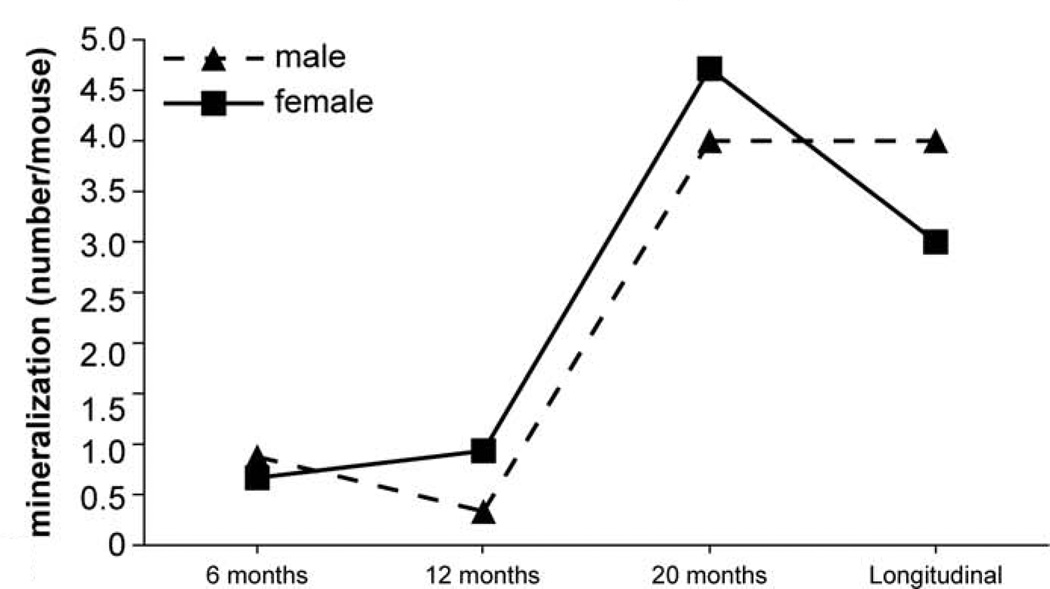
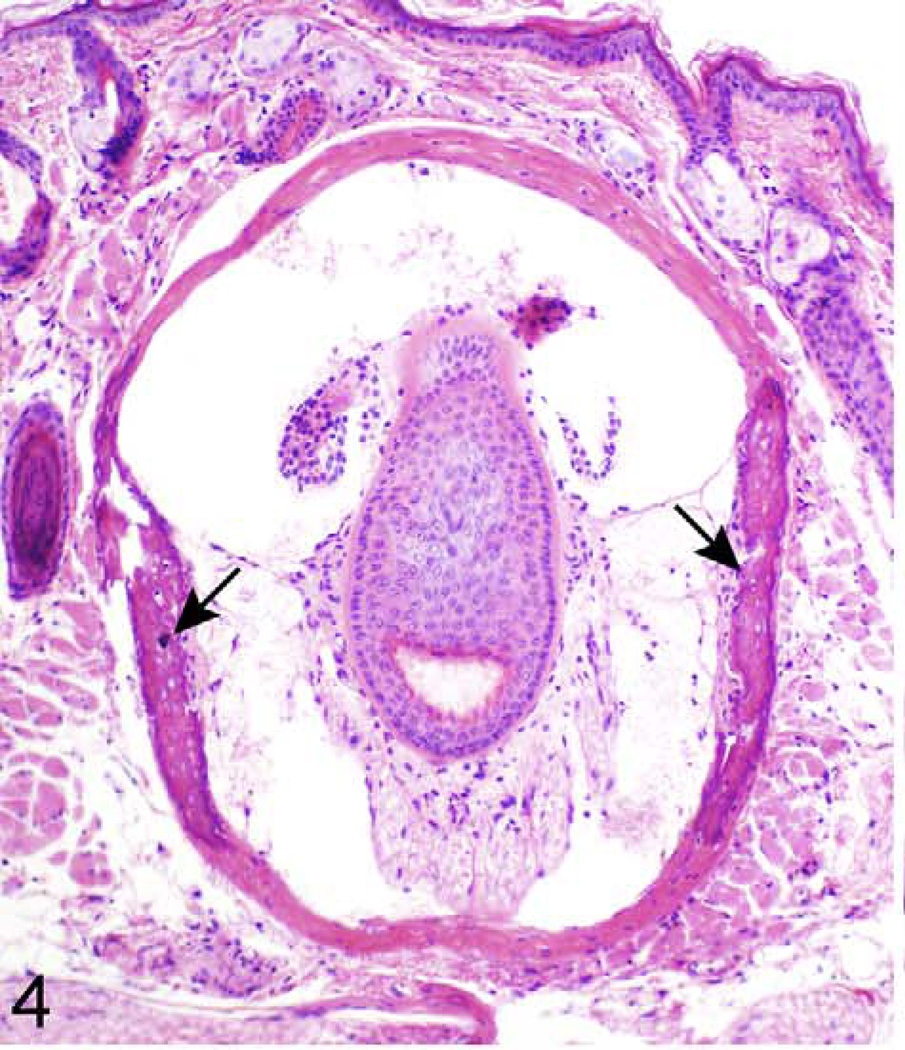
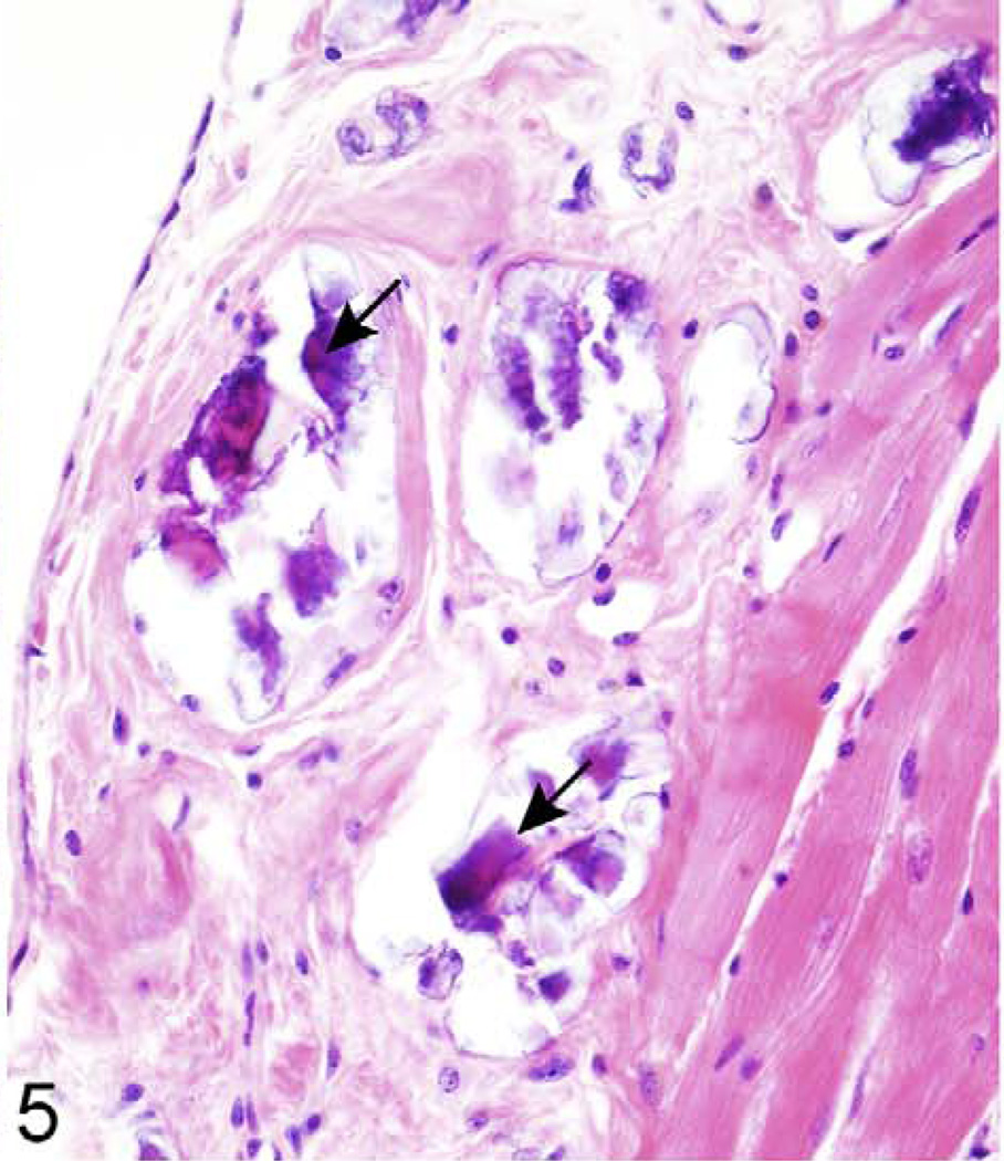
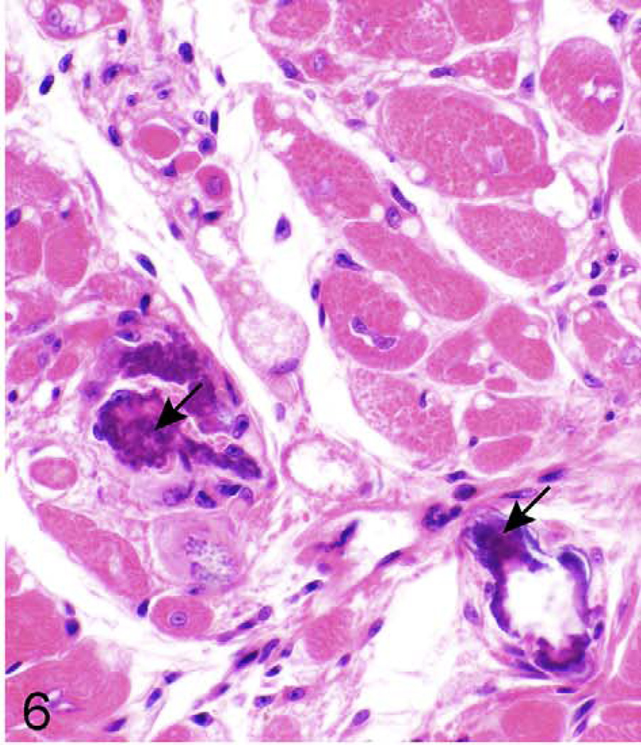
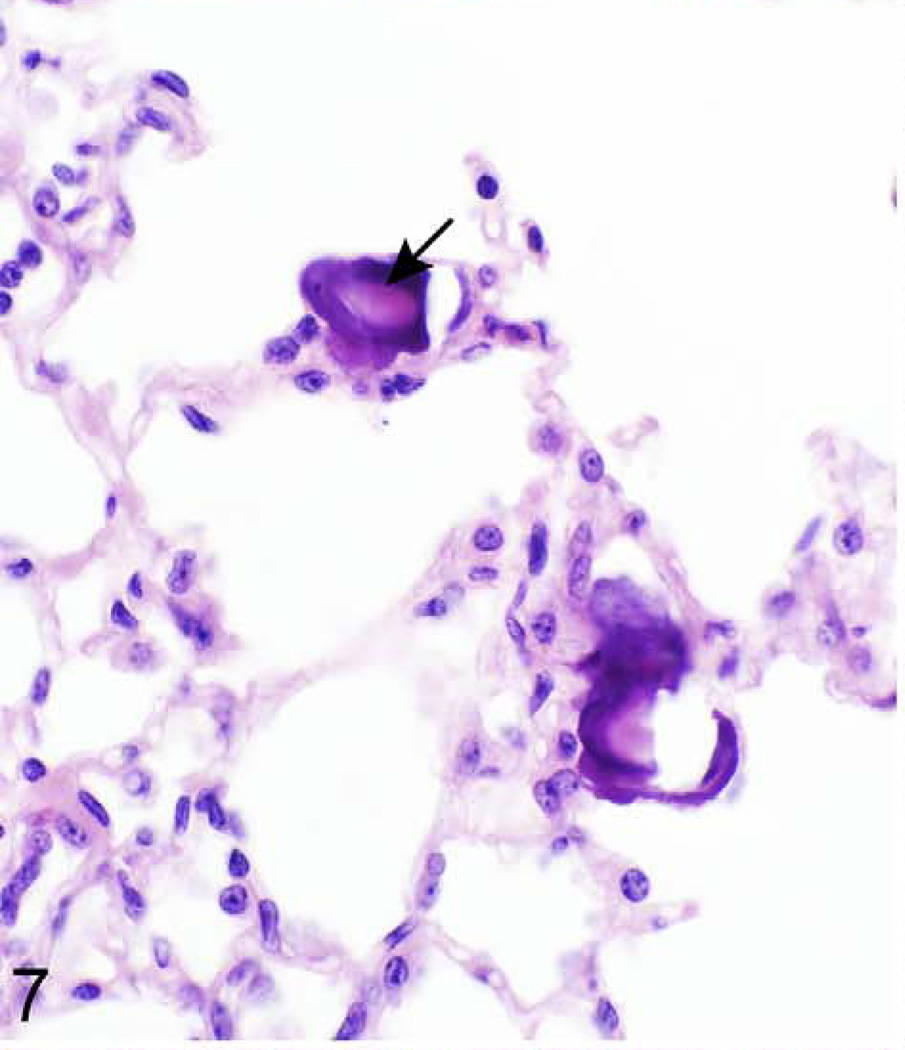
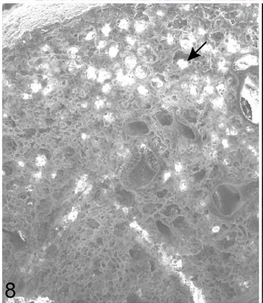
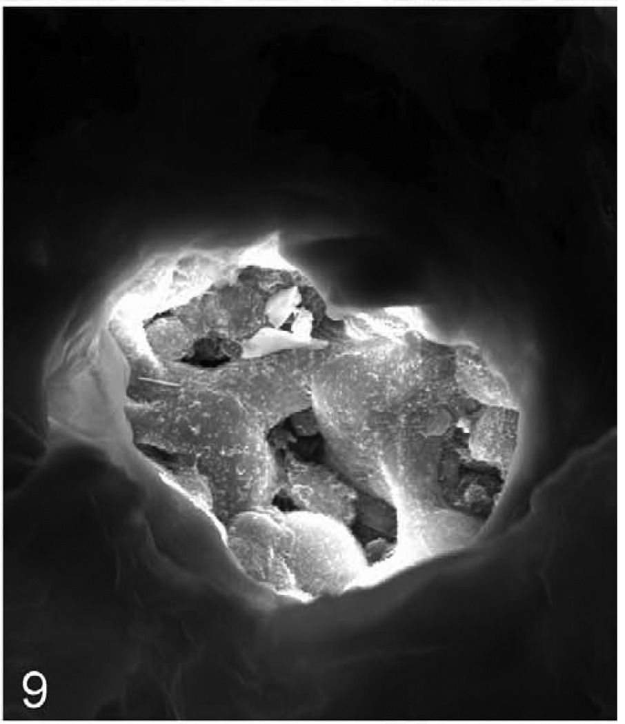
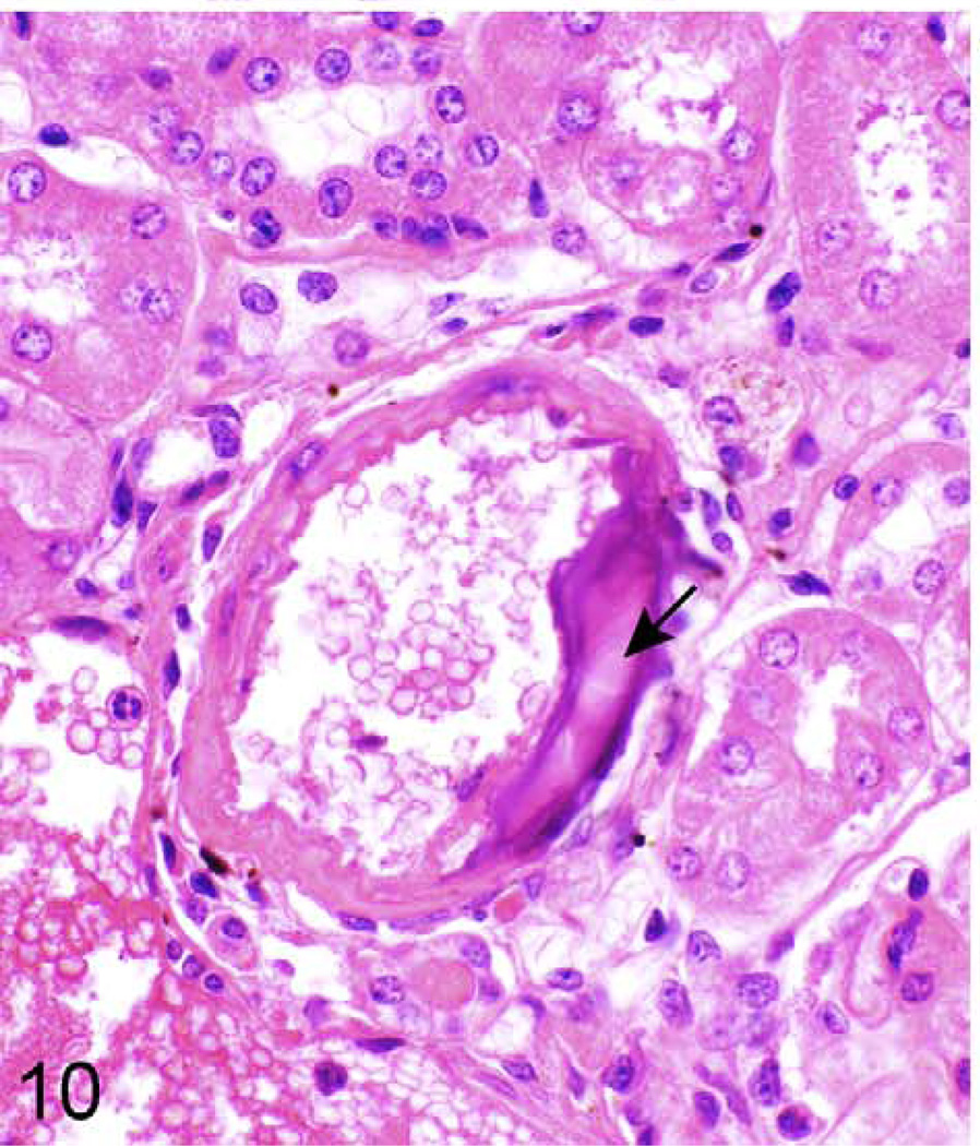
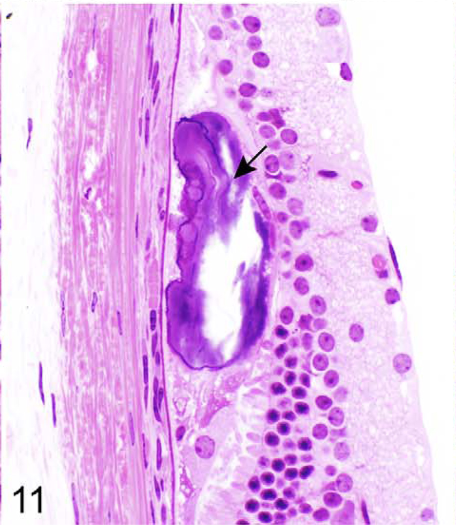
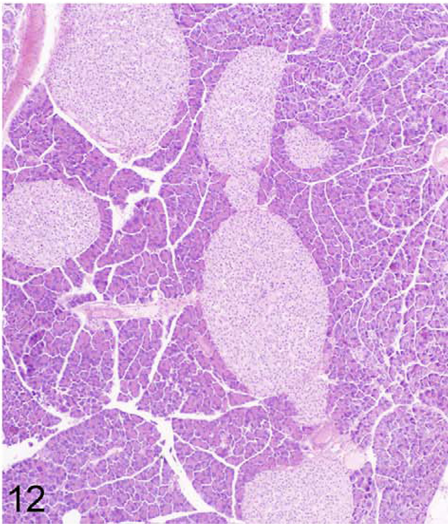
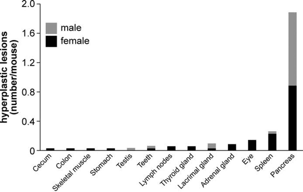
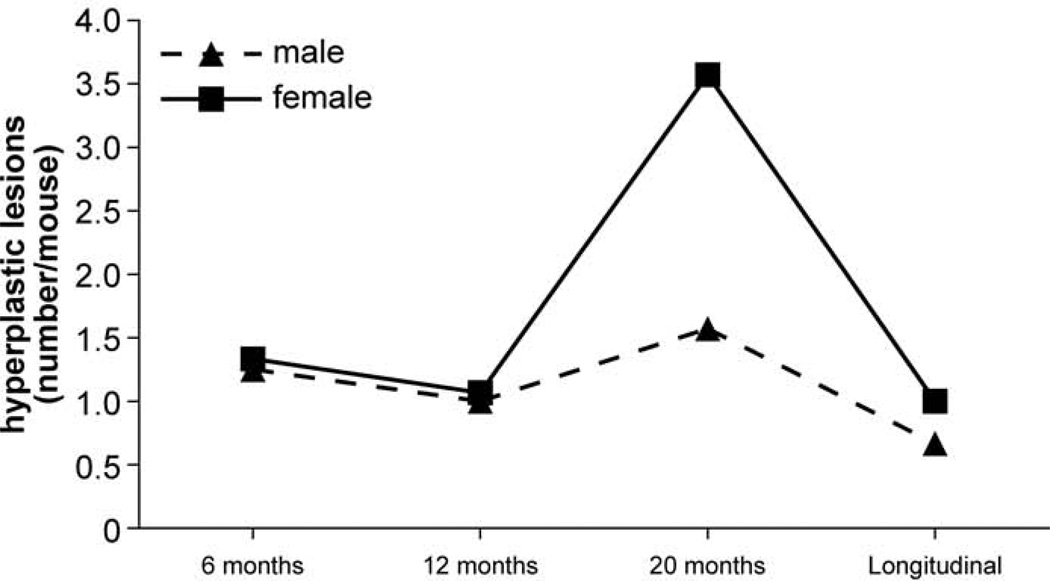
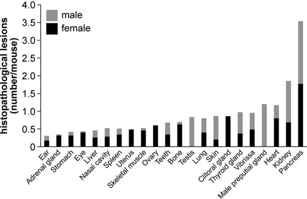
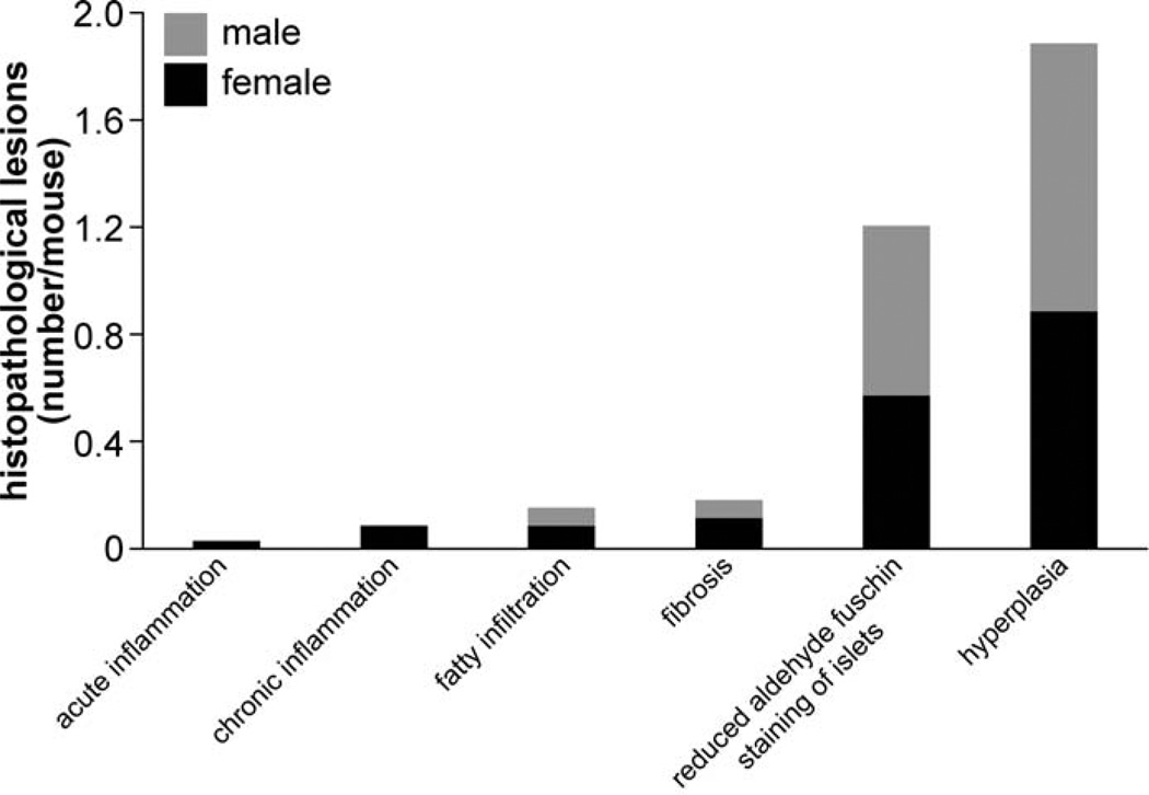
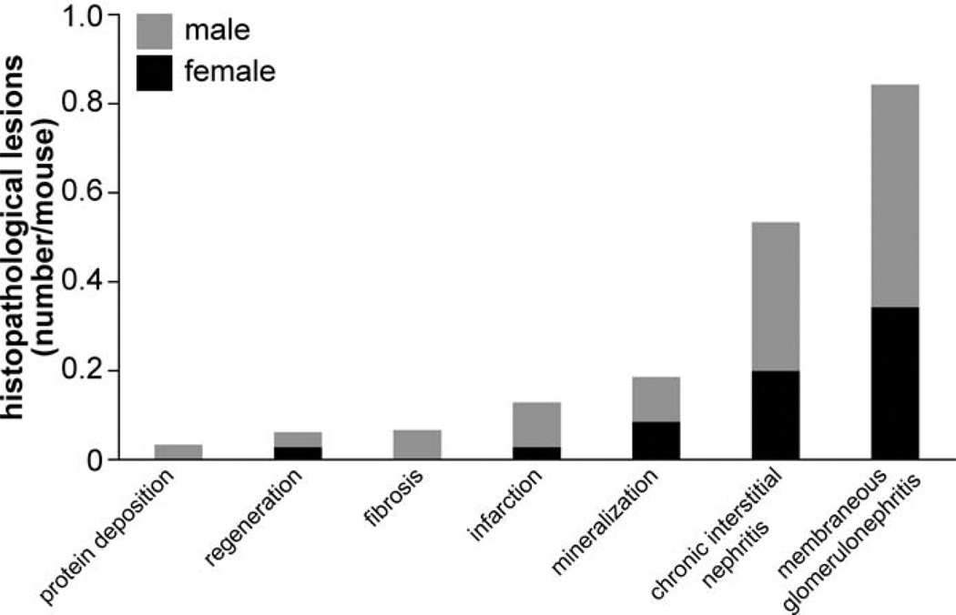
Detailed histopathological diagnoses of inbred mouse strains are important for interpreting research results and defining novel models of human diseases. The aim of this study was to histologically detect lesions affecting the KK/HlJ inbred strain. Mice were examined at 6, 12, and 20 months of age and near natural death (ie, moribund mice). Histopathological lesions were quantified by percentage of affected mice per age group and sex. Predominant lesions were mineralization, hyperplasia, and fibro-osseous lesions. Mineralization was most frequently found in the connective tissue dermal sheath of vibrissae, the heart, and the lung. Mineralization was also found in many other organs but to a lesser degree. Hyperplasia was found most commonly in the pancreatic islets, and fibro-osseous lesions were observed in several bones. The percentage of lesions increased with age until 20 months. This study shows that KK/HlJ mice demonstrate systemic aberrant mineralization, with greatest frequency in aged mice. The detailed information about histopathological lesions in the inbred strain KK/HlJ can help investigators to choose the right model and correctly interpret the experimental results.

Keywords: KK/HlJ mice; PXE model; ectopic mineralization; systemic mineralization; vibrissae dermal sheath.


Conflict of interest statement

Declaration of Conflicting Interests

The author(s) declared no potential conflicts of interest with respect to the research, authorship, and/or publication of this article.

Figures

Figure 1
Figure 1
Type and frequency of histopathological lesions across all organs. Bars show the additive number of processes for female (black) and male (gray) mice.
Figure 2
Figure 2
Frequency of mineralization lesions in different organs. Bars show the additive number of lesions for female (black) and male (gray) mice.
Figure 3
Figure 3
Frequency of mineralization lesions at different ages. The solid line represents females and dashed line represents males.
Figure 4
Figure 4
Muzzle skin, vibrissa; 624-day-old female KK/HlJ mouse, case No. 1. There is mineralization of the connective tissue sheath of vibrissae (arrows). Hematoxylin and eosin (HE).
Figure 5
Figure 5
Right ventricle, epicardium; 624-day-old female KK/HlJ mouse, case No. 2. Epicardial fibrosis and mineralization (arrows) are a prominent feature in the right ventricular free wall of the heart. HE.
Figure 6
Figure 6
Myocardium, left ventricle; 624-day old-KK/HlJ female mouse, case No. 2. Mineralization (arrows) with minimal fibrosis is evident in the heart. HE.
Figure 7
Figure 7
Lung; 624-dayold female KK/HlJ mouse, case No. 2. Multiple foci of mineralization are present within the alveolar septa (arrow). HE.
Figure 8
Figure 8
Lung; 624-day-old female KK/HlJ mouse; case No. 2. Horizontal plane of lung illustrating mineralization foci in the alveoli (arrow). Gold sputter coat, scanning electron microscopy (SEM).
Figure 9
Figure 9
Lung; 624-day-old female KK/HlJ mouse, case No. 2. Higher magnification of focus marked with an arrow in Figure 8 to illustrate the mineralization. Gold sputter coat, SEM.
Figure 10
Figure 10
Kidney (arcuate artery); 624-day-old female KK/HlJ mouse, case No. 3. Mineralization (arrow) of the arterial wall. HE.
Figure 11
Figure 11
Retina; 624-day-old female KK/HlJ mouse, case No. 4. Mineralization (arrow) at the base of the retina. HE.
Figure 12
Figure 12
Pancreas; 384-day-old male KK/HlJ mouse, case No. 5. A severe case of pancreatic islet hyperplasia. HE.
Figure 13
Figure 13
Frequency of hyperplasia across several organs. The bars show the additive number of processes for female (black) and male (gray) mice.
Figure 14
Figure 14
Frequency of hyperplasia across all organs at different ages. The solid line represents females and dashed line represents males.
Figure 15
Figure 15
Frequency of histopathological lesions in different organs. Bars show the additive number of processes for female (black) and male (gray) mice.
Figure 16
Figure 16
Frequency of histopathological lesions in pancreata. Bars show the additive number of lesions for female (black) and male (gray) mice.
Figure 17
Figure 17
Frequency of histopathological lesions in kidney. Bars show the additive number of processes for female (black) and male (gray) mice.

References

    1. Austin CP, Battey JF, Bradley A, et al. The knockout mouse project. Nat Genet. 2004;36:921–924. - PMC - PubMed
    1. Bechtold LS. Ultrastructural evaluation of mouse mutations. In: Sundberg JP, Boggess D, editors. Systematic Characterization of Mouse Mutations. Boca Raton, FL: CRC Press; 2000. pp. 121–129.
    1. Begley DA, Krupke DM, Neuhauser SB, et al. The Mouse Tumor Biology Database (MTB): a central electronic resource for locating and integrating mouse tumor pathology data. Vet Pathol. 2012;49:218–223. - PMC - PubMed
    1. Berndt A, Leme AS, Williams LK, et al. Comparison of unrestrained plethysmography and forced oscillation for identifying genetic variability of airway responsiveness in inbred mice. Physiol Genomics. 2011;43:1–11. - PMC - PubMed
    1. Berndt A, Li Q, Potter CS, et al. A single-nucleotide polymorphism in the Abcc6 gene associates with connective tissue mineralization in mice similar to targeted models for pseudoxanthoma elasticum. J Invest Dermatol. 2013;133:833–836. - PMC - PubMed

Publication types

LinkOut - more resources