

Extramammary Paget's disease of the perineum: Avoiding pitfalls in diagnosis and management
- PMID: 24009439
- PMCID: PMC3760750
- DOI: 10.1177/229255030301100406
Extramammary Paget's disease of the perineum: Avoiding pitfalls in diagnosis and management
Abstract
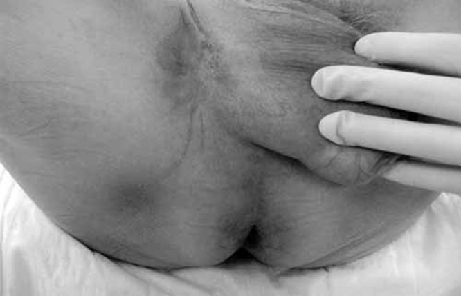
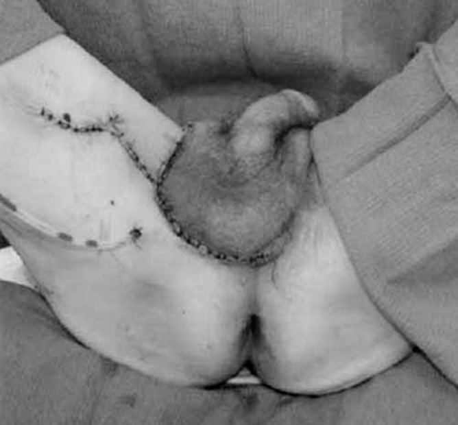
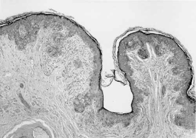
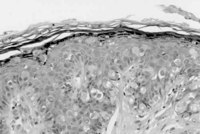
Extramammary Paget's disease (EMPD) is a rare entity, especially in the perinoscrotal region, and typically presents in elderly white patients as a pruritic white or red patch in the area of distribution of apocrine glands. Typically, it affects a single site. Since its manifestations are insidious and easily misdiagnosed, the appropriate management is delayed. Management of this problem is complex and effective treatment can not only lower recurrence rates but also provide an optimal reconstructive result. The present report describes three patients with scrotal EMPD. Based on literature search, the etiopathology, diagnosis and management of these lesions is discussed. Reconstructive options, with special emphasis on scrotal lesions, are also discussed.
La maladie de Paget extramammaire (MPEM) est une maladie rare qui se manifeste surtout dans la région périnéoscrotale et qui se présente généralement chez des personnes âgées de race blanche sous forme de tache prurigineuse rougeâtre ou blanchâtre dans la zone de distribution des glandes apocrines. D’ordinaire, elle atteint un seul foyer. Puisque ses manifestations sont insidieuses et faciles à mal diagnostiquer, sa prise en charge convenable est retardée. D’ailleurs, cette prise en charge est complexe. Toutefois, un traitement efficace peut non seulement faire chuter le taux de récidive mais également assurer une reconstruction optimale. Le présent rapport décrit trois patients atteints d’une MPEM scrotale. Compte tenu d’une recherche dans la documentation scientifique, l’étiopathologie, le diagnostic et la prise en charge de ces lésions sont abordés. Les possibilités de reconstruction, surtout axées sur les lésions scrotales, sont également examinées.
Keywords: Cutaneous pruritic patch; Extramammary Paget’s disease; Scrotal lesion.
Figures
Similar articles
-
Scrotal Extramammary Paget's Disease in an Elderly Caucasian Male.Cureus. 2022 Sep 23;14(9):e29486. doi: 10.7759/cureus.29486. eCollection 2022 Sep. Cureus. 2022. PMID: 36299959 Free PMC article.
-
A Case Report of Familial Extramammary Paget's Disease in Female Siblings.Case Rep Dermatol. 2021 Mar 22;13(1):176-183. doi: 10.1159/000514253. eCollection 2021 Jan-Apr. Case Rep Dermatol. 2021. PMID: 34703424 Free PMC article.
-
Definition, Association with Malignancy, Biologic Behavior, and Treatment of Ectopic Extramammary Paget's Disease: A Review of the Literature.J Clin Aesthet Dermatol. 2019 Aug;12(8):40-44. Epub 2019 Aug 1. J Clin Aesthet Dermatol. 2019. PMID: 31531170 Free PMC article. Review.
-
PIK3CA inhibitor treatment for metastatic scrotal extramammary Paget's disease: a case report and literature review.AME Case Rep. 2025 Apr 10;9:47. doi: 10.21037/acr-24-170. eCollection 2025. AME Case Rep. 2025. PMID: 40330932 Free PMC article.
-
Applications of photodynamic therapy in extramammary Paget's disease.Am J Cancer Res. 2023 Oct 15;13(10):4492-4507. eCollection 2023. Am J Cancer Res. 2023. PMID: 37970368 Free PMC article. Review.
Cited by
-
SkIndia Quiz 37: A Persistent Plaque in the Pubic Region.Indian Dermatol Online J. 2017 May-Jun;8(3):227-228. doi: 10.4103/2229-5178.202362. Indian Dermatol Online J. 2017. PMID: 28584770 Free PMC article. No abstract available.
-
Integrative bioinformatics approaches to map key biological markers and therapeutic drugs in Extramammary Paget's disease of the scrotum.PLoS One. 2021 Jul 22;16(7):e0254678. doi: 10.1371/journal.pone.0254678. eCollection 2021. PLoS One. 2021. Retraction in: PLoS One. 2022 Aug 31;17(8):e0273532. doi: 10.1371/journal.pone.0273532. PMID: 34292991 Free PMC article. Retracted.
References
-
- Paget J. On disease of the mammary areola preceding cancer of the mammary gland. St Bartholomew’s Hosp Rep. 1874;10:87–9.
-
- Kageyama N, Izumi AK. Bilateral scrotal extramammary Paget’s disease in a Chinese man. Int J Dermatol. 1997;36:695–7. - PubMed
-
- Zollo JD, Zeitouni NC. The Roswell Park Cancer Institute experience with extramammary Paget’s disease. Br J Dematol. 2000;142:59–65. - PubMed
-
- Heymann WR. Extramammary Paget’s disease. Clin Dermatol. 1993;11:83–7. - PubMed
-
- Merot Y, Mazoujian G, Pinkus G, et al. Extramammary Paget’s disease of the perianal and perineal regions. Evidence of apocrine derivation. Arch Dermatol. 1985;121:750–2. - PubMed
Publication types
LinkOut - more resources
Full Text Sources