Clinical Trial
. 2013;9(8):e1003590.
doi: 10.1371/journal.ppat.1003590. Epub 2013 Aug 29.

CD36 recruits α₅β₁ integrin to promote cytoadherence of P. falciparum-infected erythrocytes

Affiliations
Clinical Trial

CD36 recruits α₅β₁ integrin to promote cytoadherence of P. falciparum-infected erythrocytes

Shevaun P Davis et al. PLoS Pathog. 2013.

Abstract

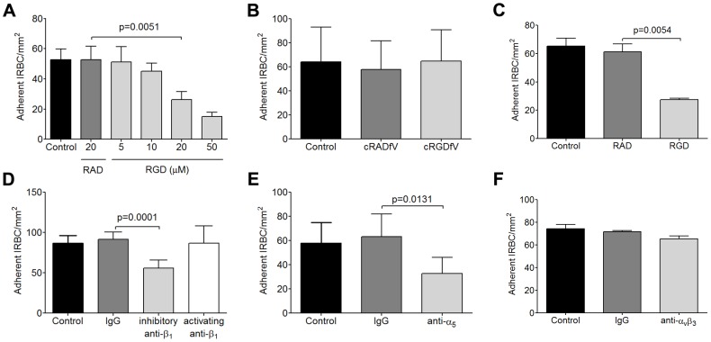
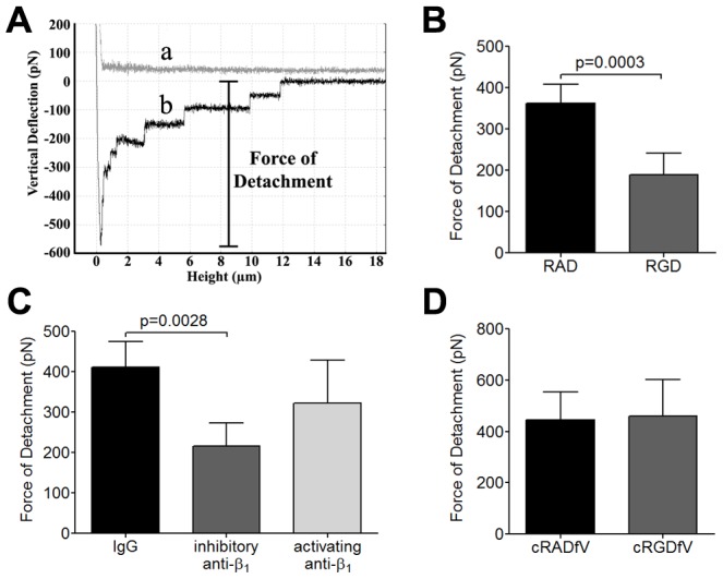
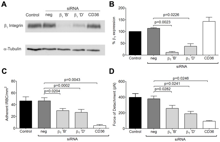
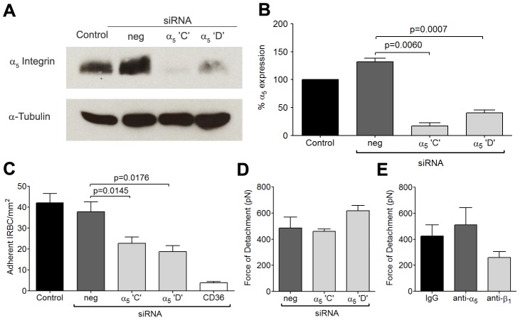
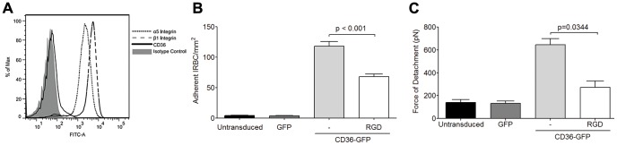
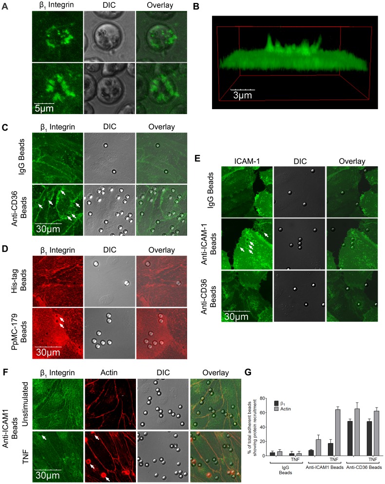
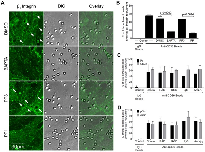
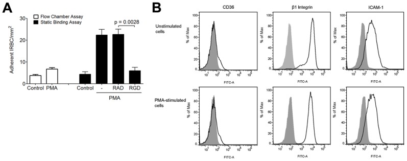
The adhesion of Plasmodium falciparum-infected erythrocytes (IRBC) to receptors on different host cells plays a divergent yet critical role in determining the progression and outcome of the infection. Based on our ex vivo studies with clinical parasite isolates from adult Thai patients, we have previously proposed a paradigm for IRBC cytoadherence under physiological shear stress that consists of a recruitment cascade mediated largely by P-selectin, ICAM-1 and CD36 on primary human dermal microvascular endothelium (HDMEC). In addition, we detected post-adhesion signaling events involving Src family kinases and the adaptor protein p130CAS in endothelial cells that lead to CD36 clustering and cytoskeletal rearrangement which enhance the magnitude of the adhesive strength, allowing adherent IRBC to withstand shear stress of up to 20 dynes/cm². In this study, we addressed whether CD36 supports IRBC adhesion as part of an assembly of membrane receptors. Using a combination of flow chamber assay, atomic force and confocal microscopy, we showed for the first time by loss- and gain-of function assays that in the resting state, the integrin α₅β₁ does not support adhesive interactions between IRBC and HDMEC. Upon IRBC adhesion to CD36, the integrin is recruited either passively as part of a molecular complex with CD36, or actively to the site of IRBC attachment through phosphorylation of Src family kinases, a process that is Ca²⁺-dependent. Clustering of β1 integrin is associated with an increase in IRBC recruitment as well as in adhesive strength after attachment (∼40% in both cases). The adhesion of IRBC to a multimolecular complex on the surface of endothelial cells could be of critical importance in enabling adherent IRBC to withstand the high shear stress in the microcirculations. Targeting integrins may provide a novel approach to decrease IRBC cytoadherence to microvascular endothelium.


Conflict of interest statement

The authors have declared that no competing interests exist.

Figures

Figure 1
Figure 1. Inhibition of cytoadherence on HDMEC by RGD peptide and anti-b1 and anti-a5 mAb.
(A) Adhesion of IRBC to endothelial monolayers pre-incubated with 20 µM RAD or 5–50 µM RGD peptide for 30 min at 37°C in 5% CO2. A 1% hematoctit suspension of IRBC from the lab-adapted parasite clone 7G8 was drawn over the monolayers at 1 dyne/cm2. Results shown are the mean number of adherent IRBC/mm2 in 4–6 microscopic fields (20×) after 7 min of infusion (n = 6). (B) Adhesion of 7G8 parasites to endothelial monolayers pre-incubated with 20 µM cRADfV or cRGDfV peptide (n = 3). (C) Adhesion of 3 clinical parasite isolates to endothelial monolayers pre-incubated with 20 µM RAD or RGD peptide (n = 10 for 3 clinical isolates each tested in 3 to 4 independent experiments). (D) Adhesion of clinical isolates to endothelial monolayers pre-incubated with control IgG1, an inhibitory anti-β1 integrin mAb TDM29 or the activating anti-β1 integrin mAb TS2/16 at 10 mg/ml (n = 3 for 3 clinical isolates each tested in 1 independent experiment). (E) Adhesion of clinical isolates to endothelial monolayers pre-incubated with control IgG1, and an inhibitory anti-α5 integrin mAb JBS5 at 10 µg/ml (n = 3 for 3 clinical isolates each tested in 1 independent experiment). (F) Adhesion of clinical isolates to endothelial monolayers pre-incubated with control IgG1, and an inhibitory anti-αvβ3 integrin mAb 23C6 at 10 µg/ml (n = 3 for 3 clinical isolates each tested in 1 independent experiment).
Figure 2
Figure 2. Inhibition of adhesive strength between IRBC and HDMEC by RGD peptide and anti-β1 mAb.
(A) Schematic representation of a typical AFM force curve which depicts a) approach of the IRBC to an endothelial monolayer and b) retraction of the IRBC. The bar indicates the force of detachment that is used as a measure of adhesive strength between IRBC and endothelium. (B) Force measurement on endothelial monolayers pre-incubated with 20 µM RAD or RGD peptide for 30 min at 37°C in 5% CO2 (n = 7). (C) Force measurement on endothelial monolayers pre-incubated with control IgG1, an inhibitory anti-β1 integrin mAb TDM29 or the activating anti-β1 integrin mAb TS2/16 at 10 µg/ml (n = 4). (D) Force measurement on endothelial monolayers pre-incubated with 20 µM cRADfV or cRGDfV peptide (n = 3). For each experiment, 2 IRBC were brought into contact with 3 HDMEC. Contact for 5 min was maintained with a constant force of 150 pN.
Figure 3
Figure 3. Inhibition of IRBC cytoadherence and adhesive force on TNF-α-stimulated HDMEC by RGD peptide.
(A) Flow cytometric analysis of the expression of CD36, ICAM-1, α5, β1 and VCAM-1 on unstimulated and TNF-α-stimulated HDMEC. TNF-α was added at 1 ng/ml for 20–24 h. Results shown are representative of at least 3 experiments. (B) Adhesion of IRBC from the parasite line 7G8 to unstimulated and TNF-α-stimulated endothelial monolayers pre-incubated with 20 µM RGD peptide or 5 µg/ml of anti-CD36 for 30 min at 37°C in 5% CO2 (n = 3). (C) Adhesion of IRBC from two clinical parasite isolates to unstimulated and TNF-α-stimulated endothelial monolayers pre-incubated with 20 µM RGD peptide, 5 µg/ml of anti-ICAM-1 or both (n = 5 with 2 clinical isolates). (D) Force of detachment measurements of 7G8 parasites on unstimulated and TNF-α-stimulated endothelial monolayers pre-incubated with 20 µM RAD or RGD peptide (n = 3). (E) Force of detachment measurements of two clinical parasites on unstimulated and TNF-α-stimulated endothelial monolayers pre-incubated with 20 µM RAD or RGD peptide (n = 3 with 2 clinical isolates).
Figure 4
Figure 4. Inhibition of cytoadherence and adhesive strength on HDMEC transfected with small interference RNA of β1 integrin.
(A) Western blot analysis of HDMEC lysates 72 h after transfection with 20 nM of negative siRNA and siRNA for β1 integrin ‘B’ and ‘D’, and CD36. Blots were probed with mAb anti-β1 integrin TS2/16 (top) and anti-α-tubulin (bottom). Results shown are representative of 3 independent experiments. (B) Densitometric analysis showing the effectiveness of the knockdown of β1 integrin (n = 3). (C) Adhesion of IRBC to β1 integrin and CD36 knock down endothelial monolayers (n = 3). (D) Force of detachment for IRBC on β1 integrin and CD36 knock down endothelial monolayers (n = 4).
Figure 5
Figure 5. Inhibition of cytoadherence but not adhesive strength on HDMEC transfected with small interference RNA of α5 integrin.
(A) Western blot analysis of HDMEC lysates 72 h after transfection with 20 nM of negative siRNA and siRNA for α5 integrin ‘C’ and ‘D, and CD36. Blots were probed with a polyclonal anti-α5 integrin antibody (top) and a monoclonal anti-α-tubulin antibody (bottom). Results shown are representative of 3 independent experiments. (B) Densitometric analysis showing the effectiveness of the knockdown of α5 integrin (n = 3). (C) Adhesion of IRBC to α5 integrin knockdown endothelial monolayers (n = 3). (D) Force of detachment for IRBC on α5 integrin knockdown endothelial monolayers (n = 2). (E) Force of detachment for IRBC on endothelial monolayers pre-incubated with the anti-α5 mAb JBS5 (n = 2). Results for (D) and (E) are shown as mean ± SD.
Figure 6
Figure 6. Adhesion of IRBC on the endothelial cell line HMEC-1.
(A) Expression of the integrins α5 and β1 but not CD36 on HMEC-1. Results are representative of 3 independent experiments. (B) Adhesion of IRBC to untransduced, GFP-transduced and CD36-GFP transduced HMEC-1 (n = 3). (C) Force of detachment for IRBC on GFP-transduced and CD36-GFP transduced HMEC-1 (n = 3).
Figure 7
Figure 7. β1 integrin recruitment by adherent IRBC, antibody- or PpMC-179 coated-beads.
HDMEC were grown to 95% confluence in ibidi VI chambers. (A) IRBC purified on a MACS separation column were added to a monolayer at 0.1% hematocrit. An FITC-labelled anti-b1integrin mAb was added at 10 µg/ml. The IRBC interaction with HDMEC was imaged in a humidified chamber with 5% CO2 at 37°C. Results are representative of 2 experiments. (B) 3D reconstruction of the cup-shaped structure formed by clustered β1 integrin on the endothelial cell membrane. (C and D) HDMEC monolayers in ibidi VI chambers were incubated with beads coated with IgG1, anti-CD36, anti-HIS or PpMC-179, the CD36 binding peptide of PfEMP1, for 30 min at 37°C in 5% CO2. The monolayers were washed 2× with HBSS to remove unbound beads prior to fixing with 1% PFA for 30 minutes at room temperature. Fixed cells were stained with an anti-β1 mAb at 10 µg/ml followed by Alexa 488-labelled anti-mouse IgG1 (C) or with a PE-labelled anti-β1 integrin mAb at 10 mg/ml (D). Results shown are representative of 3 experiments. (E) HDMEC monolayers in ibidi chambers transduced with GFP-ICAM-1 were incubated with beads coated with IgG1, anti-ICAM-1 and anti-CD36, and processed as in (C) and (D). (F) Unstimulated and TNF-α-stimulated HDMEC monolayers in ibidi chambers were incubated with beads coated with IgG1, anti-ICAM-1 and anti-CD36, and processed as in (C) and (D). The actin cytoskeleton was visualized by permeabilizing the cells for 5 min with 0.2% TX-100 prior to adding 1 µl of rhodamine-phalloidin in 60 µl HBSS to each chamber for 30 min at room temperature. Results shown are for anti-ICAM-1 coated beads only, and are representative of 3 experiments. All images were taken on an Olympus IX81 inverted confocal microscope (Center Valley, Pa) with Fluorview 1000 acquisition software using a PlanAPO 60× N.A. 1.42 oil immersion objective. White arrows indicate representative sites of β1 integrin or ICAM-1 recruitment. (G) Quantification of microscopic changes seen in (F). For every condition in every experiment, three randomly selected microscopic fields at 60× magnification were selected. Each field with 15–25 adherent beads were scanned, and adherent beads associated with protein recruitment were scored as positive. Beads partially in the field of view or rings with no adherent beads were excluded in the enumeration. Results are expressed as positive adherent beads/total adherent beads ×100% (n = 3).
Figure 8
Figure 8. β1 integrin recruitment by adherent anti-CD36 coated beads on HMEC-1.
HMEC-1 monolayers in ibidi chambers transduced with GFP-CD36 were incubated with beads coated with IgG1 or anti-CD36 for 30 min at 37°C in 5% CO2. The slides were fixed and stained as described in Figure 7 using a PE-labelled anti-β1 integrin mAb. Results shown are representative of 3 independent experiments.
Figure 9
Figure 9. Inhibition of β1 integrin recruitment by Src family kinase inhibitor and intracellular Ca2+ chelator.
(A) HDMEC monolayers were pre-incubated with PP3 or PP1 at a concentration of 10 µM for 2 hours, or DMSO or BAPTA for 30 min followed by HBSS for 30 min, at 37°C in 5% CO2. Anti-CD36 coated beads were then added to the monolayers for 30 min. After unattached beads were washed off, the monolayers were fixed and stained with Alexa 488-labeled anti-β1 integrin for 1 hr at room temperature. The images were taken as in Figure 7. Results shown are representative of 3 experiments. (B) Quantification of microscopic changes as described in Figure 7F. (C) HDMEC transduced with GFP-CD36 were either untreated, or pre-incubated with 20 µM RAD/RGD, or IgG1/inhibitory anti-β1 integrin mAb (clone TDM29) at 10 µg/ml for 30 min at 37°C in 5% CO2. After the addition of anti-CD36 coated beads for 30 min, the monolayers were fixed and stained as in (A). Quantification of 2 experiments is shown. (D) Anti-CD36 coated beads were added to peptide or antibody-treated HDMEC monolayers as in (C). The monolayers were fixed, permeabilized with 0.2%TX-100 for 5 min, and blocked with 1%BSA+0.003%TX-100 for 30 min. Monolayers were stained with a polyclonal anti-phospho-Src antibody overnight at 4°C followed by goat-anti-rabbit IgG-Alexa 488 for 1 hr at room temperature. The actin cytoskeleton was visualized by adding 1 µl of rhodamine-phalloidin in 60 µl HBSS to each chamber for 30 min at room temperature. Quantification of 2 experiments is shown.
Figure 10
Figure 10. Effect of PMA on IRBC adhesion to HMEC-1.
(A) Adhesion of IRBC on untreated HMEC-1 or HMEC-1 pre-incubated with PMA 50 ng/ml for 30 min at 37°C and 5%CO2 under flow (left) or static (right) conditions. In the static binding experiments, monolayers were pre-treated with either 50 ng/ml of PMA alone, or 100 µM RGD or RAD peptide was added after 10 min of incubation. At the end of 30 min, 1 ml of a 1% hematocrit suspension of IRBC in RPMI at 5–9% parasitemia was added and allowed to adhere for 20 min. The 35 mm dish with the HMEC-1 monolayer and IRBC was then mounted into the flow chamber system. HBSS was infused at 1 dyne/cm2, and the number of adherent cells were counted at 30 sec intervals for a total of 4 min and the mean taken (n = 3). (B) CD36, β1 integrin and ICAM-1 expression on control and PMA-treated HMEC-1 cells. Results are representative of 2 independent experiments.

Similar articles

Cited by

References

    1. Ley K, Laudanna C, Cybulsky MI, Nourshargh S (2007) Getting to the site of inflammation: the leukocyte adhesion cascade updated. Nat Rev Immunol 7: 678–689. - PubMed
    1. Legate KR, Wickström SA, Fässler R (2009) Genetic and cell biological analysis of integrin outside-in signaling. Genes Dev 23: 397–418. - PubMed
    1. Yipp BG, Anand S, Schollaardt T, Patel KD, Looareesuwan S, et al. (2000) Synergism of multiple adhesion molecules in mediating cytoadherence of Plasmodium falciparum-infected erythrocytes to microvascular endothelial cells under flow. Blood 96: 2292–2298. - PubMed
    1. Yipp BG, Hickey MJ, Andonegui G, Murray AG, Looareesuwan S, et al. (2007) Differential roles of CD36, ICAM-1, and P-selectin in Plasmodium falciparum cytoadherence in vivo. Microcirculation 14: 593–602. - PubMed
    1. Yipp BG, Robbins SM, Resek ME, Baruch DI, Looareesuwan S, et al. (2003) Src-family kinase signaling modulates the adhesion of Plasmodium falciparum on human microvascular endothelium under flow. Blood 101: 2850–2857. - PubMed

Publication types

MeSH terms

LinkOut - more resources