. 2013 Aug;13(4):133-40.
doi: 10.4110/in.2013.13.4.133. Epub 2013 Aug 26.

Mesenchymal Stem Cell Lines Isolated by Different Isolation Methods Show Variations in the Regulation of Graft-versus-host Disease

Affiliations

Mesenchymal Stem Cell Lines Isolated by Different Isolation Methods Show Variations in the Regulation of Graft-versus-host Disease

Hyun Seung Yoo et al. Immune Netw. 2013 Aug.

Erratum in

  • Immune Netw. 2014 Feb;14(1):66

Abstract

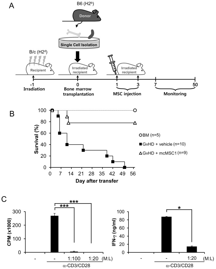
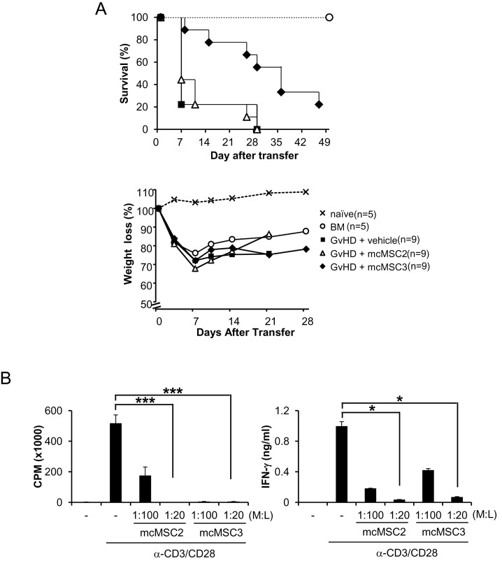
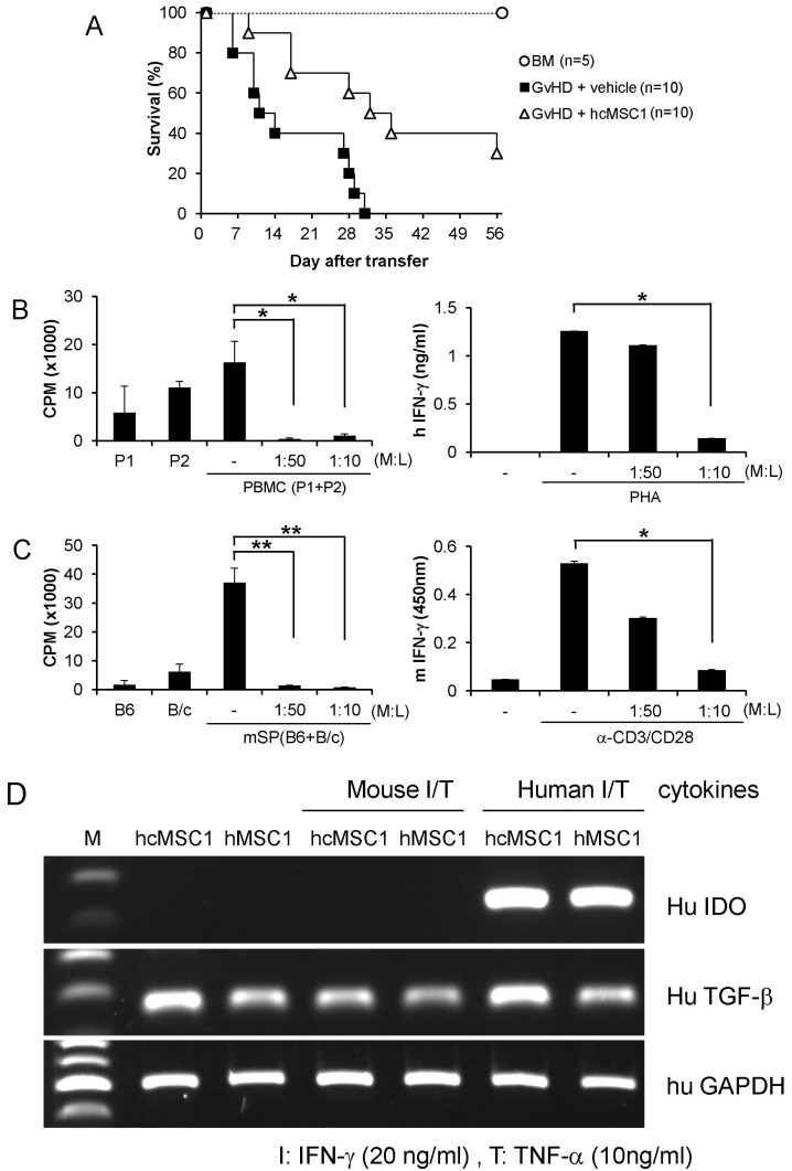
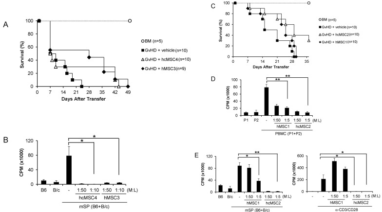
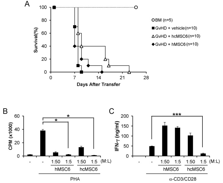
Since the discovery of the immunomodulation property of mesenchymal stem cells (MSCs) about a decade ago, it has been extensively investigated whether MSCs can be used for the treatment of immune-related diseases, such as graft-versus-host disease (GvHD). However, how to evaluate the efficacy of human MSCs for the clinical trial is still unclear. We used an MHC-mismatched model of GvHD (B6 into BALB/c). Surprisingly, the administration of the human MSCs (hMSCs) could reduce the GvHD-related mortality of the mouse recipients and xenogeneically inhibit mouse T-cell proliferation and IFN-γ production in vitro. We recently established a new protocol for the isolation of a homogeneous population of MSCs called subfractionation culturing methods (SCM), and established a library of clonal MSC lines. Therefore, we also investigated whether MSCs isolated by the conventional gradient centrifugation method (GCM) and SCM show different efficacy in vivo. Intriguingly, clonal hMSCs (hcMSCs) isolated by SCM showed better efficacy than hMSCs isolated by GCM. Based on these results, the MHC-mismatched model of GvHD may be useful for evaluating the efficacy of human MSCs before the clinical trial. The results of this study suggest that different MSC lines may show different efficacy in vivo and in vitro.

Keywords: Efficacy; IFN-γ; Mesenchymal stem cells; T-cell; graft-versus-host disease.


Conflict of interest statement

The authors have no financial conflict of interest.

Figures

Figure 1
Figure 1
In vivo efficacy model for MSC. (A) BALB/c mice were irradiated in a dose of 8.5 Gy. Twenty-four hours after irradiation, 5×10⁶ BM and 5×10⁶ spleen cells from B6 female mice were injected. On days 1 and 3 after transplantation, 5×10⁵ MSCs were injected intravenously. (B) Survival rates were monitored. (C) Lymphocytes from the spleen and lymph node were stimulated with anti-CD3 and anti-CD28 antibodies in the presence or absence of mcMSC1, and mcMSC1 was co-cultured with lymphocytes at a ratio of 1:20 or 1:100 (MSCs: lymphocytes). T cell proliferation and IFN-γ production were measured by [3H]-thymidine incorporation and ELISA, respectively. T cell proliferation and IFN-γ assays were repeated three times and similar results were obtained. L, lymphocytes; M, MSCs; BM, bone marrow, ***p<0.005, *p<0.05.
Figure 2
Figure 2
Different in vivo efficacy of mouse cMSC lines. (A) The mcMSC2 and mcMSC3 isolated from BM of C3H/HeN mice were introduced into the GvHD mice. The percentage of survival was plotted. The weight changes of the BALB/c recipients were measured over the experiment period. (B) Both MSC lines were co-cultured with lymphocytes in vitro which were stimulated with anti-CD3 and anti-CD28 antibodies. Mouse T-cell proliferation and IFN-γ production were measured by [3H]-thymidine incorporation and ELISA, respectively. T cell proliferation and IFN-γ assays were repeated three times and similar results were obtained. ***p<0.005, *p<0.05.
Figure 3
Figure 3
In vivo efficacy of human cMSCs. (A) The hcMSC1 isolated from the BM of healthy donors were injected into the GvHD mice. The percentage of survival was plotted. (B) PBMCs from 2 different donors were co-cultured with hcMSC1 in indicated doses for 5 days. Human T-cell proliferation was measured by [3H]-thymidine incorporation. PBMC were stimulated with PHA for 48 h in the presence of hcMSC1, and IFN-γ production were measured in culture media by ELISA. (C) The hcMSC1 were co-cultured with lymphocytes from 2 different mouse strains for 5 days. Mouse T-cell proliferation was measured by [3H]-thymidine incorporation. BALB/c lymphocytes were stimulated with anti-CD3 and anti-CD28 antibodies for 48 h in the presence of hcMSC1, and IFN-γ production were measured in culture media by ELISA. T cell proliferation and IFN-γ assays were repeated twice. (D) The hcMSC1 and hMSC1 were stimulated either mouse or human recombinant IFN-γ and TNF-α for 24 h, and the mRNA expressions of human IDO, TGF-β, and GAPDH were measured by RT-PCR. *p<0.05, **p<0.01.
Figure 4
Figure 4
In vivo efficacy of human MSC lines isolated from different donors. (A) The hMSC3 and hcMSC4 isolated from the BM of 2 different donors by GCM and SCM, respectively, were injected into the GvHD mice. The percentage of survival was plotted. (B) Both human MSCs were co-cultured with lymphocytes from 2 different mouse strains for 5 days. Mouse T-cell proliferation was measured by [3H]-thymidine incorporation. (C) The hMSC1 and hcMSC2 isolated from BM of 2 other donors by GCM and SCM, respectively, were injected into the GvHD mice. The percentage of survival was plotted. (D) PBMCs from 2 different donors were co-cultured with hMSC1 or hcMSC2 in indicated doses for 5 days. Human T-cell proliferation was measured by [3H]-thymidine incorporation. (E) The hMSC1 or hcMSC2 were co-cultured with lymphocytes from 2 different mouse strains for 5 days. BALB/c lymphocytes were stimulated with anti-CD3 and anti-CD28 antibodies for 3 days in the presence of either hMSC1 or hcMSC2. Mouse T-cell proliferation was measured by [3H]-thymidine incorporation. T cell proliferation assays were repeated twice and similar results were obtained. *p<0.05, **p<0.01
Figure 5
Figure 5
In vivo efficacy of human MSC lines isolated from a single donor. (A) The hMSC6 and hcMSC6 isolated from the BM of a single donor by GCM and SCM, respectively, were injected into the GvHD mice. The percentage of survival was plotted. (B) PBMCs were stimulated with PHA for 3 days in the presence of either hMSC6 or hcMSC6. Human T-cell proliferation was measured by [3H]-thymidine incorporation. (C) BALB/c lymphocytes were stimulated with anti-CD3 and anti-CD28 antibodies for 48 h in the presence of either hMSC6 or hcMSC6. IFN-γ productions were measured in culture media by ELISA. T cell proliferation and IFN-γ assays were repeated twice and similar results were obtained. *p<0.05, ***p<0.005

References

    1. Goker H, Haznedaroglu IC, Chao NJ. Acute graft-vs-host disease: pathobiology and management. Exp Hematol. 2001;29:259–277. - PubMed
    1. Menillo SA, Goldberg SL, McKiernan P, Pecora AL. Intraoral psoralen ultraviolet A irradiation (PUVA) treatment of refractory oral chronic graft-versus-host disease following allogeneic stem cell transplantation. Bone Marrow Transplant. 2001;28:807–808. - PubMed
    1. Deeg HJ. How I treat refractory acute GVHD. Blood. 2007;109:4119–4126. - PMC - PubMed
    1. Rager A, Frey N, Goldstein SC, Reshef R, Hexner EO, Loren A, Luger SM, Perl A, Tsai D, Davis J, Vozniak M, Smith J, Stadtmauer EA, Porter DL. Inflammatory cytokine inhibition with combination daclizumab and infliximab for steroid-refractory acute GVHD. Bone Marrow Transplant. 2011;46:430–435. - PMC - PubMed
    1. Schub N, Günther A, Schrauder A, Claviez A, Ehlert C, Gramatzki M, Repp R. Therapy of steroid-refractory acute GVHD with CD52 antibody alemtuzumab is effective. Bone Marrow Transplant. 2011;46:143–147. - PubMed