. 2013 Aug 30:7:156.
doi: 10.3389/fnins.2013.00156. eCollection 2013.

Central CRF neurons are not created equal: phenotypic differences in CRF-containing neurons of the rat paraventricular hypothalamus and the bed nucleus of the stria terminalis

Affiliations

Central CRF neurons are not created equal: phenotypic differences in CRF-containing neurons of the rat paraventricular hypothalamus and the bed nucleus of the stria terminalis

Joanna Dabrowska et al. Front Neurosci. .

Abstract

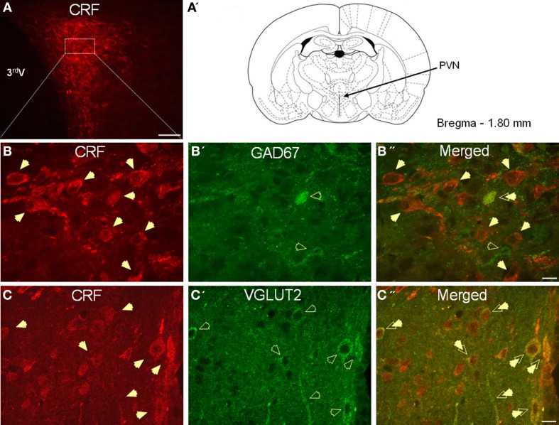
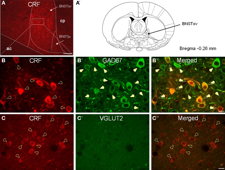
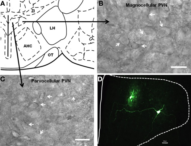
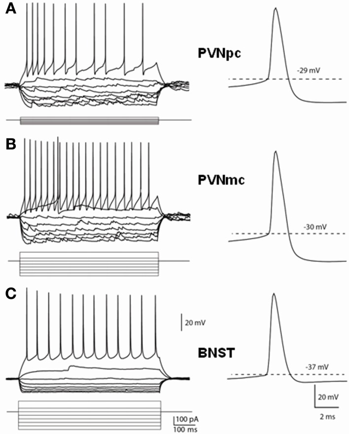
Corticotrophin-releasing factor (CRF) plays a key role in initiating many of the endocrine, autonomic, and behavioral responses to stress. CRF-containing neurons of the paraventricular nucleus of the hypothalamus (PVN) are classically involved in regulating endocrine function through activation of the stress axis. However, CRF is also thought to play a critical role in mediating anxiety-like responses to environmental stressors, and dysfunction of the CRF system in extra-hypothalamic brain regions, like the bed nucleus of stria terminalis (BNST), has been linked to the etiology of many psychiatric disorders including anxiety and depression. Thus, although CRF neurons of the PVN and BNST share a common neuropeptide phenotype, they may represent two functionally diverse neuronal populations. Here, we employed dual-immunofluorescence, single-cell RT-PCR, and electrophysiological techniques to further examine this question and report that CRF neurons of the PVN and BNST are fundamentally different such that PVN CRF neurons are glutamatergic, whereas BNST CRF neurons are GABAergic. Moreover, these two neuronal populations can be further distinguished based on their electrophysiological properties, their co-expression of peptide neurotransmitters such as oxytocin and arginine-vasopressin, and their cognate receptors. Our results suggest that CRF neurons in the PVN and the BNST would not only differ in their response to local neurotransmitter release, but also in their action on downstream target structures.

Keywords: BNST; CRF; GAD67; Oxytocin; PVN; VGLUT2; vasopressin.


Figures

Figure 1
Figure 1
(A) Photomicrograph showing high somatodendritic immunoreactivity of CRF in the parvocellular neurons of the PVN (magnification 10×, scale bar 10 μ, 3rd V- third ventricle). (B–B”): Photomicrographs showing non-overlapping somatodendritic immunoreactivity of CRF (B, red, closed arrows) and GAD67 (B', green, open arrows) in the PVN (B”, merged). Occasionally, CRF-positive cells demonstrate immunolabeling for GAD67 as indicated by double arrow. (C–C”) In contrast, CRF-immunoreactive neurons (B, red) are highly co-localized with VGLUT2-positive neurons (B', green) in the parvocellular PVN (merged arrows, magnification 63×, scale bar 10 μm).
Figure 2
Figure 2
(A) Photomicrograph showing high somatodendritic immunoreactivity of CRF in the BNSTALG (magnification 10×, scale bar 100 μ, ac, anterior commissure; cp, caudate putamen; BNSTov, oval nucleus of the BNST; BNSTju, juxtacapsular nucleus of the BNST). (B–B”): Photomicrographs showing high-level co-localization of CRF (B, red) and GAD67 (B', green) in the BNSTALG (B”, merged, double arrows). Virtually all CRF-immunoreactive neurons co-express GAD67, but numerous GAD67-positive neurons do not co-localize CRF in the BNSTALG. (C–C”) In contrast, CRF-immunoreactive neurons (B, red, open arrows) do not co-localize VGLUT2 (C', green) in the BNSTALG (magnification 63×, scale bar 10 μm).
Figure 3
Figure 3
Localization of parvocellular and magnocellular neurons in the PVN. (A) A schematic diagram showing the recording sites from the PVN neurons. (B,C) Representative images showing the cytoarchitecture of magnocellular and parvocellular neurons under DIC illumination. (D) Immunofluoroscence image showing two anatomically reconstructed PVN neurons, one parvocellular and one magnocellular. Scale bar 25 μm.
Figure 4
Figure 4
Distinct membrane properties of putative CRF neurons in the PVN and type III CRF neurons in the BNSTALG. Representative traces demonstrate the membrane responses of these neurons to membrane current injections and typical spikes. (A) PVNpc neurons have extremely high input resistance and high threshold for spike firing. When hyperpolarized, these neurons exhibited an inward rectification current. No outward rectification was observed during depolarization current injections. Right trace shows a typical spike that has high firing threshold and a prominent fAHP. (B) PVNmc neurons have smaller input resistance than that of PNVpc neurons and also have high threshold for spike firing. Single spike trace also shows this neuron has a high spike threshold and prominent fAHP. (C) In comparison to the PVN CRF neurons, CRF neurons in the BNST have lower input resistances, lower spike threshold and smaller fAHP. PVNmc, PVNpc represent magnocellular and parvocellular neurons of PVN respectively.

Similar articles

Cited by

References

    1. Adan R. A., Van Leeuwen F. W., Sonnemans M. A., Hoffman G., Verbalis J. G., Burbach J. P. (1995). The rat oxytocin receptor. cDNA cloning and immunocytochemical localization in brain, pituitary, mammary gland and uterus. Adv. Exp. Med. Biol. 395, 345–346 - PubMed
    1. Arima H., Aguilera G. (2000). Vasopressin and oxytocin neurones of hypothalamic supraoptic and paraventricular nuclei co-express mRNA for Type-1 and Type-2 corticotropin-releasing hormone receptors. J. Neuroendocrinol. 12, 833–842 10.1046/j.1365-2826.2000.00528.x - DOI - PubMed
    1. Arima H., House S. B., Gainer H., Aguilera G. (2001). Direct stimulation of arginine vasopressin gene transcription by cAMP in parvocellular neurons of the paraventricular nucleus in organotypic cultures. Endocrinology 142, 5027–5030 10.1210/en.142.11.5027 - DOI - PubMed
    1. Bali B., Erdelyi F., Szabo G., Kovacs K. J. (2005). Visualization of stress-responsive inhibitory circuits in the GAD65-eGFP transgenic mice. Neurosci. Lett. 380, 60–65 10.1016/j.neulet.2005.01.014 - DOI - PubMed
    1. Bosch O. J., Pfortsch J., Beiderbeck D. I., Landgraf R., Neumann I. D. (2010). Maternal behaviour is associated with vasopressin release in the medial preoptic area and bed nucleus of the stria terminalis in the rat. J. Neuroendocrinol. 22, 420–429 10.1111/j.1365-2826.2010.01984.x - DOI - PubMed

LinkOut - more resources