. 2013 Aug 28:7:135.
doi: 10.3389/fncel.2013.00135. eCollection 2013.

Low micromolar Ba(2+) potentiates glutamate transporter current in hippocampal astrocytes

Affiliations

Low micromolar Ba(2+) potentiates glutamate transporter current in hippocampal astrocytes

Ramil Afzalov et al. Front Cell Neurosci. .

Abstract

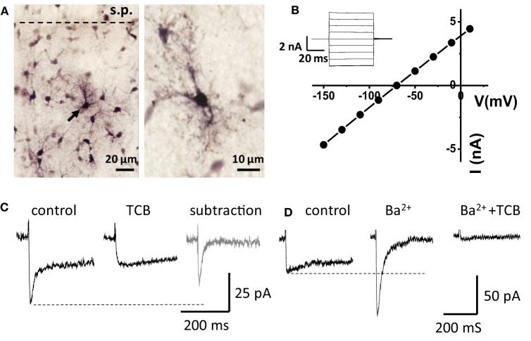
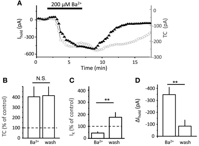
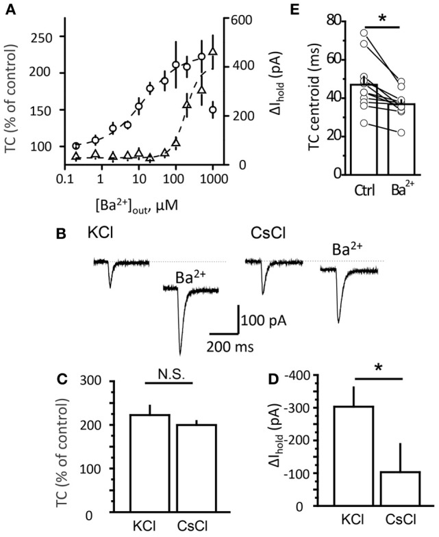
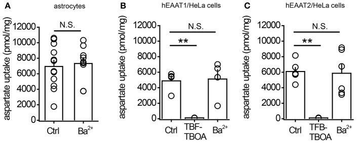
Glutamate uptake, mediated by electrogenic glutamate transporters largely localized in astrocytes, is responsible for the clearance of glutamate released during excitatory synaptic transmission. Glutamate uptake also determines the availability of glutamate for extrasynaptic glutamate receptors. The efficiency of glutamate uptake is commonly estimated from the amplitude of transporter current recorded in astrocytes. We recorded currents in voltage-clamped hippocampal CA1 stratum radiatum astrocytes in rat hippocampal slices induced by electrical stimulation of the Schaffer collaterals. A Ba(2+)-sensitive K(+) current mediated by inward rectifying potassium channels (Kir) accompanied the transporter current. Surprisingly, Ba(2+) not only suppressed the K(+) current and changed holding current (presumably, mediated by Kir) but also increased the transporter current at lower concentrations. However, Ba(2+) did not significantly increase the uptake of aspartate in cultured astrocytes, suggesting that increase in the amplitude of the transporter current does not always reflect changes in glutamate uptake.

Keywords: astrocytes; barium; glutamate transporters; glutamate uptake; hippocampus.


Figures

Figure 1
Figure 1
Recordings from “passive” astrocytes reveal sensitivity of glutamate transporter current to Ba2+. (A) Astrocytes in hippocampal CA1 stratum radiatum (stained with biocytin) s.p.—stratum pyramidale. Arrow indicates a patched astrocyte. (B) I-V relation of the patched astrocyte. Inset: membrane currents in response to voltage steps. (C) Astrocytic current induced by electrical stimulation of Schaffer collaterals in control and in the presence of glutamate transporter current blockers (TCB). Gray trace—“pure” transporter current obtained by subtracting the current in TCB from the control current. (D) Ba2+ (200 μM) abolishes the slow K+ component of the complex current and increases the amplitude of fast transporter current, which is consequently blocked by TCB (Ba2+ + TCB).
Figure 2
Figure 2
Time-course of Ba2+ effect on transporter current and K+ current. (A) Bath-applied 200 μ M BaCl2 produces downward shift in Ihold (black triangles), and increase in transporter current (TC, empty circles) in a single cell. (B) Summary graph of mean normalized transporter current (TC, n = 5) in the presence of Ba2+ and after 5 min of washout (wash). (C) Summary graph of normalized K+ current (n = 5) in the presence of Ba2+ and after 10 min of washout (wash) (D) Summary graph of mean Δ Ihold (n = 5) in the presence of Ba2+ and after 10 min of washout (wash). Error bars—SEM; **P < 0.01; N.S.—non significant; One-Way ANOVA post-hoc Tukey test.
Figure 3
Figure 3
Potentiation of transporter current is independent of Kir blockade. (A) Dose-response curve of TC (circles) and Δ Ihold (triangles) changes. (B) Transporter current (TC) elicited by local glutamate uncaging in control and in 200 μM Ba2+. The downward shift in the holding current was less pronounced in Cs+-loaded cells (CsCl). The TC amplitude increased to a similar degree in both cases. (C,D) Mean Ba2+ effect on transporter current (TC, B) and Ihold (Δ Ihold, C) for CsCl- and KCl-based intracellular solutions. (E) Transporter current centroids in control and in 20 μM Ba2+. Error bars—SEM. *P < 0.05; N.S.—non significant; paired (E) and unpaired (C,D) t-test.
Figure 4
Figure 4
1μM Ba2+ does not potentiate aspartate uptake by cultured astrocytes or by HeLa cells expressing glutamate transporters. (A) Summary graph of aspartate uptake by cultured astrocytes in control conditions (Ctrl) or in the presence of 1 μM BaCl2 (Ba2+). (B) Summary graph of aspartate uptake by HeLa cells expressing EAAT1 in control conditions (Ctrl), in the presence of 2 μM TFB-TBOA, or in the presence of 1 μM BaCl2 (Ba2+). (C) Summary graph of aspartate uptake by HeLa cells expressing EAAT2 in control conditions (Ctrl), in the presence of 2 μM TFB-TBOA, or in the presence of 1 μM BaCl2 (Ba2+). Error bars—SEM. **P < 0.01; N.S.—non significant; unpaired t-test.

References

    1. Barbour B., Brew H., Attwell D. (1991). Electrogenic uptake of glutamate and aspartate into glial cells isolated from the salamander (Ambystoma) retina. J. Physiol. (Lond.) 436, 169–193 - PMC - PubMed
    1. Bergles D. E., Diamond J. S., Jahr C. E. (1999). Clearance of glutamate inside the synapse and beyond. Curr. Opin. Neurobiol. 9, 293–298 10.1016/S0959-4388(99)80043-9 - DOI - PubMed
    1. Bergles D. E., Jahr C. E. (1997). Synaptic activation of glutamate transporters in hippocampal astrocytes. Neuron 19, 1297–1308 10.1016/S0896-6273(00)80420-1 - DOI - PubMed
    1. Bergles D. E., Jahr C. E. (1998). Glial contribution to glutamate uptake at Schaffer collateral-commissural synapses in the hippocampus. J. Neurosci. 18, 7709–7716 - PMC - PubMed
    1. Bergles D. E., Tzingounis A. V., Jahr C. E. (2002). Comparison of coupled and uncoupled currents during glutamate uptake by GLT-1 transporters. J. Neurosci. 22, 10153–10162 - PMC - PubMed