. 2013 Aug 30:7:137.
doi: 10.3389/fncel.2013.00137. eCollection 2013.

β1- and β3- voltage-gated sodium channel subunits modulate cell surface expression and glycosylation of Nav1.7 in HEK293 cells

Affiliations

β1- and β3- voltage-gated sodium channel subunits modulate cell surface expression and glycosylation of Nav1.7 in HEK293 cells

Cédric J Laedermann et al. Front Cell Neurosci. .

Abstract

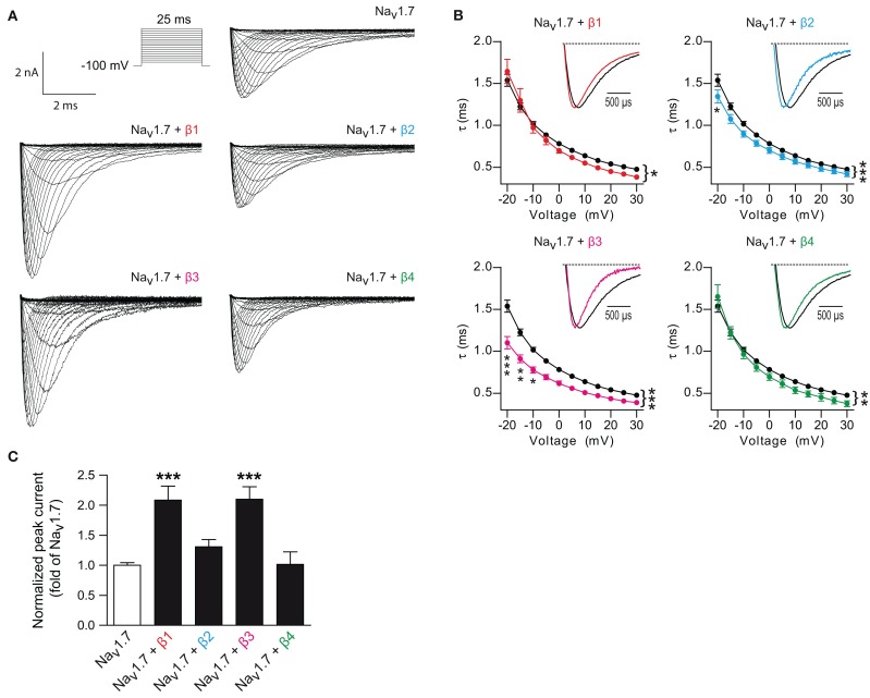
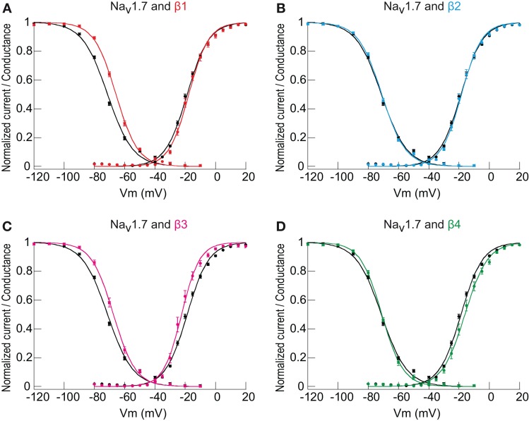
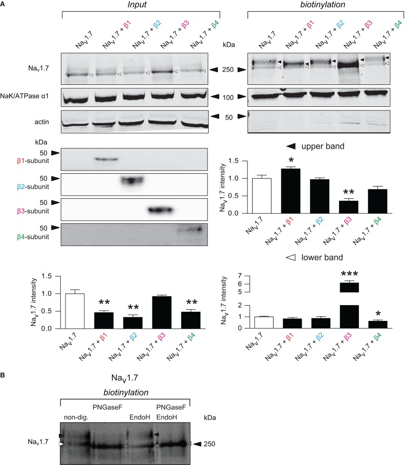
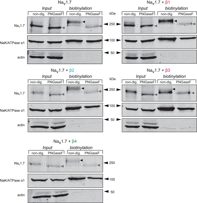
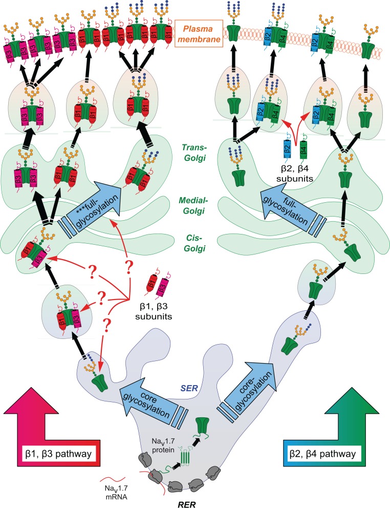
Voltage-gated sodium channels (Navs) are glycoproteins composed of a pore-forming α-subunit and associated β-subunits that regulate Nav α-subunit plasma membrane density and biophysical properties. Glycosylation of the Nav α-subunit also directly affects Navs gating. β-subunits and glycosylation thus comodulate Nav α-subunit gating. We hypothesized that β-subunits could directly influence α-subunit glycosylation. Whole-cell patch clamp of HEK293 cells revealed that both β1- and β3-subunits coexpression shifted V ½ of steady-state activation and inactivation and increased Nav1.7-mediated I Na density. Biotinylation of cell surface proteins, combined with the use of deglycosydases, confirmed that Nav1.7 α-subunits exist in multiple glycosylated states. The α-subunit intracellular fraction was found in a core-glycosylated state, migrating at ~250 kDa. At the plasma membrane, in addition to the core-glycosylated form, a fully glycosylated form of Nav1.7 (~280 kDa) was observed. This higher band shifted to an intermediate band (~260 kDa) when β1-subunits were coexpressed, suggesting that the β1-subunit promotes an alternative glycosylated form of Nav1.7. Furthermore, the β1-subunit increased the expression of this alternative glycosylated form and the β3-subunit increased the expression of the core-glycosylated form of Nav1.7. This study describes a novel role for β1- and β3-subunits in the modulation of Nav1.7 α-subunit glycosylation and cell surface expression.

Keywords: Navs β-subunits; biophysical properties; glycosylation; trafficking; voltage-gated sodium channels (Navs).


Figures

Figure 1
Figure 1
β-subunits regulate Nav1.7 currents. (A) Typical whole-cell Na+ currents of HEK293 cells transfected with Nav1.7 alone or Nav1.7 co-expressed with individual β-subunits elicited with a typical current-voltage protocol. (B) Voltage-dependence of current decay of Nav1.7 alone compared to Nav1.7 with each individual β-subunit. Inset: Normalized representative current traces of Nav1.7 elicited by test pulses at 0 mV. Co-transfection of the β1- (p = 0.011, n = 39), β2- (p < 0.0001, n = 25), β3- (p < 0.0001, n = 16), and β4-subunit (p = 0.006, n = 13) decreased the time constant decay as compared to Nav1.7 alone (n = 81). Two-Ways ANOVA and Bonferroni post-hoc tests. Data are expressed as mean ± s.e.m. (C) INa densities from HEK293 cells transfected with Nav1.7 alone or co-transfected with individual β-subunits. β1- (n = 55) and β3-subunits (n = 34), but not β2- (n = 67) nor β4-subunits (n = 27), increased the Nav1.7 current densities. p < 0.0001 for β1- and β3-subunits with One-Way ANOVA followed by Bonferroni post-hoc tests. Data are expressed as mean ± s.e.m. and were normalized to Nav1.7 alone for each experiments. Values can be found in Table 1. *p < 0.05, **p < 0.01 and ***p < 0.001.
Figure 2
Figure 2
The influence of β1- and β3-subunits on Nav1.7 voltage-dependence of activation and inactivation. (A–D) Normalized currents for both activation and steady-state inactivation (see Materials and Methods) are plotted against the test potential. Each panel compares Nav1.7 alone to Nav1.7 co-expressed with individual β-subunits. A minor but significant effect on the V½of activation was observed for β3-subunit (n = 17, hyperpolarizing shift, p < 0.05) co-expression as compared to Nav1.7 alone (n = 81). β1- (n = 39), β2- (n = 25) and β4-subunits (n = 13) did not modify the V½of activation of Nav1.7. The effect on the V½ of inactivation was highly significant for β1- (n = 51, depolarizing shift with p < 0.0001) and β3-subunit (n = 18, depolarizing shift with p < 0.0001) compared to Nav1.7 alone (n = 92); whereas β2- (n = 43) and β4-subunits (n = 20) had no effect. Individual points are the mean ± s.e.m. of the normalized current at each voltage point. The smooth curves are Boltzmann fits whose equations give both the V½of activation and inactivation (midpoints) and their associated slope factors (see Materials and Methods). The V½ of steady-state activation and inactivation comparing Nav1.7 alone vs. the co-expression with each subunit were tested by One-Way ANOVA followed by Bonferroni's multiple comparison tests. Values and statistics can be found in Table 1.
Figure 3
Figure 3
Only β1-subunit significantly increases Nav1.7 recovery from inactivation (RFI). (A–D) RFI from HEK293 cells transfected with Nav1.7 alone or co-transfected with individual β-subunits. Smooth curves were fitted incorporating a geometric weight to arrive at the final curve (no equation) and the t½ was calculated by interpolation on the x-axis from a linear relation between the 2 points juxtaposing half recovery (y1 < 0.5 < y2, see Materials and Methods). Only when co-expressed with β1-subunit (n = 27, p < 0.01) was Nav1.7 RFI significantly faster as compared to Nav1.7 alone (n = 72). β2- (n = 34), β3- (n = 16), and β4-subunits (n = 19) did not significantly alter RFI when co-expressed with Nav1.7. Individual points are the mean ± s.e.m. of the normalized current at each time point. Non-parametric One-Way analysis of variance (Kruskal-Wallis test) with Dunn post-hoc tests to compare each subunit co-expressed with Nav1.7 vs. Nav1.7 alone.
Figure 4
Figure 4
Nav1.7 transcripts upon β-subunit co-transfection. Bar graph showing transcriptional levels of Nav1.7 in control conditions (co-transfected with empty vector) over Nav1.7 levels when cells were co-transfected with each individual β-subunit. Transcripts were normalized to GAPDH and run in triplicate. Data represent the mean ± s.e.m., n = 3 independent transfections for every condition.
Figure 5
Figure 5
β1- and β3-subunit mediate differential forms of Nav1.7 whose expression is increased at the membrane. (A) Representative western blot of a biotinylation assay with total lysate (input, left) and cell surface (biotinylation, right) fractions from HEK293 cells transiently transfected with Nav1.7 alone, or co-expressed with each individual β-subunit and the associated quantifications. Input: Nav1.7 is detected in two forms: a fast migrating band (~250 kDa, that will be referred to as lower band) that consist mostly of the Nav1.7 immunoreactive signal and a slow migrating band (~280 kDa, that will be referred to as upper band). β1- (p = 0.006), β2- (p = 0.003), and β4-subunits (p = 0.009) significantly decreased Nav1.7 expression, whereas the β3-subunit had no effect (p = 0.570). Because the upper band was below the sensitivity threshold, both bands were quantified together. Biotinylation: Nav1.7 membrane protein is detected in three forms. When expressed alone, one lower band (white triangle, ~250 kDa) and one upper band (black triangle, ~280 kDa) were present (for identification of these bands, see Panel B). When the β1-subunit is co-expressed, the upper band was clearly shifted into an intermediate migrating band (~260 kDa) with increased expression (p = 0.047). β2- and β4-subunits revealed the same pattern as when Nav1.7 was transfected alone and did not change its expression, except for the small decrease of the lower band when the β4-subunit is co-transfected (p = 0.020). The β3-subunit clearly increased Nav1.7 immunoreactivity of the lower band (p < 0.0001). For input and biotinylation fractions, actin and the α1-subunit of NaK-ATPase were used as biotin leakiness and loading controls, respectively. Data represent mean ± s.e.m, n = 4 independent experiments. Student's unpaired t-test, each condition being compared with Nav1.7. *p < 0.05, **p < 0.01 and ***p < 0.001. (B) Representative western blot and identification of glycosylation state of Nav1.7 in biotinylated fraction from HEK293 cells transiently transfected with Nav1.7. EndoH only cleaves the lower band of biotinylated Nav1.7, demonstrating that this band represents the core-glycosylated form of the channel. The upper band is digested by PNGaseF, demonstrating that it corresponds to fully-glycosylated form of the channel. PNGaseF can also digest the core-glycosylated form of Nav1.7.
Figure 6
Figure 6
The different forms of Nav1.7 observed with β-subunits are due to differential glycosylation patterns. Western blot of a biotinylation assay followed by deglycosylation with total lysate and cell surface fractions from HEK293 cells transiently transfected with Nav1.7 alone, or co-expressed with each individual β-subunit. Samples were non-treated or treated with Peptide: N-Glycosidase F (PNGaseF) to remove glycosylated residues of the protein. The total lysate Nav1.7 band (black/white triangle) was slightly shifted to an apparent lower molecular weight (gray triangle) when treated with PNGaseF. In the biotinylation fraction, the pattern of Nav1.7 glycosylation by the β-subunits was the same as in Figure 4.(black and white triangles). When treated with PNGaseF, all the different bands shifted to the lower band of the same apparent molecular weight, irrespective of the β-subunit co-expressed.
Figure 7
Figure 7
Proposed scheme of the different intracellular pathways of α-subunits depending on the presence of different β-subunits. After synthesis in the rough endoplasmic reticulum (RER), α-subunits are rapidly folded and undergo a first step of glycosylation in the smooth endoplasmic reticulum (SER). It is known that N-acetlyglucosamine and oligosaccharide chains are bound on Asp residues of the protein, a process known as core-glycosylation. Newly synthesized glycoproteins are then translocated into the Golgi network, where they are subject to a second step of more complex glycosylation, involving many different enzymes. Once matured, proteins eventually translocate to the plasma membrane. The present findings suggest that core-glycosylated proteins can also be found anchored at the membrane. By some yet undefined mechanism, the β1-subunit interferes with the second glycosylation step (***full-glycosylation on the scheme) which leads to a modification of the glycosylation pattern of the α-subunits. The β1-subunit enhances the core-glycosylated form of Nav1.7. This suggests that the β1- and β3-subunits already interact with the α-subunits before the step of full-glycosylation of the channel occurring in the Golgi network. By enhancing the differential glycosylation pattern of Nav1.7, it can be proposed that the β1- and β3-subunits promote stabilization of the channel at the plasma membrane. On the contrary, it is likely that the β2- and β4-subunits, which have no effect on the glycosylation nor the anchoring, only briefly interact with the α-subunits before translocation of the channel to the plasma membrane. For sake of simplicity, the shown glycosylation patterns are arbitrary. Nav1.7 is depicted as being “freely” expressed in ER/Golgi and membrane networks for easier interpretation of the scheme. However, Nav1.7 is embedded in the membranes of the different organelles.

Similar articles

Cited by

References

    1. Bennett E., Urcan M. S., Tinkle S. S., Koszowski A. G., Levinson S. R. (1997). Contribution of sialic acid to the voltage dependence of sodium channel gating. a possible electrostatic mechanism. J. Gen. Physiol. 109, 327–343 10.1085/jgp.109.3.327 - DOI - PMC - PubMed
    1. Brackenbury W. J., Isom L. L. (2011). Na channel beta subunits: overachievers of the ion channel family. Front. Pharmacol. 2:53 10.3389/fphar.2011.00053 - DOI - PMC - PubMed
    1. Catterall W. A. (2000). From ionic currents to molecular mechanisms: the structure and function of voltage-gated sodium channels. Neuron 26, 13–25 10.1016/S0896-6273(00)81133-2 - DOI - PubMed
    1. Coward K., Jowett A., Plumpton C., Powell A., Birch R., Tate S., et al. (2001). Sodium channel beta 1 and beta 2 subunits parallel SNS/PN3 alpha-subunit changes in injured human sensory neurons. Neuroreport 12, 483–488 10.1097/00001756-200103050-00012 - DOI - PubMed
    1. Cronin N. B., O'reilly A., Duclohier H., Wallace B. A. (2005). Effects of deglycosylation of sodium channels on their structure and function. Biochemistry 44, 441–449 10.1021/bi048741q - DOI - PubMed