Review
. 2013 Aug 30:4:233.
doi: 10.3389/fphys.2013.00233. eCollection 2013.

Cell volume regulation in epithelial physiology and cancer

Affiliations
Review

Cell volume regulation in epithelial physiology and cancer

Stine F Pedersen et al. Front Physiol. .

Abstract

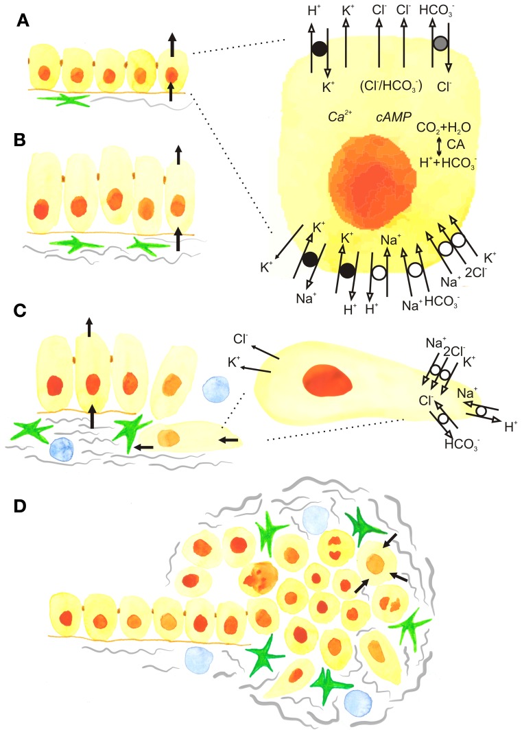
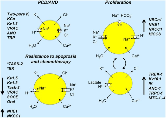
The physiological function of epithelia is transport of ions, nutrients, and fluid either in secretory or absorptive direction. All of these processes are closely related to cell volume changes, which are thus an integrated part of epithelial function. Transepithelial transport and cell volume regulation both rely on the spatially and temporally coordinated function of ion channels and transporters. In healthy epithelia, specific ion channels/transporters localize to the luminal and basolateral membranes, contributing to functional epithelial polarity. In pathophysiological processes such as cancer, transepithelial and cell volume regulatory ion transport are dys-regulated. Furthermore, epithelial architecture and coordinated ion transport function are lost, cell survival/death balance is altered, and new interactions with the stroma arise, all contributing to drug resistance. Since altered expression of ion transporters and channels is now recognized as one of the hallmarks of cancer, it is timely to consider this especially for epithelia. Epithelial cells are highly proliferative and epithelial cancers, carcinomas, account for about 90% of all cancers. In this review we will focus on ion transporters and channels with key physiological functions in epithelia and known roles in the development of cancer in these tissues. Their roles in cell survival, cell cycle progression, and development of drug resistance in epithelial cancers will be discussed.

Keywords: Cl− channels; K+ channels; breast cancer; drug resistance; pancreatic cancer; secretion; stroma; tumour microenvironment.


Figures

Figure 1
Figure 1
The development of epithelial cancer and roles of ion transport and cell volume. (A) Normal secreting epithelium showing net movements of ions and fluid across the basolateral and luminal membranes. Black arrows show the movement of ions and fluid across cell membranes. The insert shows the detailed model of a cell with basic ion channels and transporters that operate, for example, in pancreatic ducts, but are also applicable to other secreting epithelia. The luminal Cl channels include CFTR and TMEM16A/ANO1, as well as a SLC26 family Cl/HCO3 exchanger. The basolateral membrane contains the Na+/K+/2Cl transporter NKCC1, SLC7 family Na+-HCO3 cotransporters (NBCs), and the Na+/H+ exchanger NHE1. The epithelium also expresses H+/K+ pumps, as well as several types of K+ channels such as IK-KCa3.1, BK-KCa1.1, KCNQ1 and voltage-activated K+ channels, some of which may be expressed on both luminal and basolateral membranes. The major Ca2+ and cAMP signalling pathways are not elaborated and for simplicity, Ca2+ -channels/transporters and aquaporins are not included. (B) Carcinogenesis and postulated dysregulation of cells volume and secretion. Increased activity of fibroblastic cells, such as cancer associated fibroblasts (CAFs) and pancreatic stellate cells (PSCs) (green). (C) The epithelial-to-mesenchymal (EMT) transition showing cells that loose apico-basal polarity and the appearance of some ion transporters from the lumimal membrane in the rear of the cells and some from the basolateral membrane in the leading edge, contributing to driving cell migration. (D) Progression to cancer, showing tumor with extensive fibrosis (gray), fibrogenic cells (green) and immune cells (blue). (Blood vessels are not shown). In the center of the tumor cells may die, while surrounding cells are proliferating and cell volume and corresponding ion transport is up-regulated (see the text).
Figure 2
Figure 2
Ion channels and transporters and cell volume changes associated in normal and cancer cells. Cell sizes refer to expected cell volume changes and lengths of arrows on cells indicates up- or down-regulation or ion transporters/channels. Resistance to apoptosis is associated with down-regulation of several channels and inhibition of some channels (asterisks) induces resistance to apoptosis. In proliferation, several transporters and channels are up-regulated and over-expressed in cancer (see text). The right part of the figure shows ion transporters and channels that would lead to cell volume increase and those in the lower part indicate those that would lead to cell volume decrease. Large arrows next to named ion channels/transporters indicate their up- or down-regulation in cancer. Chronic activation of ion transport may lead to cell death. Dynamic activation or suppression of ion transport/cell volume with specific signals, in time or in given cells may lead to cancer development and progression.

References

    1. Anbari K., Schultz R. M. (1993). Effect of sodium and betaine in culture media on development and relative rates of protein synthesis in preimplantation mouse embryos in vitro. Mol. Reprod. Dev. 35, 24–28 10.1002/mrd.1080350105 - DOI - PubMed
    1. Bachmann O., Heinzmann A., Mack A., Manns M. P., Seidler U. (2007). Mechanisms of secretion-associated shrinkage and volume recovery in cultured rabbit parietal cells. Am. J. Physiol. Gastrointest. Liver Physiol. 292, G711–G717 10.1152/ajpgi.00416.2006 - DOI - PubMed
    1. Bachmann O., Juric M., Seidler U., Manns M. P., Yu H. (2011). Basolateral ion transporters involved in colonic epithelial electrolyte absorption, anion secretion and cellular homeostasis. Acta Physiol. (Oxf). 201, 33–46 10.1111/j.1748-1716.2010.02153.x - DOI - PubMed
    1. Bartosova M., Parkkila S., Pohlodek K., Karttunen T. J., Galbavy S., Mucha V., et al. (2002). Expression of carbonic anhydrase IX in breast is associated with malignant tissues and is related to overexpression of c-erbB2. J. Pathol. 197, 314–321 10.1002/path.1120 - DOI - PubMed
    1. Becchetti A. (2011). Ion channels and transporters in cancer. 1. Ion channels and cell proliferation in cancer. Am. J. Physiol. Cell Physiol. 301, C255–C265 10.1152/ajpcell.00047.2011 - DOI - PubMed