Co-Infection with Cytomegalovirus and Helicobacter pylori in a Child with Ménétrier's Disease
- PMID: 24010116
- PMCID: PMC3760702
- DOI: 10.5223/pghn.2013.16.2.123
Co-Infection with Cytomegalovirus and Helicobacter pylori in a Child with Ménétrier's Disease
Abstract
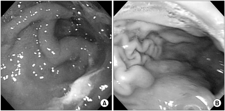
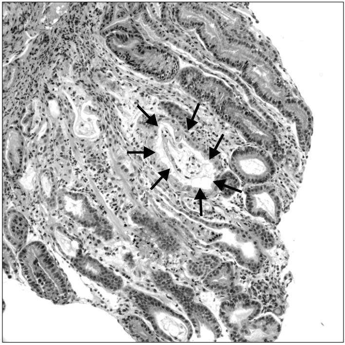
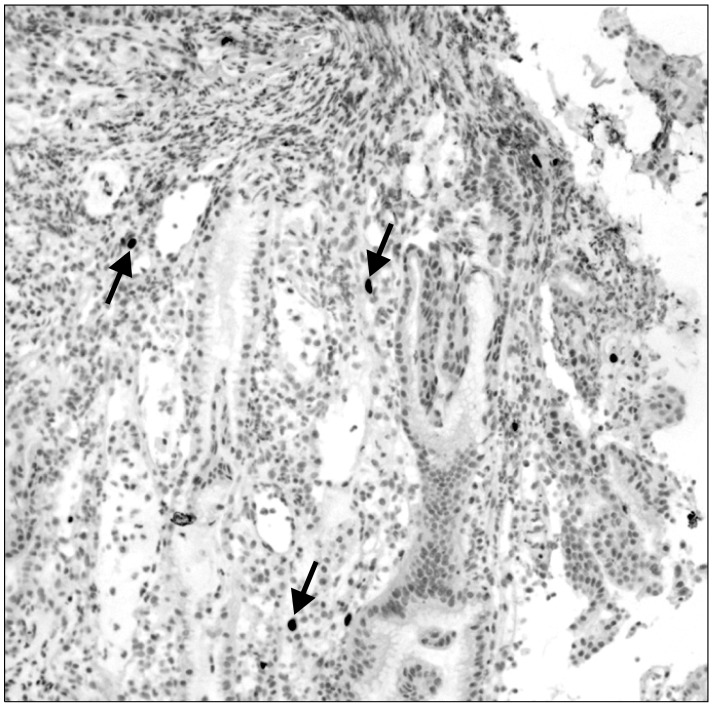
Ménétrier's disease is a rare protein-losing gastropathy characterized by hypertrophic gastric fold, foveolar hyperplasia, and hypoproteinemia with resulting peripheral edema. It is clinically evident as nonspecific gastrointestinal symptoms, including abdominal discomfort, nausea and vomiting, abdominal pain, weight loss, diarrhea, and edema. Pediatric Ménétrier's disease usually has an insidious onset and progressive, chronic clinical course and it spontaneously resolves in weeks or months. The pathogenesis of Ménétrier's disease is not clearly understood. Ménétrier's disease is thought to be associated with some gastric infections. But the cause of Ménétrier's disease is unknown, an association with cytomegalovirus (CMV) and Helicobacter pylori has been suggested. In Korea, We present the first a case of pediatric Ménétrier's disease with positive evidence of CMV and H. pylori.
Keywords: Cytomegalovirus; Helicobacter pylori; Ménétrier's disease.
Conflict of interest statement
No potential conflict of interest relevant to this article was reported.
Figures
Similar articles
-
A case of Menetrier's disease without Helicobacter pylori or hypoalbuminemia.Int J Surg Case Rep. 2015;17:58-60. doi: 10.1016/j.ijscr.2015.10.025. Epub 2015 Oct 27. Int J Surg Case Rep. 2015. PMID: 26551554 Free PMC article.
-
Ménétrier's Disease and Its Atypical Presentation in Four Siblings.Cureus. 2022 Oct 27;14(10):e30759. doi: 10.7759/cureus.30759. eCollection 2022 Oct. Cureus. 2022. Retraction in: Cureus. 2023 Oct 11;15(10):r78. doi: 10.7759/cureus.r78. PMID: 36320788 Free PMC article. Retracted.
-
Curative resection with endoscopic submucosal dissection of early gastric cancer in Helicobacter pylori-negative Ménétrier's disease: A case report.World J Gastroenterol. 2022 Feb 7;28(5):594-601. doi: 10.3748/wjg.v28.i5.594. World J Gastroenterol. 2022. PMID: 35316958 Free PMC article.
-
Menetrier's disease associated with Helicobacter pylori: three cases with sonographic findings and a literature review.Ann Trop Paediatr. 2011;31(2):141-7. doi: 10.1179/146532811X13006353133876. Ann Trop Paediatr. 2011. PMID: 21575319 Review.
-
Protein-losing gastropathy associated with cytomegalovirus in two sisters - Case reports and review of the literature.Arch Pediatr. 2019 May;26(4):232-235. doi: 10.1016/j.arcped.2019.03.005. Epub 2019 Apr 4. Arch Pediatr. 2019. PMID: 30954365 Review.
Cited by
-
Diagnosis and Management of Ménétrier Disease in Children: A Case Series Review.Pediatr Gastroenterol Hepatol Nutr. 2021 Jan;24(1):109-117. doi: 10.5223/pghn.2021.24.1.109. Epub 2021 Jan 8. Pediatr Gastroenterol Hepatol Nutr. 2021. PMID: 33505900 Free PMC article.
-
An unusual association of Ménétrier's disease with a gastric bezoar.BMJ Case Rep. 2015 Feb 16;2015:bcr2014207087. doi: 10.1136/bcr-2014-207087. BMJ Case Rep. 2015. PMID: 25687706 Free PMC article.
-
The Unmasking of Cytomegalovirus as an Accomplice to Helicobacter pylori-Induced Severe Acute Gastroenteritis in a Healthy Host.ACG Case Rep J. 2023 Oct 26;10(10):e01181. doi: 10.14309/crj.0000000000001181. eCollection 2023 Oct. ACG Case Rep J. 2023. PMID: 37899956 Free PMC article.
-
Epstein-Barr Virus and Helicobacter Pylori Co-Infection in Non-Malignant Gastroduodenal Disorders.Pathogens. 2020 Feb 6;9(2):104. doi: 10.3390/pathogens9020104. Pathogens. 2020. PMID: 32041355 Free PMC article. Review.
-
Cytomegalovirus Infections in Patients Treated With Immune Checkpoint Inhibitors for Solid Malignancies.Open Forum Infect Dis. 2023 Mar 28;10(4):ofad164. doi: 10.1093/ofid/ofad164. eCollection 2023 Apr. Open Forum Infect Dis. 2023. PMID: 37065986 Free PMC article. Review.
References
-
- Baker A, Volberg F, Sumner T, Moran R. Childhood Ménétrier's disease: four new cases and discussion of the literature. Gastrointest Radiol. 1986;11:131–134. - PubMed
-
- Canan O, Ozçay F, Bilezikçi B. Ménétrier's disease and severe gastric ulcers associated with cytomegalovirus infection in an immunocompetent child: a case report. Turk J Pediatr. 2008;50:291–295. - PubMed
-
- Ménétrier P. Des polyadenomes gastriques et de leurs rapports avec le cancer de I'estomac. Arch Physiol Norm Pathol. 1888;32:236–262.
-
- Scharschmidt BF. The natural history of hypertrophic gastrophy (Ménétrier's disease). Report of a case with 16 year follow-up and review of 120 cases from the literature. Am J Med. 1977;63:644–652. - PubMed
-
- Moon K, Kim C, Kim K. Ménétrier's disease. Korean J Intern Med. 1967;10:733–737.
Publication types
LinkOut - more resources
Full Text Sources
Other Literature Sources