. 2013 Sep 3;105(5):1143-50.
doi: 10.1016/j.bpj.2013.07.036.

Phagocytosis dynamics depends on target shape

Affiliations

Phagocytosis dynamics depends on target shape

Debjani Paul et al. Biophys J. .

Abstract

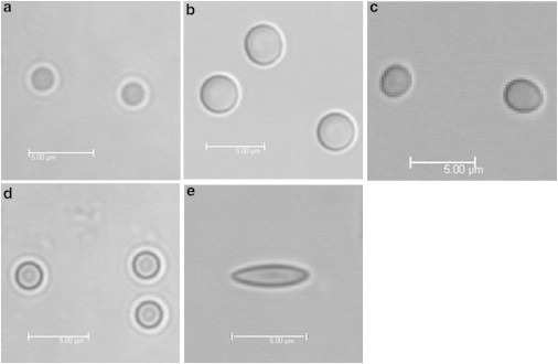
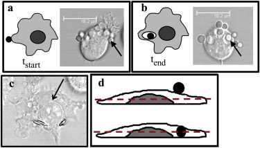
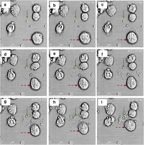
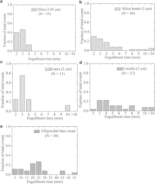
A complete understanding of phagocytosis requires insight into both its biochemical and physical aspects. One of the ways to explore the physical mechanism of phagocytosis is to probe whether and how the target properties (e.g., size, shape, surface states, stiffness, etc.) affect their uptake. Here we report an imaging-based method to explore phagocytosis kinetics, which is compatible with real-time imaging and can be used to validate existing reports using fixed and stained cells. We measure single-event engulfment time from a large number of phagocytosis events to compare how size and shape of targets determine their engulfment. The data shows an increase in the average engulfment time for increased target size, for spherical particles. The uptake time data on nonspherical particles confirms that target shape plays a more dominant role than target size for phagocytosis: Ellipsoids with an eccentricity of 0.954 and much smaller surface areas than spheres were taken up five times more slowly than spherical targets.


Figures

Figure 1
Figure 1
Optical micrographs of the different particles used in this study. (a) A 1.85-μm diameter silica sphere; (b) 3-μm diameter silica sphere; (c) ∼3-μm diameter dead conidia of Aspergillus fumigatus (slightly ovoid); (d) 2-μm diameter latex sphere; and (e) ∼1.5 × 1.5 × 5 μm latex ellipsoid. The dimensions of the ellipsoid indicate principal-axis lengths. All scale bars correspond to 5 μm.
Figure 2
Figure 2
Measurement of single-event engulfment time. Start of phagocytosis (tstart) is schematically defined as the time point when the particle (solid) makes irreversible contact with the cell membrane (a). A silica bead (indicated by the solid arrow) is seen to make contact with the RAW cell membrane. Engulfment (tend) is indicated either by the formation of a complete phagosome around the particle (b) or when the bead and the outline of the cell nucleus (shown by the solid arrow) are in focus in the same z-frame (c); ellipsoidal beads internalized by a RAW cell are seen in the panel. Z-stack imaging is used to determine whether a bead is actually ingested or is simply lying on top of the cell (d). The bead (solid) is considered to be engulfed when it lies in the same focal plane (dashed line) as the nucleus (dark shaded).
Figure 3
Figure 3
Time-lapse images from a typical phagocytosis event to illustrate the start and end points of phagocytosis. Only those frames that were in focus during z-stack data acquisition are shown here. The timestamps on the frames indicate the time elapsed since the start of image acquisition (t = 0). The phagocytosed bead (uncoated silica) is indicated by a solid (green) arrow in all the frames. The cell in question is indicated by a dashed (red) arrow. (ac) The cell membrane protrudes toward the bead without making contact. (d) The bead is seen to be in contact with the cell membrane for the first time in this frame. Therefore, we designate this time point as the start time (tstart: 20 min 10 s). (eg) Membrane ruffles usually accompanying phagocytosis are seen to form. (h) The phagosome starts forming, but the engulfment of the bead is not yet complete. (i) A complete phagosome boundary can be seen around the bead in this frame. Therefore, we call this time point as the end time (tend: 25 min 10 s). The single-event engulfment time (Δt = tendtstart) for this particular phagocytosis event is 5 min.
Figure 4
Figure 4
Histograms showing the distribution of engulfment time for targets of different sizes, shapes, and materials. Each plot has been normalized with respect to the total number of phagocytosis events (N) measured with the particular target. The spread in the distribution for each target results from the different phagocytic capabilities of the RAW cells in the particular population. (a) 1.85-μm silica spheres, (b) 3-μm silica spheres, (c) 2-μm latex spheres, (d) 3-μm conidia, and (e) ellipsoidal latex beads. Irrespective of size or material, >90% of the spherical targets are ingested within 10 min of making the first contact with the RAW cells, whereas only ∼10% of the ellipsoids are internalized in that period.

Similar articles

Cited by

References

    1. Cabeen M.T., Jacobs-Wagner C. Bacterial cell shape. Nat. Rev. Microbiol. 2005;3:601–610. - PubMed
    1. Aderem A., Underhill D.M. Mechanisms of phagocytosis in macrophages. Annu. Rev. Immunol. 1999;17:593–623. - PubMed
    1. Burke B., Lewis C.E., editors. The Macrophage. Oxford University Press; New York: 2002.
    1. Underhill D.M., Goodridge H.S. Information processing during phagocytosis. Nat. Rev. Immunol. 2012;12:492–502. - PMC - PubMed
    1. Schäfer G., Jacobs M., Brown G.D. Non-opsonic recognition of Mycobacterium tuberculosis by phagocytes. J. Innate Immun. 2009;1:231–243. - PMC - PubMed

Publication types