. 2013 Sep 8:14:20.
doi: 10.1186/1471-2199-14-20.

Pseudo attP sites in favor of transgene integration and expression in cultured porcine cells identified by Streptomyces phage phiC31 integrase

Affiliations

Pseudo attP sites in favor of transgene integration and expression in cultured porcine cells identified by Streptomyces phage phiC31 integrase

Yanzhen Bi et al. BMC Mol Biol. .

Abstract

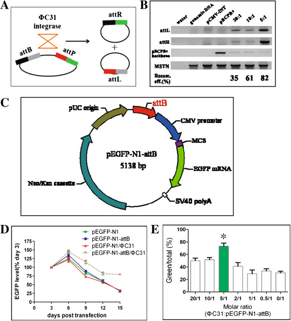
Phage PhiC31 integrase integrates attB-containing plasmid into pseudo attP site in eukaryotic genomes in a unidirectional site-specific manner and maintains robust transgene expression. Few studies, however, explore its potential in livestock. This study aims to discover the molecular basis of PhiC31 integrase-mediated site-specific recombination in pig cells. We show that PhiC31 integrase can mediate site-specific transgene integration into the genome of pig kidney PK15 cells. Intramolecular recombination in pig PK15 cell line occurred at maximum frequency of 82% with transiently transfected attB- and attP-containing plasmids. An optimal molar ratio of pCMV-Int to pEGFP-N1-attB at 5:1 was observed for maximum number of cell clones under drug selection. Four candidate pseudo attP sites were identified by TAIL-PCR from those cell clones with single-copy transgene integration. Two of them gave rise to higher integration frequency occurred at 33%. 5' and 3' junction PCR showed that transgene integration mediated by PhiC31 integrase was mono-allelic. Micro- deletion and insertion were observed by sequencing the integration border, indicating that double strand break was induced by the recombination. We then constructed rescue reporter plasmids by ABI-REC cloning of the four pseudo attP sites into pBCPB + plasmid. Transfection of these rescue plasmids and pCMV-Int resulted in expected intramolecular recombination between attB and pseudo attP sites. This proved that the endogenous pseudo attP sites were functional substrates for PhiC31 integrase-mediated site-specific recombination. Two pseudo attP sites maintained robust extracellular and intracellular EGFP expression. Alamar blue assay showed that transgene integration into these specific sites had little effect on cell proliferation. This is the first report to document the potential use of PhiC31 integrase to mediate site-specific recombination in pig cells. Our work established an ideal model to study the position effect of identical transgene located in diverse chromosomal contexts. These findings also form the basis for targeted pig genome engineering and may be used to produce genetically modified pigs for agricultural and biomedical uses.


Figures

Figure 1
Figure 1
PhiC31 integrase is active in pig cells. (A) Mechanism of action of PhiC31 integrase-mediated site-specific recombination. The integrase catalyzes the intramolecular recombination between attB and attP sites within a single plasmid, producing two mini-circle plasmids with hybrid attR and attL sites, respectively. (B) PhiC31 integrase is functional in pig cells. Transfection of pig PK15 cells were performed with three different ratios of PhiC31 integrase plasmid to reporter pBCPB+. Resultant attL and attR sites were examined by PCR. The indicated recombination efficiency was calculated by measuring signal intensity in Visioncapture with the following formula: recombination efficiency = Intensity of attL/Intensity of (attL and pBCPB + backbone). A 5:1 ratio had 82% recombination efficiency. (C) The map of pEGFP-N1-attB (5138 bp). A 400 bp attB sequence was sub-cloned into the AseI site of pEGFP-N1, resulting in the site-specific integration reporter pEGFP-N1-attB. (D) EGFP expression level in a time lapse. The y-axis shows the expression levels and x-axis shows days after transfection. Four groups of plasmids (pEGFP-N1, pEGFP-N1-attB, pEGFP-N1/PhiC31 and pEGFP-N1-attB/PhiC31) were transfected into PK15 cells. EGFP expression level was quantified by measuring the extracellular EGFP by a Glomax Multi Jr fluorescence detector. IGF-I was detected by ELISA and used as an internal control for data normalization. pEGFP-N1-attB/PhiC31 maintained high EGFP level in comparison to other settings. (E) An optimal molar ratio of PhiC31 and pEGFP-C1-attB plasmids produced more cell clones expressing EGFP. Various ratios of PhiC31 and pEGFP-N1-attB plasmids were transfected into PK15 cells and cell clones were selected by G418. The y-axis shows % of green to total clones and x-axis shows different molar ratios. A 5:1 ratio produced more cell clones expressing EGFP, significant higher than other settings, including random integration (0/1 ratio). *, p < 0.01.
Figure 2
Figure 2
Isolation of pig pseudo attP sites. (A) Screening of transgenic cell lines with single-copy transgene integration by quantitative PCR. 34 transgenic cell lines expressing EGFP (shown in x-axis) were screened for single-copy transgene integration within PK15 cellular genome. This assay demonstrated that 11 cell lines harbored a single-copy pEGFP-N1-attB. The other 23 cell lines had distinct copy numbers of pEGFP-C1-attB, varying from 2 copies to 15 copies. (B) Isolation of integration sites. Left, microscopic images of four transgenic cell clones under fluorescence field (10×). Middle, TAIL-PCR was performed to isolate the integration sites. Right, Specific PCR products were TA cloned, sequenced and aligned by BALT against pig genome (UCSC). Red arrow represents pig genome sequence, and green arrow represents pEGFP-N1-attB sequence. The lanes shown as 1st, 2nd and 3rd represent the first, second and third round TAIL-PCR products fractionized by 1.5% agarose gel. M is 1 kb DNA molecular marker (Fermentas).
Figure 3
Figure 3
Confirmation of integration sites and allele analysis. (A) attR and attL detection by junction PCR. Integration site and its neighboring sequences were retrieved from UCSC Pig Genome. Primers were designed across the junction site. PCR analysis revealed that pEGFP-N1-attB had been integrated into the sites identified by TAIL-PCR. In addition, all the 4 integration events occurred to one chromosome as demonstrated, implying that the transgene is integrated as single-allele at the 4 pseudo attP sites. (B) Micro insertion or deletion at integration site. PCR products in Figure 3A were TA cloned and sequenced to analyze the integration fidelity. Micro insertion and deletion were observed in the integration sites, implying that DNA strand breakage and repair occurred during recombination. (C) Representative examples of micro insertion or deletion at integration site. For attR hybrid sequence of 5156 pseudo attP site, four nucleotides “ACCC” were inserted between PK15 cellular genome and transgene sequence. For attR of 2015 pseudo attP site, two nucleotides of attB were lost.
Figure 4
Figure 4
Candidate pig pseudo attP sites can reconstitute site-specific recombination in a functional rescue assay. (A) Scheme of reporter plasmid construction. Pig pseudo attP sites were cloned into pBCPB+ plasmid to replace the wild-type attP site by ABI-REC. The resultant plasmid was named p’BCPB+. This resulted in 4 p’BCPB+ plasmids, i.e. 5113, 5156, 1015, and 2015, respectively. p’BCPB+ was used to reconstitute the attB-pseudo attP recombination with the presence of PhiC31 integrase. For the ABI-REC details, please refer to additional files. (B) Rescue assay. PhiC31 integrase plasmid, pBCPB+, and 4 p’BCPB+ were transfected into PK15 cells. 48 h post transfection, genomic DNA was extracted and used for PCR detection of attR hybrid site with attR-F and attR-R primers. Pig endogenous MSTN gene (500 bp) was used as internal control to normalize the quantity of genomic DNA template. Primers attL-F and attR-R (555 bp) were used to quantify the amount of pBCPB+. (C) Recombination efficiency. The VisionCapture tool was used to quantify the density of the PCR product in (B). Recombination efficiency was calculated with the following formulation: Amount of attR/(amount of attR + amount of pBCPB+). It shows that the recombination efficiency of wild-type attP and attB sites was up to 80%. Pig 5113 pseudo attP site has a recombination efficiency of 60%, higher than the other 3 pig pseudo attP sites.
Figure 5
Figure 5
Pig pseudo attP sites are in favor of robust transgene expression. (A) Extracellular EGFP expression. Medium containing extracellular EGFP was harvested for EGFP fluorescence and IGF-I detection. IGF-I was internal control for input normalization. IGF-I concentration was measured by a porcine ELISA kit. Sp, the supernatant of PK15 cell line as negative control. (B) Intracellular EGFP level by western blot. Transgenic cells were harvested and quantified by a BCA protein quantification kit. Anti-EGFP antibody was used to detect the intracellular EGFP expression, and anti-β-actin antibody was used as internal control. MW of EGFP and pig β-actin is 27 kD and 43 kD, respectively. They are shown by arrows. PK15 cell lysate was used as negative control. The intensity of the bands was quantified by VisionCapture and converted into Prism to create the bar graph. (C) Cell proliferation assay. 103 cells were seeded in a 96-well plate in triplicate. 10 μl Alamarblue indicator (Gibco) was added into the medium (with a final concentration of 10% v/v). The plate was incubated for additional 5 hours. The optical densities were measured at 570 nm and 630 nm in a micro-plate reader. Reduction percentage was calculated according to the formula provided in the instruction. Such a procedure was repeated in continuous 15 days. The reduction of four time points (day1, day5, day10 and day15) was indicated. Fresh medium was used as blank control. PK15 is the wild-type cell line. R1, R2 and R3 are randomly integrated transgenic cell lines.

Similar articles

Cited by

References

    1. Gama Sosa MA, De Gasperi R, Elder GA. Animal transgenesis: an overview. Brain Struct Funct. 2010;214:91–109. doi: 10.1007/s00429-009-0230-8. - DOI - PubMed
    1. Wilson MH, Coates CJ, George AL Jr. PiggyBac transposon-mediated gene transfer in human cells. Mol Ther. 2007;15:139–145. doi: 10.1038/sj.mt.6300028. - DOI - PubMed
    1. Hoffmann A, Kessler B, Ewerling S, Weppert M, Vogg B, Ludwig H, Stojkovic M, Boelhauve M, Brem G, Wolf E, Pfeifer A. Efficient transgenesis in farm animals by lentiviral vectors. EMBO Rep. 2003;4:1054–1060. doi: 10.1038/sj.embor.7400007. - DOI - PMC - PubMed
    1. Jakobsen JE, Li J, Kragh PM, Moldt B, Lin L, Liu Y, Schmidt M, Winther KD, Schyth BD, Holm IE, Vajta G, Bolund L, Callesen H, Jørgensen AL, Nielsen AL, Mikkelsen JG. Pig transgenesis by Sleeping Beauty DNA transposition. Transgenic Res. 2011;20:533–545. doi: 10.1007/s11248-010-9438-x. - DOI - PubMed
    1. Carlson DF, Garbe JR, Tan W, Martin MJ, Dobrinsky JR, Hackett PB, Clark KJ, Fahrenkrug SC. Strategies for selection marker-free swine transgenesis using the Sleeping Beauty transposon system. Transgenic Res. 2011;20:1125–1137. doi: 10.1007/s11248-010-9481-7. - DOI - PubMed

Publication types