Factors influencing the cell adhesion and invasion capacity of Mycoplasma gallisepticum
- PMID: 24011130
- PMCID: PMC3847126
- DOI: 10.1186/1751-0147-55-63
Factors influencing the cell adhesion and invasion capacity of Mycoplasma gallisepticum
Abstract
Background: The cell invasiveness of Mycoplasma gallisepticum, the causative agent of respiratory disease in chickens and infectious sinusitis in turkeys, may be a substantial factor in the well-known chronicity of these diseases and in the systemic spread of infection. To date, not much is known about the host factors and mechanisms involved in promotion or obstruction of M. gallisepticum adherence and/or cell invasion.In the current study, the influence of extracellular matrix (ECM) proteins such as fibronectin, collagen type IV and heparin, as well as plasminogen/plasmin, on the adhesion and cell invasion levels of M. gallisepticum to chicken erythrocytes and HeLa cells was investigated in vitro. Two strains, Rhigh and Rlow, which differ in their adhesion and invasion capacity, were analyzed by applying a modified gentamicin invasion assay. Binding of selected ECM molecules to M. gallisepticum was proven by Western blot analysis.
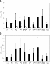
Results: Collagen type IV, fibronectin, and plasminogen exerted positive effects on adhesion and cell invasion of M. gallisepticum, with varying degrees, depending on the strain used. Especially strain Rhigh, with its highly reduced cell adhesion and invasion capabilities seemed to profit from the addition of plasminogen. Western and dot blot analyses showed that Rhigh as well as Rlow are able to adsorb horse fibronectin and plasminogen present in the growth medium. Depletion of HeLa cell membranes from cholesterol resulted in increased adhesion, but decreased cell invasion.
Conclusion: ECM molecules seem to play a supportive role in the adhesion/cell invasion process of M. gallisepticum. Cholesterol depletion known to affect lipid rafts on the host cell surface had contrary effects on cell adherence and cell invasion of M. gallisepticum.
Figures


Similar articles
-
Characterization of Mycoplasma gallisepticum pyruvate dehydrogenase alpha and beta subunits and their roles in cytoadherence.PLoS One. 2018 Dec 10;13(12):e0208745. doi: 10.1371/journal.pone.0208745. eCollection 2018. PLoS One. 2018. PMID: 30532176 Free PMC article.
-
Mycoplasma gallisepticum invades chicken erythrocytes during infection.Infect Immun. 2008 Jan;76(1):71-7. doi: 10.1128/IAI.00871-07. Epub 2007 Oct 22. Infect Immun. 2008. PMID: 17954728 Free PMC article.
-
Identification of fibronectin-binding proteins in Mycoplasma gallisepticum strain R.Infect Immun. 2006 Mar;74(3):1777-85. doi: 10.1128/IAI.74.3.1777-1785.2006. Infect Immun. 2006. PMID: 16495551 Free PMC article.
-
The expression of GapA and CrmA correlates with the Mycoplasma gallisepticum in vitro infection process in chicken TOCs.Vet Res. 2022 Sep 2;53(1):66. doi: 10.1186/s13567-022-01085-2. Vet Res. 2022. PMID: 36056451 Free PMC article.
-
Avian mycoplasmosis (Mycoplasma gallisepticum).Rev Sci Tech. 2000 Aug;19(2):425-42. Rev Sci Tech. 2000. PMID: 10935272 Review.
Cited by
-
Analysis of Paracoccidioides secreted proteins reveals fructose 1,6-bisphosphate aldolase as a plasminogen-binding protein.BMC Microbiol. 2015 Feb 27;15:53. doi: 10.1186/s12866-015-0393-9. BMC Microbiol. 2015. PMID: 25888027 Free PMC article.
-
Characterization of Mycoplasma gallisepticum pyruvate dehydrogenase alpha and beta subunits and their roles in cytoadherence.PLoS One. 2018 Dec 10;13(12):e0208745. doi: 10.1371/journal.pone.0208745. eCollection 2018. PLoS One. 2018. PMID: 30532176 Free PMC article.
-
Gga-miR-101-3p Plays a Key Role in Mycoplasma gallisepticum (HS Strain) Infection of Chicken.Int J Mol Sci. 2015 Dec 2;16(12):28669-82. doi: 10.3390/ijms161226121. Int J Mol Sci. 2015. PMID: 26633386 Free PMC article.
-
Diet Driven Differences in Host Tolerance Are Linked to Shifts in Global Gene Expression in a Common Avian Host-Pathogen System.Mol Ecol. 2025 Jun;34(12):e17793. doi: 10.1111/mec.17793. Epub 2025 May 12. Mol Ecol. 2025. PMID: 40356059 Free PMC article.
-
DnaK Functions as a Moonlighting Protein on the Surface of Mycoplasma hyorhinis Cells.Front Microbiol. 2022 Mar 3;13:842058. doi: 10.3389/fmicb.2022.842058. eCollection 2022. Front Microbiol. 2022. PMID: 35308339 Free PMC article.
References
-
- Lo SC, Dawson MS, Wong DM, Newton PB 3rd, Sonoda MA, Engler WF, Wang RY, Shih JW, Alter HJ, Wear DJ. Identification of Mycoplasma incognitus infection in patients with AIDS: an immunohistochemical, in situ hybridization and ultrastructural study. Am J Trop Med Hyg. 1989;41:601–616. - PubMed
-
- Lo SC, Hayes MM, Kotani H, Pierce PF, Wear DJ, Newton PB 3rd, Tully JG, Shih JW. Adhesion onto and invasion into mammalian cells by Mycoplasma penetrans: a newly isolated mycoplasma from patients with AIDS. Mod Pathol. 1993;6:276–280. - PubMed
Publication types
MeSH terms
LinkOut - more resources
Full Text Sources
Other Literature Sources

