. 2014 Jan;16(1):95-114.
doi: 10.1111/cmi.12186. Epub 2013 Sep 9.

The Toxoplasma gondii calcium-dependent protein kinase 7 is involved in early steps of parasite division and is crucial for parasite survival

Affiliations

The Toxoplasma gondii calcium-dependent protein kinase 7 is involved in early steps of parasite division and is crucial for parasite survival

Juliette Morlon-Guyot et al. Cell Microbiol. 2014 Jan.

Abstract

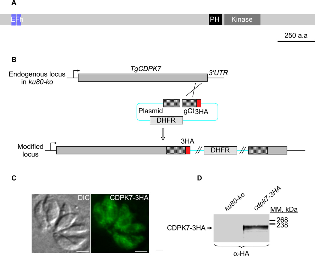
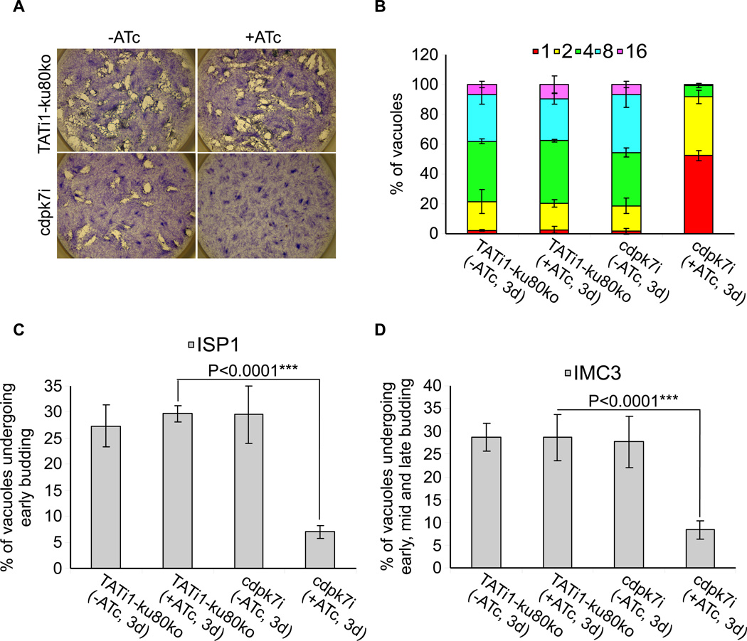
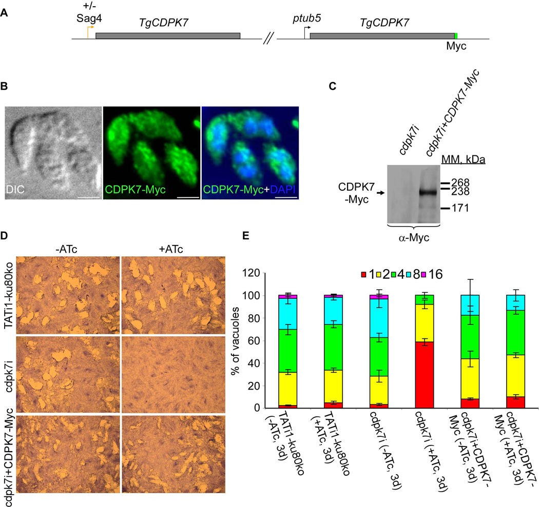
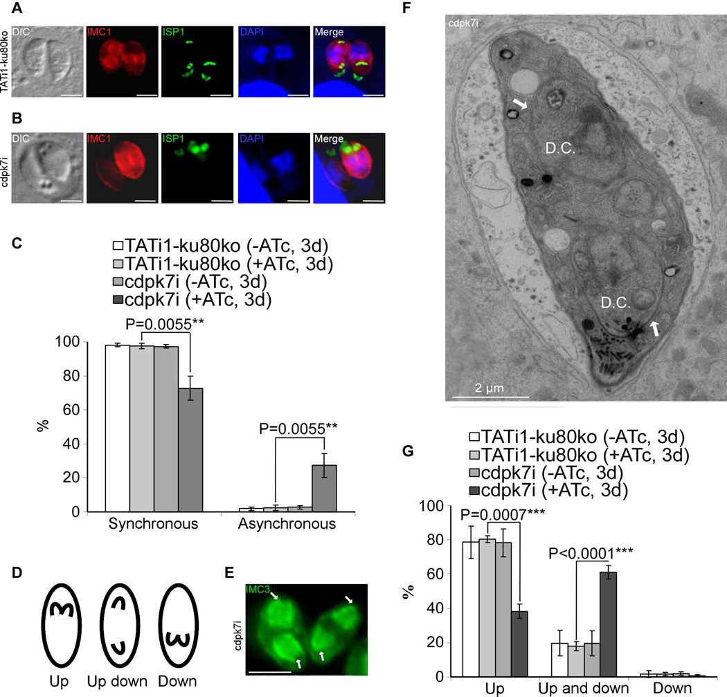
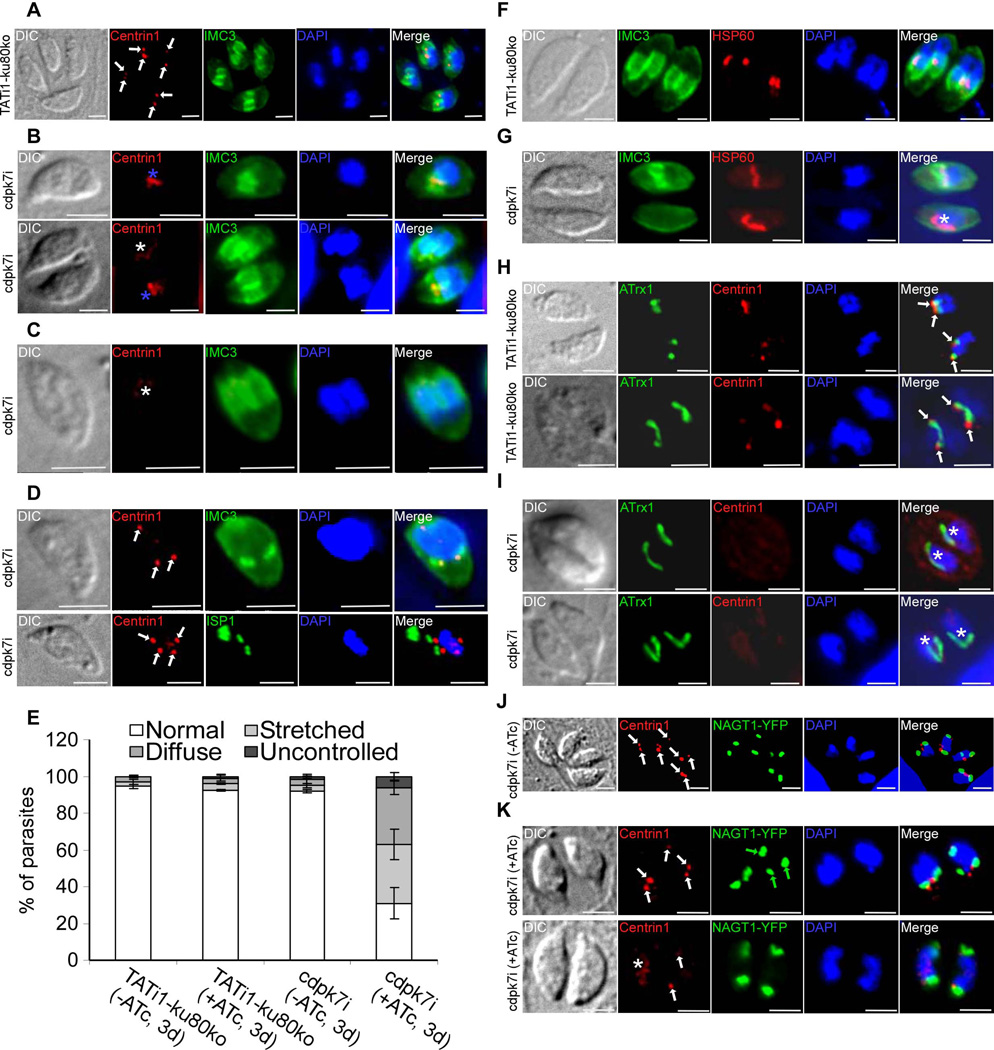
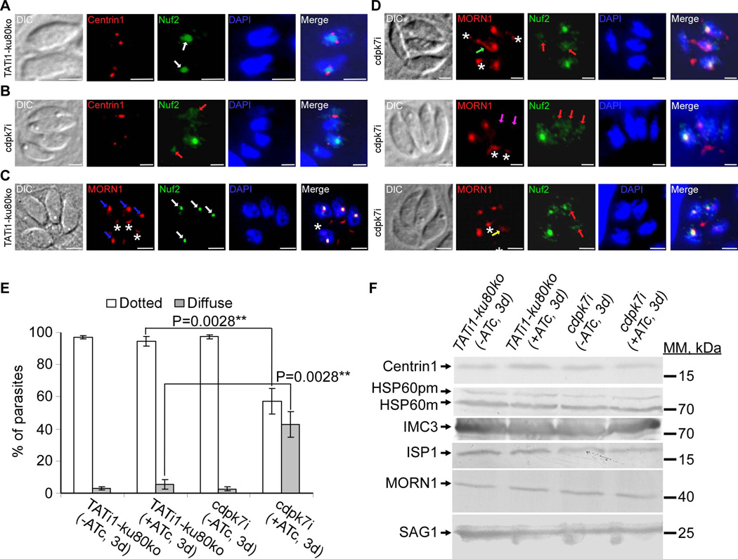
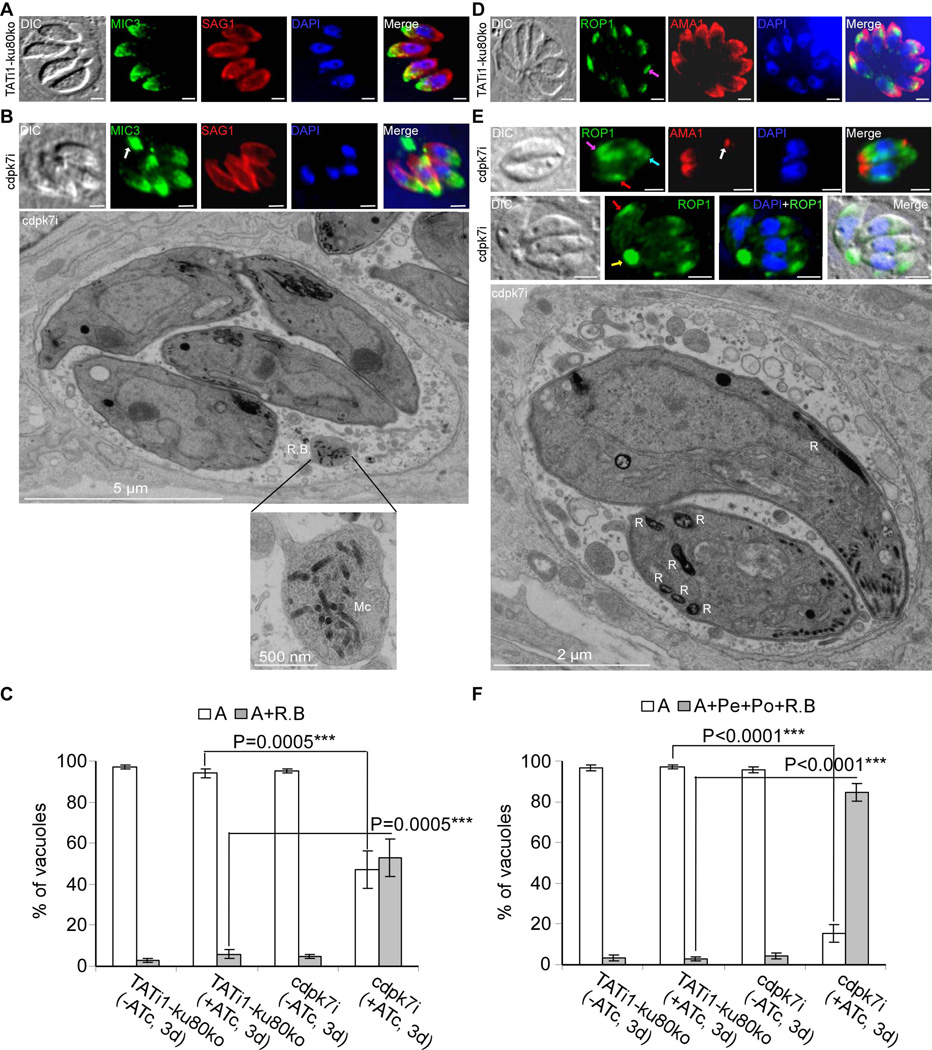
Apicomplexan parasites express various calcium-dependent protein kinases (CDPKs), and some of them play essential roles in invasion and egress. Five of the six CDPKs conserved in most Apicomplexa have been studied at the molecular and cellular levels in Plasmodium species and/or in Toxoplasma gondii parasites, but the function of CDPK7 was so far uncharacterized. In T. gondii, during intracellular replication, two parasites are formed within a mother cell through a unique process called endodyogeny. Here we demonstrate that the knock-down of CDPK7 protein in T. gondii results in pronounced defects in parasite division and a major growth deficiency, while it is dispensable for motility, egress and microneme exocytosis. In cdpk7-depleted parasites, the overall DNA content was not impaired, but the polarity of daughter cells budding and the fate of several subcellular structures or proteins involved in cell division were affected, such as the centrosomes and the kinetochore. Overall, our data suggest that CDPK7 is crucial for proper maintenance of centrosome integrity required for the initiation of endodyogeny. Our findings provide a first insight into the probable role of calcium-dependent signalling in parasite multiplication, in addition to its more widely explored role in invasion and egress.


Figures

Figure 1
Figure 1. Expression and localization of TgCDPK7 in tachyzoites
(A) Schematic representation of Toxoplasma gondii CDPK7 (TgCDPK7) showing the domains of homology to EF hands, the pleckstrin homology (PH) and the kinase domains. All domains were predicted using SMART EMBL. Scale bar represents 250 aa. (B) Insertion of three HA epitope tags at the C-terminus of TgCDPK7, by single homologous recombination at the 3’ of the corresponding gene (knock-in in RH-ku80ko strain). (C) IFA performed on intracellular transgenic parasites using anti-HA antibodies. Scale bars represent 2 µm. (D) Western blot analysis performed on transgenic or RH-ku80ko parasite lysates probed with anti-HA antibodies. TgCDPK7-3HA is found at the expected molecular mass (220 kDa).
Figure 2
Figure 2. TgCDPK7 conditional knock-in by promoter exchange strategy
(A) Schematic representation of the strategy used to replace the endogenous promoter of TgCDPK7 with the tetracycline inducible promoter. The DHFR-tetO7-SAG4NtgCDPK7 plasmid contains the dihydrofolate reductase (DHFR) gene (in blue) and the N-terminal genomic coding sequence of TgCDPK7 (in grey, 1455 bp) under the control of the inducible tetO7SAG4 promoter (Orange arrow). Black arrows represent the primers used for PCR analysis and the length of the PCR fragments generated is indicated. To study the regulation of TgCDPK7i gene, we inserted by single homologous recombination at the 3’ of the gene, a sequence coding for three HA epitope tags at the C-terminus of the corresponding TgCDPK7i protein. (B) PCR analysis performed on Tgcdpk7i, showing that single homologous recombination occurred. Genomic DNA from TATi1-ku80ko parasites was used as negative control. (C) Western blot analysis of TATi1-ku80ko and cdpk7i-3HA strains in presence or in absence of ATc. Parasites were treated for 1 or 2 or 3 days (d) with ATc. SAG1 was used as loading control. (D) Down regulation of TgCDPK7-3HA in the cdpk7i strain as shown by IFA with anti-HA antibodies 3 days after ATc treatment. Scale bars represent 2 µm. (E) Quantification of the percentage of vacuoles showing positive HA staining upon treatment with ATc at days 1 or 2 or 3 respectively using anti-HA antibodies.
Figure 3
Figure 3. Phenotypic consequences of TgCDPK7 depletion in cdpk7i strain
(A) Plaque assay performed on HFF monolayer infected with TATi1-ku80ko or cdpk7i parasites pretreated first during 48 hours with ATc. After 7 days ± ATc, the HFF were stained with Giemsa. (B) Intracellular growth of TATi1-ku80ko and cdpk7i cultivated in presence or absence of ATc for 48 hours and allowed to invade new HFF cells. Numbers of parasites per vacuole (X axis) were counted 24 hours after inoculation. The percentages of vacuoles containing varying numbers of parasites are represented on the Y-axis. Values are means ± SD for three independent experiments. (C and D) Endodyogeny assay performed on TATi-ku80ko or cdpk7i strains cultivated in presence or absence of ATc for 48 hours and allowed to invade new HFF cells. Numbers of vacuoles showing the formation of newly formed buds (Y axis) were counted 24 hours after inoculation using anti-ISP1 or anti-IMC3 antibodies. Values are means ± SD for three independent experiments. Statistical significance was evaluated using the student’s t test. ***P<0.0001 (C, ISP1), ***P<0.0001 (D, IMC3).
Figure 4
Figure 4. Functional complementation of the cdpk7i strain
(A) The scheme depicts the strategy used for complementation of cdpk7i parasites with CDPK7-Myc controlled by Tgtub5 promoter. (B) cdpk7i parasites stably expressing CDPK7-Myc were grown in presence of ATc during 3 days and visualized by IFA using anti-Myc antibodies. (C) Constitutive expression of integrated CDPK7-Myc in cdpk7i strain was confirmed by western blotting using anti-Myc antibodies. (D) Plaque assays were carried out by infecting HFF monolayers with TATi1-ku80ko or cdpk7i or cdpk7i stably expressing CDPK7-Myc for 7 days ± ATc. (E) Intracellular growth of TATi1-ku80ko, cdpk7i and cdpk7i stably expressing CDPK7-Myc cultivated in presence or absence of ATc for 48 hours and allowed to invade new HFF cells. Numbers of parasites per vacuole (X axis) were counted 24 hours after inoculation. The percentages of vacuoles containing varying numbers of parasites are represented on the Y-axis. Values are means ± SD for three independent experiments.
Figure 5
Figure 5. cdpk7i mutant parasites display asynchronous division and reveal that TgCDPK7 contributes to the close positioning of daughter cells during division
(A) and (B): IFA analysis of vacuoles containing 2 parasites undergoing division after 3 days of ATc treatment. (A): IFA of representative TATi1-ku80ko vacuole which was compared to IFA of representative cdpk7i vacuole, (B). (A) and (B): anti-IMC1 (in red) and anti-ISP1 (in green) antibodies, respectively, were used to detect the nascent buds. In (A) TATi1-ku80ko vacuole exhibits synchronous IMC buds, while in the cdpk7i vacuole shown in (B), the formation of IMC buds appears only in one parasite. (C) Scoring of synchronous and asynchronous dividing parasites containing vacuoles by IFA, using anti-IMC3 antibodies. Statistical significance was evaluated using the student’s t test. **P=0.0055 (synchronous), **P=0.0055 (asynchronous). (D) A scheme showing the three directions adopted by the IMC buds during parasite division. (E) IFA of representative cdpk7i parasites showing an up/down division (with anti-IMC3 antibodies staining), one nascent IMC grows towards the apical end, while the second buds towards the posterior pole of the parasite (white arrows). (F) Electron micrograph of a dividing cdpk7i parasite (treated with ATc during 3 days). Two daughters are assembled within a mother cell in up/down orientation (white arrows). D.C.: daughter cells. (G) Scoring of daughter cell orientation during parasite division by IFA using anti-ISP1 antibodies. Interference with TgCDPK7 function favoured the up/down topology whereas wild-type or untreated parasites adopted mainly the up topology. Scale bars represent 2 µm. Statistical significance was evaluated using the student’s t test. ***P=0.0007 (up), ***P<0.0001 (up and down).
Figure 6
Figure 6. cdpk7i mutant parasites display centrosomal defects, reveal a plastid elongation independent of centrin 1 protein and IMC buds and show the dependency of the Golgi positioning and number with that of centrosome duplication
A) to (D): IFA analysis of parasites undergoing division after 3 days of ATc treatment. (A) to (D): anti-centrin1 and anti-IMC3 or anti-ISP1 antibodies were used to stain the centrosome and daughter buds, respectively. (A) A representative TATi1-ku80ko vacuole containing 4 parasites is shown. The duplicated centrosomes (in red) in each parasite are marked with a white arrow. Each newly formed IMC (in green) encapsulates one centrosome. (B) to (D): Representative examples of cdpk7i parasites showing centrosomes or centrin 1 staining defects. (B) A stretched centrosome (blue asterisks) which is located in between two newly formed daughter buds (upper image) or partitioned without being split between the nascent IMCs (lower image) is shown. (C) A diffused centrin 1 staining (white asterisk) is shown. (D) Two parasites in division showing an abnormal number of centrosomes per nucleus (white arrows) (3 in the upper image and 4 in the lower picture). (E) Scoring of centrosome defects during parasite division by IFA using anti-centrin1 antibodies. Interference with TgCDPK7 function led to stretched centrosomes, affected the distribution of centrin 1 protein and impaired the regular number of centrosome duplication. Data are mean values ± SD for three independent experiments. (F) to (I): IFA analysis of vacuoles containing 2 parasites undergoing division after 3 days of ATc treatment. (F) and (H): IFAs of representative TATi1-ku80ko vacuoles which were compared to IFAs of representative cdpk7i vacuoles, (G) and (I). (F) and (G): anti-IMC3 and anti-HSP60 antibodies were used to stain the daughter buds and the apicoplast, respectively. (F): a TATi1-ku80ko vacuole with newly formed IMC (in green) encapsulating each apicoplast. (G) A cdpk7i vacuole exhibiting the formation of IMC buds in only one parasite out of two. In the parasite lacking the 2 IMC buds, the apicoplast adopts its elongated shape and lies on the nucleus (white asterisk). (H) and (I): anti-centrin 1 and anti-ATrx1 antibodies were used to stain the centrosome and the apicoplast, respectively. (H) The ends of the plastid in dividing apicoplasts of the TATi1-ku80ko parasites are consistently associated with the parasite’s centrosomes. This association is maintained even after apicoplast division (white arrows). (I) Two representative cdpk7i vacuoles showing the diffused staining of centrin 1 protein. In the parasite lacking a concentrated centrin 1 dotted-like staining, the apicoplast adopts its elongated shape and lies on the nucleus (white asterisk). (J) and (K): anti-centrin 1 antibodies and NAGT1-YFP were used to detect the centrosome and the Golgi, respectively. (J) The two centrosomes (white arrows) are found at the inner ends of the newly-divided Golgi (in green). (K) Two representative cdpk7i vacuoles treated with ATc showing an abnormal numbers of centrosomes and Golgi per nucleus (white and green arrows, upper image), and a diffuse staining of centrin 1 protein (white asterisk) or two centrosomes in up and down position (white arrows) (lower image). Scale bars represent 2 µm.
Figure 7
Figure 7. TgCDPK7 knockdown affects the distribution of MORN1 and Nuf2 proteins
(A) to (D): IFA analysis of parasites undergoing division after 3 days of ATc treatment. (A) and (C): IFAs of representative TATi1-ku80ko parasites which were compared to IFAs of representative cdpk7i mutant parasites, (B) and (D). (A) and (B): anti-centrin1 (in red) and anti-Nuf2 (in green) antibodies were used to stain the centrosome and kinetochore respectively. (A) Two TATi1-ku80ko vacuoles were shown with each containing 1 parasite undergoing division. The kinetochore (in green) in each parasite is marked with a white arrow. TgNuf2 protein which is a component of the kinetochore complex is concentrated in the parasite nucleus and is flanked by two centrosomes during division (white arrows). (B) A cdpk7i vacuole showing the typical dotted-like staining of Nuf2 protein surrounded by a duplicated centrosome in only 1 parasite out of 4. In the parasites displaying a stretched centrosome or an undetectable centrin 1 labelling, the Nuf2 protein was found diffused throughout the nucleus (pink arrows). (C) and (D): anti-Myc (TATi1-ku80ko and cdpk7i parasites express a Myc tagged MORN1 protein) and anti-Nuf2 antibodies were used to label the spindle pole and kinetochore sub-cellular structures respectively. (C) A TATi1-ku80ko vacuole containing 4 parasites is shown. The kinetochore (in green) in each parasite is marked with a white arrow. TgNuf2 protein (white arrows) shows a partial colocalisation with MORN1 protein which is a component of the spindle pole structure located at the nuclear envelope of the parasite (blue arrows). The MORN1 protein marked also the basal end of the parasites (white asterisks). (D) IFA showed abnormal MORN1 protein distribution at the spindle pole in cdpk7i parasites displaying a diffused Nuf2 protein (pink arrows). The MORN1 protein was distributed in the form of a pipe (green arrow), was found undetectable (the two white arrows) or diffused (yellow arrow). Scale bars represent 2 µm. (E) Scoring of Nuf2 protein distribution defect by IFA using anti-Nuf2 antibodies. Data are mean values ± SD for three independent experiments. Statistical significance was evaluated using the student’s t test. **P=0.0028 (dotted), **P=0.0028 (diffuse). (F) Western blot analyses performed on mutant or TATi1-ku80ko parasite lysates probed with anti-centrin1 or anti-HSP60 or anti-IMC3 or anti-ISP1 or anti-Myc or anti-SAG1 antibodies. pm: pre-mature; m: mature.
Figure 8
Figure 8. TgCDPK7 knockdown accumulates micronemes in residual bodies and impairs rhoptries positioning to the apical end of the parasite
(A), (B), (D) and (E): IFA analysis of parasites after 3 days of ATc treatment. Scale bars represent 2 µm. (A) TATi1-ku80ko parasites showed a normal apical staining of MIC3 protein (in green). The SAG1 protein localizes at the parasite plasma membrane (in red). (B) cdpk7i parasites showed accumulation of micronemes in residual bodies (white arrow). Thin section electron micrographs were taken from cdpk7i that had grown for a total of 3 days in presence of ATc. A vacuole with 4 parasites is presented showing a residual body containing intact micronemes (Mc). Some micronemes and rhoptries were also found correctly localized to the parasite apical pole. Scale bar, 5 µm. (C) Scoring of vacuoles showing accumulation of MIC3 protein in residual bodies by IFA using anti-MIC3 antibodies. Data are mean values ± SD for three independent experiments. Statistical significance was evaluated using the student’s t test. ***P=0.0005 (A), ***P=0.0005 (A+ R.B). A: apical, R.B: residual body. (D) TATi1-ku80ko parasites showed a normal apical staining of ROP1 (a rhoptry marker, in green) (pink arrows) and AMA1 proteins (a microneme marker, in red). (E) In the cdpk7i parasites, rhoptries were dispersed at the periphery (red arrows), at the posterior ends (blue arrow) and in residual bodies (yellow arrow). Thin section electron micrographs depict rhoptry organelles (R) at the periphery or at the posterior ends of cdpk7i parasites that had grown for a total of 3 days with ATc. Scale bar, 2 µm. (F) Scoring of vacuoles showing abnormal distribution of ROP1 protein at the periphery, at the posterior ends and in residual bodies by IFA using anti-ROP1 antibodies. Data are mean values ± SD for three independent experiments. Statistical significance was evaluated using the student’s t test. ***P<0.0001 (A), ***P<0.0001 (A+ Pe + Po + R.B). A: apical, Pe: periphery, Po: Posterior ends, R.B: residual body.

Similar articles

Cited by

References

    1. Achbarou A, Mercereau-Puijalon O, Autheman JM, Fortier B, Camus D, Dubremetz JF. Characterization of microneme proteins of Toxoplasma gondii. Mol Biochem Parasitol. 1991;47:223–233. - PubMed
    1. Agrawal S, van Dooren GG, Beatty WL, Striepen B. Genetic evidence that an endosymbiont-derived endoplasmic reticulum-associated protein degradation (ERAD) system functions in import of apicoplast proteins. J Biol Chem. 2009;284:33683–33691. - PMC - PubMed
    1. Ahmed A, Gaadhe K, Sharma GP, Kumar N, Neculai M, Hui R, et al. Novel insights into the regulation of malarial calcium-dependent protein kinase 1. FASEB J. 2012;26:3212–3221. - PMC - PubMed
    1. Anderson-White B, Beck JR, Chen CT, Meissner M, Bradley PJ, Gubbels MJ. Cytoskeleton assembly in Toxoplasma gondii cell division. Int Rev Cell Mol Biol. 2012;298:1–31. - PMC - PubMed
    1. Anderson-White BR, Ivey FD, Cheng K, Szatanek T, Lorestani A, Beckers CJ, et al. A family of intermediate filament-like proteins is sequentially assembled into the cytoskeleton of Toxoplasma gondii. Cell Microbiol. 2011;13:18–31. - PMC - PubMed

Publication types

LinkOut - more resources