. 2013 Sep 8;3(1):59.
doi: 10.1186/1869-5760-3-59.

Longitudinal spectral domain optical coherence tomography changes in eyes with intraocular lymphoma

Affiliations

Longitudinal spectral domain optical coherence tomography changes in eyes with intraocular lymphoma

Hyun Soo Jang et al. J Ophthalmic Inflamm Infect. .

Abstract

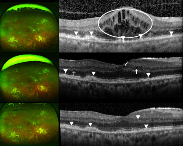
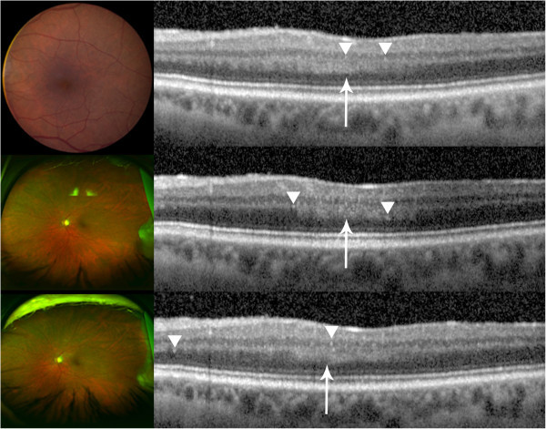
Background: Cases of patients with primary intraocular lymphoma (PIOL) were retrospectively analyzed to describe the longitudinal intra-retinal morphological changes in PIOL as visualized on images obtained by spectral domain optical coherence tomography (SD-OCT).

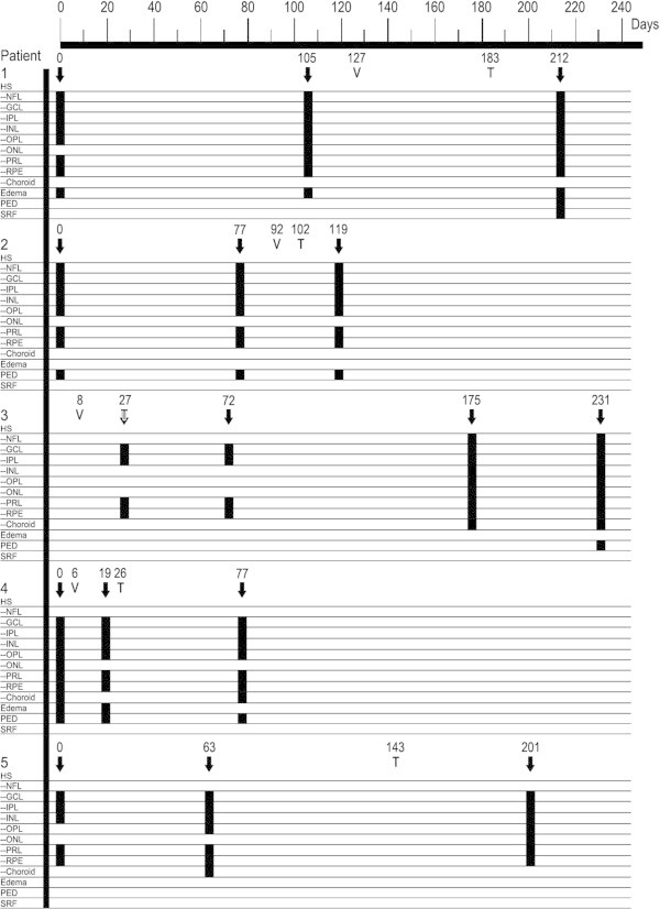
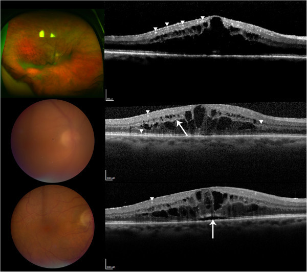
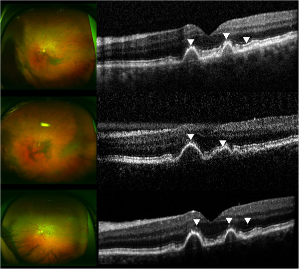
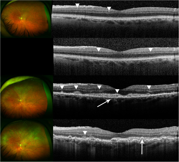
Results: In a retrospective case series, Heidelberg Spectralis SD-OCT images obtained in the longitudinal evaluation of patients with biopsy-proven PIOL were analyzed and assessed. The images were graded for the presence of macular edema (ME), pigment epithelial detachment (PED), subretinal fluid (SRF), and hyperreflective signals. SD-OCT scans of five eyes from five patients were assessed. Patients showed signs of inflammation, such as ME and SRF, which were resolved with treatments in some cases. Hyperreflective signals were found in all eyes in the form of nodules or bands across the retina, with the highest frequency of appearance in the ganglion cell layer, inner plexiform layer, photoreceptor layer, and retinal pigment epithelium; such signals increased with the progression of PIOL.

Conclusion: SD-OCT may be employed to monitor the progression of PIOL. Hyperreflective signals on OCT may correspond with increase in disease activities, along with other findings such as ME, PED, and SRF.


Figures

Figure 1
Figure 1
Longitudinal SD-OCT finding. Day 0 corresponds to the baseline visit for patients 1 to 4 and the first visit of the latest recurrence of intraocular lymphoma for patient 5. V indicates the date of the diagnostic vitrectomy. T indicates the initiation of therapy (systemic methotrexate and/or rituximab). SD-OCT images taken on the days marked with downward arrows were graded for ME, PED, SRF, and HS in retinal layers with positive finding being indicated by a black box.
Figure 2
Figure 2
Patient 1. (Top) Day 0, there are multiple hyperreflective signals in the inner retina, indicated by the arrowheads. ME is present. The Optos® wide-angle fundus image shows sheets of cells. (Center) Day 105, examples of hyperreflective signals are pointed by the arrowheads. The arrowhead on the right points to thickening of the OPL. The arrow indicates hyperreflective signals in OPL. Slight disruption of the RPE is pointed by the bottom arrowhead. Color fundus shows significant vitritis. (Bottom) Day 212, the arrow points to elevation of RPE due to an accumulation of SRF. A nodular hyperreflective signal is pointed by the arrowhead. The view of the fundus is clear following vitrectomy.
Figure 3
Figure 3
Patient 2. (Top) Day 0, multiple nodular hyperreflective signals in the outer retina are indicated by the arrowheads. The rightmost arrowhead points to hyperreflective signals at PRL and RPE level. RPE disruption is present due to drusen. There are sheets of vitreous cells shown in the Optos® wide-angle image. (Center) Day 77, arrowheads indicate hyperreflective changes at PRL and RPE. Severe vitritis as shown in the left fundus contributes to the noisy SD-OCT scan. Optos® wide-angle image shows increased vitreous inflammation. (Bottom) Day 119, arrowheads point to nodular hyperreflective signals that may be lymphomatous infiltrates. The view of the fundus is clearer post-vitrectomy.
Figure 4
Figure 4
Patient 3. (Top) Day 27, the image appears normal with few abnormal hyperreflective signals that may indicate infiltrates as indicated by the arrowheads. Optos® wide-angle image shows clear vitreous following vitrectomy. (Center, upper) Day 72, the retina remains unchanged with a few hyperreflective signals as indicated by the arrowheads. No fundus image was available. (Center, lower) Day 175, evident hyperreflective signals are pointed by arrowheads. Layers are distorted, and RPE is disrupted as shown by the arrow. Inner segment/outer segment interface is no longer visible. There are new yellow lesions seen in the Optos® wide-angle image. A creamy lesion in the fovea is evident. (Bottom) Day 231, an increased amount of hyperreflective signals is found throughout the retina, and the overall reflectivity has also increased. RPE disruption has developed into pigment epithelial detachment as indicated by the arrow. Hyperreflective foci are found in the choroid as well. Optos® wide-angle image shows an increased degree of infiltration.
Figure 5
Figure 5
Patient 4. (Top) Day 0, macular edema with intracystic changes is circled. Arrowheads indicate hyperreflective signals at the PRL and RPE. The OCT scan was done before vitrectomy; there is significant noise in the scan. Optos® wide-angle image is from Day 14, following vitrectomy. (Center) Day 19, the arrows indicate intraretinal fluids. Lesions at the PRL and RPE are pointed by the arrowheads. Optos® wide-angle image from the same day shows stable retina. (Bottom) Day 77, hyperreflective signals at the RPE pointed by the arrowheads have arguably thickened. Hyperreflective signals are also seen in the inner retina. An arrowhead points to an example in the inner nuclear layer. No changes are seen in the Optos® wide-angle image from the same day.
Figure 6
Figure 6
Patient 5. Unusually thick OPL is noted by an arrow and hyperreflective signals are indicated with arrowheads. (Top) Day 0, color fundus photograph is from a visit that took place a month before Day 0; no significant change in the fundus examination was noted in between the two visits. (Center) Day 63, an increase in hyperreflective signals is seen, which correlates to the increase in disease activity. (Bottom) Day 201, an increase in hyperreflective signals was noted, which may correlate to a significant increase in disease activity, manifested also by increased hypopyon, on this visit.

References

    1. Bardenstein DS. Intraocular lymphoma. Cancer Control. 1998;3(4):317–325. - PubMed
    1. Choi JY, Kafkala C, Foster CS. Primary intraocular lymphoma: a review. Semin Ophthalmol. 2006;3(3):125–133. doi: 10.1080/08820530500350498. doi:10.1080/08820530500350498. - DOI - PubMed
    1. Davis JL. Diagnosis of intraocular lymphoma. Ocul Immunol Inflamm. 2004;3(1):7–16. doi: 10.1076/ocii.12.1.7.28072. - DOI - PubMed
    1. Rothova A, Ooijman F, Kerkhoff F, Van Der Lelij A, Lokhorst HM. Uveitis masquerade syndromes. Ophthalmology. 2001;3(2):386–399. doi: 10.1016/S0161-6420(00)00499-1. - DOI - PubMed
    1. Velez G, de Smet MD, Whitcup SM, Robinson M, Nussenblatt RB, Chan CC. Iris involvement in primary intraocular lymphoma: report of two cases and review of the literature. Surv Ophthalmol. 2000;3(6):518–526. doi: 10.1016/S0039-6257(00)00118-1. - DOI - PubMed

LinkOut - more resources