. 2013 Sep 6:13:10.
doi: 10.1186/1471-5945-13-10.

Toll-like receptor 2 activation and comedogenesis: implications for the pathogenesis of acne

Affiliations

Toll-like receptor 2 activation and comedogenesis: implications for the pathogenesis of acne

Joanne Louise Selway et al. BMC Dermatol. .

Abstract

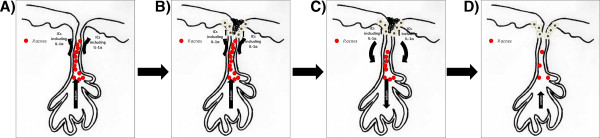
Background: Acne is a common disorder of the human pilosebaceous unit, yet the mechanisms underlying hyperkeratinisation and subsequent inflammation (comedogenesis) remain to be determined, although cutaneous pathogens are implicated. Previously, it was reported that the release of the cytokine interleukin-1α (IL-1α) by keratinocytes of the sebaceous duct was pivotal in the life cycle of the comedone, mediating both its development and its spontaneous resolution. Toll-like receptors are a family of molecules that recognise pathogen associated molecular patterns (PAMPs) presented by microorganisms, initiating a signalling cascade terminating in the release of antimicrobial compounds and cytokines.

Methods: We used ex vivo sebaceous gland and primary monolayer keratinocyte culture, alongside ELISAs, immunohistochemistry, Western blotting and RT-PCR to investigate the contribution of TLR activation to acne pathogenesis.

Results: We found TLR2 to be expressed in basal and infundibular keratinocytes, and sebaceous glands, and its activation provoked the release of IL-1α from primary human keratinocytes in vitro. The exposure of microdissected human sebaceous glands to PAMPs specific for TLR2 in vitro resulted in a pattern of IL-1α like cornification after seven days of exposure.

Conclusions: TLR activation and secretion of IL-1α from keratinocytes may be initiating steps in comedogenesis and, therefore, critical to the pathophysiology of acne.


Figures

Figure 1
Figure 1
TLR Expression in vitro and in vivo. A) TLR2 expression was assessed by RT-PCR in cDNA prepared from HaCaT keratinocytes (first lane), NHEK (second lane) or freshly-isolated human sebaceous glands (third lane, representative of ten different individuals). A no-template cDNA control was included (fourth lane). B) TLR2 antisera were used to evaluate receptor localisation in skin isolated from ten individuals and representative expression patterns are shown, with and without a blocking peptide (pep). Images were captured at 10 × original magnification.
Figure 2
Figure 2
TLR activation results in IL-1α release and NF-κB activation. A) Preconfluent NHEK were treated with PGN in the presence or absence of a TLR2 neutralising antibody, and levels of IL-1α release determined by ELISA. B) Three separate cultures of preconfluent NHEK were harvested following 90 min exposure to PGN, and IκB expression relative to β-actin was determined by Western blotting.
Figure 3
Figure 3
TLR activation promotes hypercornification in isolated human sebaceous glands maintained ex vivo. A) Histological sections of freshly-isolated and IL-1α treated sebaceous glands are shown, along with an example of a comedone in situ. B) Morphology representative of 3 experiments showing sebaceous glands treated with vehicle only (control), TLR2 (LTA and PGN) or TLR4 agonists (LPS), or TLR agonists pre-incubated with a blocking antibody (block). All images are shown at 10 × original magnification.
Figure 4
Figure 4
A model of TLR activation and the life-cycle of acne. A)P. acnes colonises the sebaceous gland and stimulates infundibular keratinocytes to release inflammatory cytokines (ICs) including IL-1α via TLR activation. B) ICs, including IL-1α, stimulate hypercornification, thus formation of the comedone. C) IC secretion, including IL-1α, is also associated with a reduction in lipogenesis in basal sebocytes, thereby reducing the sebum levels and starving the P. acnes of nutrients. D) Reduction in P. acnes levels reduces IC secretion, resolution of the comedone and restoration of sebocyte maturation. Adapted from Downie MM et al.[3].

References

    1. Ingham E, Eady EA, Goodwin CE, Cove JH, Cunliffe WJ. Pro-inflammatory levels of interleukin-1 alpha-like bioactivity are present in the majority of open comedones in acne vulgaris. J Invest Dermatol. 1992;98:895–901. doi: 10.1111/1523-1747.ep12460324. - DOI - PubMed
    1. Guy R, Green MR, Kealey T. Modeling acne in vitro. J Invest Dermatol. 1996;106:176–182. doi: 10.1111/1523-1747.ep12329907. - DOI - PubMed
    1. Downie MM, Sanders DA, Kealey T. Modelling the remission of individual acne lesions in vitro. Br J Dermatol. 2002;147:869–878. doi: 10.1046/j.1365-2133.2002.04946.x. - DOI - PubMed
    1. Wolff K, Stingl G. The Langerhans cell. J Invest Dermatol. 1983;80(Suppl):17s–21s. - PubMed
    1. Lemaitre B, Nicolas E, Michaut L, Reichhart JM, Hoffmann JA. The dorsoventral regulatory gene cassette spatzle/Toll/cactus controls the potent antifungal response in Drosophila adults. Cell. 1996;86:973–983. doi: 10.1016/S0092-8674(00)80172-5. - DOI - PubMed

Publication types