. 2013 Sep 4;79(5):903-16.
doi: 10.1016/j.neuron.2013.06.035.

The EBAX-type Cullin-RING E3 ligase and Hsp90 guard the protein quality of the SAX-3/Robo receptor in developing neurons

Affiliations

The EBAX-type Cullin-RING E3 ligase and Hsp90 guard the protein quality of the SAX-3/Robo receptor in developing neurons

Zhiping Wang et al. Neuron. .

Abstract

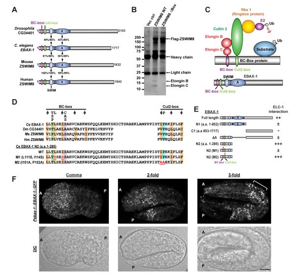
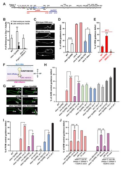
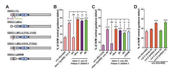
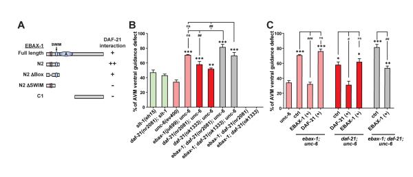
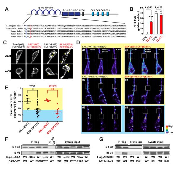
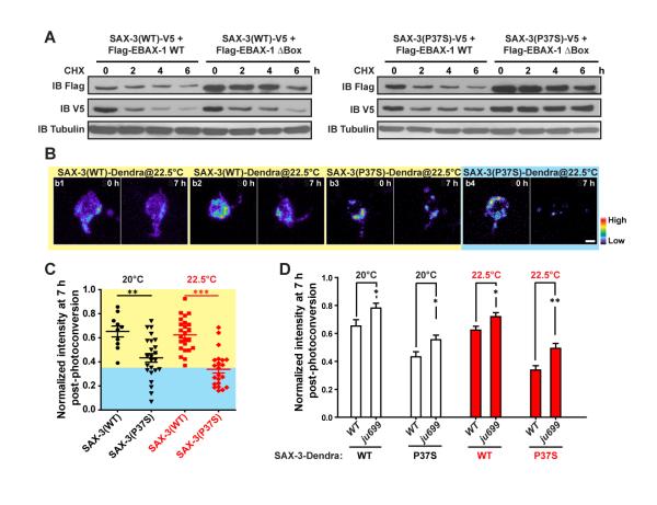
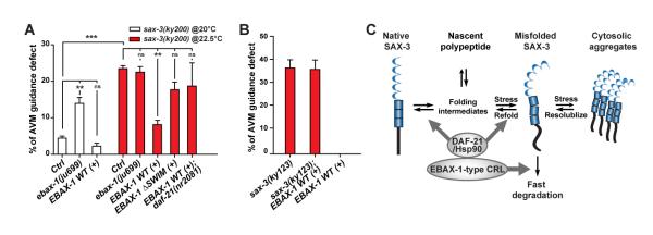
Although protein quality control (PQC) is generally perceived as important for the development of the nervous system, the specific mechanisms of neuronal PQC have remained poorly understood. Here, we report that C. elegans Elongin BC-binding axon regulator (EBAX-1), a conserved BC-box protein, regulates axon guidance through PQC of the SAX-3/Robo receptor. EBAX-1 buffers guidance errors against temperature variations. As a substrate-recognition subunit in the Elongin BC-containing Cullin-RING ubiquitin ligase (CRL), EBAX-1 also binds to DAF-21, a cytosolic Hsp90 chaperone. The EBAX-type CRL and DAF-21 collaboratively regulate SAX-3-mediated axon pathfinding. Biochemical and imaging assays indicate that EBAX-1 specifically recognizes misfolded SAX-3 and promotes its degradation in vitro and in vivo. Importantly, vertebrate EBAX also shows substrate preference toward aberrant Robo3 implicated in horizontal gaze palsy with progressive scoliosis (HGPPS). Together, our findings demonstrate a triage PQC mechanism mediated by the EBAX-type CRL and DAF-21/Hsp90 that maintains the accuracy of neuronal wiring.


Figures

Figure 1
Figure 1. EBAX-1 is a Conserved BC-box Protein Serving as a Substrate Recognition Subunit in a Cullin-RING E3 Ligase Complex (CRL)
(A) Schematic diagrams of Drosophila, C. elegans, mouse and human homologs of EBAX-1. The EBAX family of proteins contain a BC-box (purple), a Cul2-box (green) and a zinc-finger SWIM domain (light blue) at the N-terminus followed by several conserved regions, the largest of which is marked as domain A. Numbers indicate percentages of identity and similarity, respectively. (B) Endogenous human Elongin B and Elongin C interact with the mouse homolog of EBAX-1 (ZSWIM8). HEK293T cells stably expressing Flag-tagged ZSWIM8 WT, ZSWIM8 ΔBox mutant or an empty vector control were subjected to immunoprecipitation with mouse anti-Flag antibodies. The immunoprecipitants were separated by SDS-PAGE and revealed by silver staining. (C) Schematic illustration of a BC box-type Cullin-RING E3 ubiquitin ligase (CRL). (D) Alignment of the BC-box and Cul2-box of EBAX-1 homologs and mutants. Amino acids that are conserved in more than 2 sequences are highlighted: orange (aliphatic residues), green (Thr or Pro), purple (Ala or Ser), and blue (Pro). The consensus BC-box and Cul2-box sequences are indicated above the alignment (Mahrour et al., 2008). Essential amino acids for Elongin B/C and CUL2 interactions (marked in red and underlined) were mutated in EBAX-1 M1 (L111S, I114S) and M2 (I151A, P152A) mutants. (E) Interactions between C. elegans ELC-1 and EBAX-1 full-length protein or mutants in yeast two-hybrid (Y2H) assays. ++ and +++, intermediate and strong interaction; ±, weak interaction; −, no interaction. (F) EBAX-1 is expressed in the embryonic nervous system. Representative Pebax-1::EBAX-1::GFP fluorescence and DIC images from the comma stage, the 2-fold stage and the 3-fold stage are shown. The anterior (A) and posterior (P) ends of embryos are marked on images. The bracket in the right panel indicates developing anterior neurons. Scale, 10 μm.
Figure 2
Figure 2. EBAX-1 Regulates the Ventral Axon Guidance of AVM and PVM Neurons
(A) Illustration of the genomic structure of ebax-1. In the ebax-1(ju699) allele, a region of 1548 bp covering the SWIM domain and the domain A is deleted. In the ebax-1(tm2321) allele, a region of 551 bp in exon 5 is deleted. (B) ebax-1 mutants show mild egg-laying (Egl) defects. The Egl phenotype can be fully rescued by ebax-1 genomic DNA drive by a pan-neuronal promoter (Prgef-1::EBAX-1), suggesting EBAX-1 functions in neurons that control the egg-laying behavior. Data are shown as means (SD); statistics, t-test. **p<0.01 and ##p<0.01 relative to WT. (C) Morphological defects of HSN axons labeled by Punc-86::myr::GFP(kyIs262) in WT and ebax-1(ju999) animals. Starting from the L2 stage, the HSN axon migrates ventrally, and then extends along the ventral nerve cord. At the L4 stage, the axon forms synapses with the VC4 motor neuron and vulva muscles (c1). A significant proportion of ebax-1(ju699) animals showed various HSN guidance defects, including parallel or directionless axon growth (c3-c4), or axons reaching the ventral cord but missing the vulva midline (marked by asterisk in c2). Scale, 5 μm. (D) Genetic interactions between ebax-1 and the unc-6/unc-40 and slt-1/sax-3 pathways in HSN guidance. The ebax-1 mutation enhanced the guidance defects in both unc-6(ev400) and sax-3(ky123) mutant backgrounds. Note that in the unc-6 mutants, HSN neurons already showed highly penetrant guidance defects; loss of ebax-1 increased the penetrance of defects to 100%. Data represent means ± SEM. *p<0.05 and ***p<0.001 relative to indicated controls; t-test. (E) The effects of EBAX-1 on HSN guidance defects at 25°C. The guidance errors increased in wild-type animals at 25°C. The guidance defects in WT was further increased by the ebax-1(ju699) mutation but suppressed by overexpression of EBAX-1. *p<0.05 and **p<0.01 relative to WT. (F) Schematic illustration of guidance signals for the ventral growth of AVM, PVM and HSN axons. The repellent guidance cue SLT-1/Slit is secreted by dorsal muscles, and the attractive guidance cue UNC-6/Netrin is secreted by the ventral nerve cord. Growth cones expressing SAX-3/Robo and UNC-40/DCC receptors are guided to grow ventrally. (G) Representative images of AVM neurons in WT, ebax-1(ju699), unc-6(ev400), and ebax-1(ju699); unc-6(ev400) mutants. ALMR is another touch neuron adjacent to AVM. Scale, 20 μm. (H-I) Quantitative analysis of AVM (H) and PVM (I) guidance defects in various genetic backgrounds. EVA-1 is a co-receptor of SAX-3 (Fujisawa et al., 2007). Data represent means ± SEM. *p<0.05, **p<0.01 and ***p<0.001 relative to indicated controls; t-test. (J) Rescue and mosaic analysis of Pebax-1::EBAX-1 in unc-6; ebax-1 and unc-40; ebax-1 mutants. The numbers of scored animals are shown in the bar graph. Data represent means ± SEM. ##p<0.01 and ***p<0.001 relative to indicated controls; one-way ANOVA.
Figure 3
Figure 3. The EBAX-1-Containing CRL is Important for AVM Guidance
(A) Illustration of EBAX-1 wild-type and mutant constructs. Dashed lines indicate deleted regions in EBAX-1 ΔBox and ΔSWIM mutants. Asterisks mark the BC-box and Cul2-Box with amino acid substitutions in EBAX-1 M1 and M2 mutants, respectively. (B-C) Quantitative analysis of AVM guidance defects rescued by the EBAX-1 WT, ΔBox, M1, M2, or ΔSWIM mutant driven by the ebax-1 promoter in unc-6 (B) or unc-40 (C) mutant animals. Data combined from 2-4 transgenic lines of each EBAX-1 construct are shown. Data represent means ± SEM. ***p<0.001 and ###p<0.001 relative to indicated controls; ns, not significant; one-way ANOVA. (D) The effect of touch neuron-specific cul-2 RNAi on AVM guidance in the unc-6(ev400) mutants. cul-2 dsRNAi animals expressed both Pmec-7-driven cul-2 sense and antisense RNA strands. cul-2 sense control animals only expressed the sense RNA strand. Two independent RNAi sequences (dsRNAi #1 and #2) were used. Data represent means ± SEM. ***p<0.001; one-way ANOVA.
Figure 4
Figure 4. DAF-21/Hsp90 Interacts with EBAX-1 and Regulates AVM Guidance
(A) Interactions between DAF-21 and the EBAX-1 WT or truncated mutants in Y2H assays. + and ++, intermediate and strong interaction; –, no interaction. (B) Genetic effects of daf-21 and ebax-1 mutants on AVM guidance. (C) Rescue analyses of daf-21 and ebax-1 transgenes in double and triple mutants. EBAX-1 (+) and DAF-21 (+), overexpression of Pebax-1::EBAX-1 WT and Pdaf-21::DAF-21 WT in indicated mutant backgrounds. Data in (B) and (C) represent means ± SEM. *p<0.05, **p<0.01, and ***p<0.001 relative to the unc-6 single mutant; #p<0.05, ##p<0.01, and ###p<0.001 relative to indicated controls; one-way ANOVA.
Figure 5
Figure 5. EBAX-1 Preferentially Interacts with a Temperature-Sensitive and Misfolding-Prone Mutant of SAX-3
(A) Sequence alignment of the Ig1 domain of C. elegans SAX-3, Drosophila Robo and human Robo1, 2 and 3. In the sax-3(ky200) mutant, a conserved Pro37 (marked in red and underlined) is mutated to Ser37. In human Robo3, an I66L mutation (marked in blue and underlined) was found in patients with HGPPS (Jen et al., 2004). (B) Quantification of AVM guidance defects in the sax-3(ky200) temperature sensitive (ts) and sax-3(ky123) null mutants at permissive (20°C) and restrictive (22.5°C) temperatures. ***p<0.001 relative to the indicated control; t-test. (C) Subcellular localization of Pmec-7::SAX-3(WT)::GFP and Pmec-7::SAX-3(P37S)::GFP at 20°C and 22.5°C. L1-L2 animals expressing Pmec-7::SAX-3::GFP were imaged for AVM and ALM soma. Yellow arrows indicate cytosolic aggregates of SAX-3(P37S)::GFP. Scale, 2 μm. (D) FRAP analysis of Pmec-7::SAX-3(WT)::GFP (d1-d2) and Pmec-7::SAX-3(P37S)::GFP (d3-d6) in ALM neurons at the late L1 stage. The fluorescence in regions with strong SAX-3(P37S) aggregates barely recovered after photobleaching (d5-d6), while regions with less aggregation showed a faster recovery rate (d3-d4). The fluorescence intensity is shown as a pseudo-color scale. Scale, 2 μm. (E) Comparison of the fluorescence recovery rates of SAX-3(WT)::GFP and SAX-3(P37S)::GFP after photobleaching at 20 and 22.5°C. The yellow area indicates the range of SAX-3(WT) samples; the blue area covers mutant samples with slower recovery rates than WT samples. Data shown as scatterplot with means ± SEM. *p<0.05, and **p<0.01; t-test. (F) Co-immunoprecipitation of Flag-EBAX-1(WT or ΔBox) and SAX-3(WT or P37S)-V5 from HEK293T cell lysates. Immunoprecipitants were subjected to SDS-PAGE separation and immunoblotting (IB) with mouse anti-Flag and mouse anti-V5 antibodies. (G) Co-immunoprecipitation of Flag-mouse ZSWIM8 (WT) and human Robo3 (WT or I66L)-V5 from HEK293T cell lysates.
Figure 6
Figure 6. EBAX-1 Promotes the Degradation of Misfolded SAX-3
(A) Pulse chase of SAX-3(WT) and SAX-3(P37S) in the presence of EBAX-1 WT or ΔBox. HEK293T cells expressing indicated constructs were treated with 1 μM cycloheximide (CHX) for 0, 2, 4, or 6 hours (h) before collection. An equal amount of protein from each sample was loaded to SDS-PAGE and immunoblotted with mouse anti-Flag, V5 and α-tubulin antibodies. (B) Representative images of Dendra-tagged SAX-3(WT) and SAX-3(P37S) in AVM neurons at 0 and 7 h after photoconversion. SAX-3(P37S) showed a faster degradation rate (b3-b4) than SAX-3(WT) (b1-b2). The fluorescence intensity is indicated as a pseudocolor scale. Scale, 2 μm. (C) Comparison of SAX-3(WT)::Dendra and SAX-3(P37S)::Dendra fluorescence intensity 7 h after photoconversion at 20°C and 22.5°C. The yellow area indicates the range of SAX-3(WT) samples and the blue area covers mutant samples with faster degradation rates than WT samples. (D) The effects of ebax-1(ju699) on the degradation of SAX-3(WT)::Dendra and SAX-3(P37S)::Dendra in AVM neurons. Data represent means ± SEM. *p<0.05, **p<0.01 and ***p<0.001 relative to indicated controls; t-test.
Figure 7
Figure 7. EBAX-1 and DAF-21/Hsp90 Facilitate the Folding and Degradation of Misfolded SAX-3
(A) The effects of ebax-1 and daf-21 on AVM guidance in the sax-3(ky200) mutant background. Control (ctrl), the sax-3(ky200) mutant alone. EBAX-1 WT (+) and EBAX-1 ΔSWIM (+), overexpression of Pebax-1::EBAX-1 WT and Pebax-1::EBAX-1 ΔSWIM in the sax-3 mutants. Data represent means ± SEM. **p<0.01 and ***p<0.001 relative to indicated controls; one-way ANOVA. (B) EBAX-1 overexpression has no effect in sax-3(ky123) null mutants. (C) Model of the EBAX-1-mediated triage mechanism for SAX-3 quality control during axon guidance. The amount of non-native SAX-3 caused by translational errors, environmental stress or genetic mutations is restricted by collaborative efforts between EBAX-1 and DAF-21/Hsp90. On one hand, EBAX-1 recruits DAF-21/Hsp-90 to promote the folding and refolding of non-native SAX-3. On the other hand, EBAX-1 degrades irreparable proteins through its connection to the CRL complex.

Similar articles

Cited by

References

    1. Birnby DA, Link EM, Vowels JJ, Tian H, Colacurcio PL, Thomas JH. A transmembrane guanylyl cyclase (DAF-11) and Hsp90 (DAF-21) regulate a common set of chemosensory behaviors in Caenorhabditis elegans. Genetics. 2000;155:85–104. - PMC - PubMed
    1. Brenner S. The genetics of Caenorhabditis elegans. Genetics. 1974;77:71–94. - PMC - PubMed
    1. Buchberger A, Bukau B, Sommer T. Protein quality control in the cytosol and the endoplasmic reticulum: brothers in arms. Mol Cell. 2010;40:238–252. - PubMed
    1. Burga A, Casanueva MO, Lehner B. Predicting mutation outcome from early stochastic variation in genetic interaction partners. Nature. 2011;480:250–253. - PubMed
    1. Claessen JH, Kundrat L, Ploegh HL. Protein quality control in the ER: balancing the ubiquitin checkbook. Trends Cell Biol. 2012;22:22–32. - PMC - PubMed

Publication types