

Systemic diagnostic testing in patients with apparently isolated uveal coloboma
- PMID: 24012100
- PMCID: PMC4167417
- DOI: 10.1016/j.ajo.2013.06.037
Systemic diagnostic testing in patients with apparently isolated uveal coloboma
Abstract
Purpose: To investigate the frequency and types of systemic findings in patients with apparently isolated uveal coloboma.
Design: Cross-sectional observational study.
Methods: setting: Single-center ophthalmic genetics clinic. study population: Ninety-nine patients with uveal coloboma seen at the National Eye Institute. observational procedure: Results of audiology testing, echocardiogram, brain magnetic resonance imaging, renal ultrasound, and total spine radiographs. main outcome measure: Prevalence of abnormal findings on systemic testing.
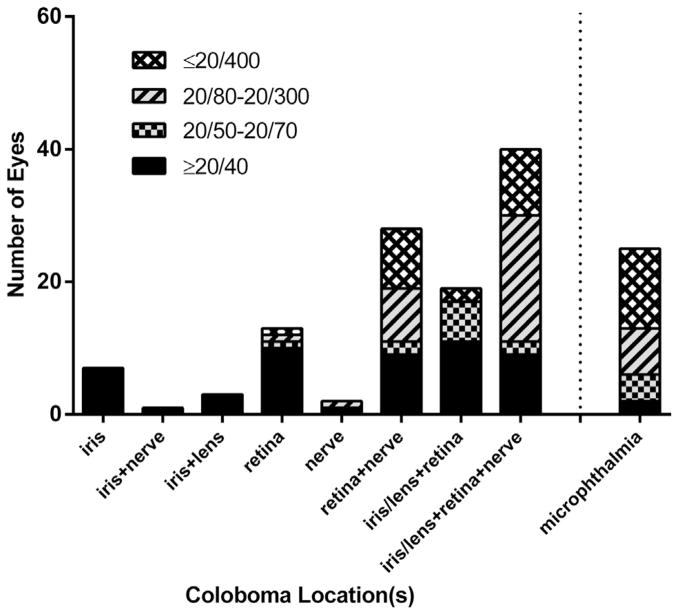
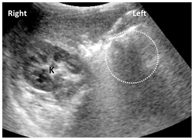
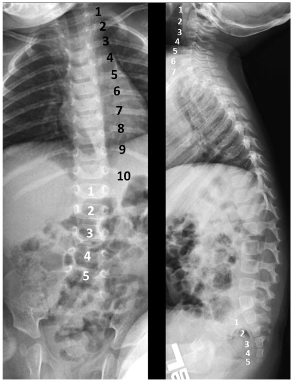
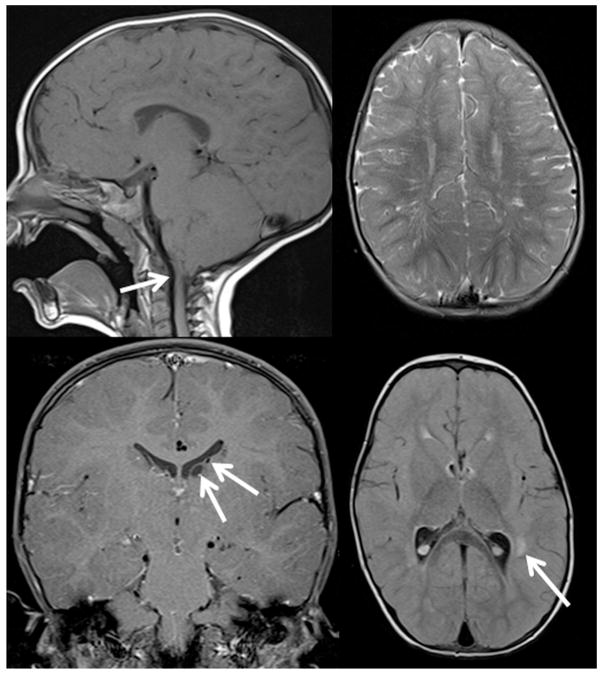
Results: Uveal coloboma affected only the anterior segment in 8 patients, only the posterior segment in 23 patients, and both anterior and posterior segments in 68 patients. Best-corrected visual acuity (BCVA) of eyes with coloboma was ≥20/40 in 45% of eyes; 23% of eyes had BCVA of ≤20/400. The majority of patients (74%) had good vision (>20/60) in at least 1 eye. Ten of the 19 patients (53%) who underwent echocardiography had abnormalities, with ventral septal defects being the most prevalent. Abnormal findings were observed in 5 of 72 patients (7%) who had a renal ultrasound and in 5 of 29 patients (17%) who underwent a brain MRI. Audiology testing revealed abnormalities in 13 of 75 patients (17%), and spine radiographs showed anomalies in 10 of 77 patients (13%). Most findings required no acute intervention.
Conclusions: Although some patients with coloboma had evidence of extraocular abnormalities, the majority of findings on routine clinical examination did not require acute intervention, but some warranted follow-up. Results from the systemic evaluation of patients with coloboma should be interpreted with caution and in view of their clinical context.
Published by Elsevier Inc.
Conflict of interest statement
ALL AUTHORS HAVE COMPLETED AND SUBMITTED THE ICMJE FORM FOR DISCLOSURE OF POTENTIAL CONFLICTS OF INTEREST and none were reported. This work was supported by the intramural research programs of the National Eye Institute, National Institute of Diabetes and Digestive and Kidney Diseases, National Institute on Deafness and Other Communication Disorders, and the Clinical Center of the National Institutes of Health, U.S. Department of Health and Human Services, Bethesda, Maryland, USA. Contributions of authors: design of study (N.H., D.B., D.L.L., E.H.B., S.H., C.C.B., J.B.K., B.P.B.); acquisition of data (B.P.B., N.H., D.B., T.G., E.L.D., W.Z., D.L.L., E.H.B., S.H., C.C.B., J.B.K., T.M.B., I.H.M., B.J.B., B.P.B.); interpretation of data (N.H., D.B., T.G., B.P.B.); preparation of manuscript (N.H., B.P.B.); review and approval of manuscript (N.H., W.Z., D.L.L., E.H.B., S.H., C.C.B., J.B.K., T.M.B., I.H.M., B.J.B., B.P.B.).
Figures
References
-
- Chang L, Blain D, Bertuzzi S, Brooks BP. Uveal coloboma: clinical and basic science update. Curr Opin Ophthalmol. 2006;17(5):447–470. - PubMed
-
- Stoll C, Alembik Y, Dott B, Roth MP. Congenital eye malformations in 212,479 consecutive births. Ann Genet. 1997;40(2):122–128. - PubMed
-
- Stoll C, Alembik Y, Dott B, Roth MP. Epidemiology of congenital eye malformations in 131,760 consecutive births. Ophthalmic Paediatr Genet. 1992;13(3):179–186. - PubMed
Publication types
MeSH terms
Grants and funding
LinkOut - more resources
Full Text Sources
Other Literature Sources
Medical
