Review
. 2013 Oct;31(10):594-605.
doi: 10.1016/j.tibtech.2013.06.005. Epub 2013 Sep 6.

Understanding of dopant-induced osteogenesis and angiogenesis in calcium phosphate ceramics

Affiliations
Review

Understanding of dopant-induced osteogenesis and angiogenesis in calcium phosphate ceramics

Susmita Bose et al. Trends Biotechnol. 2013 Oct.

Abstract

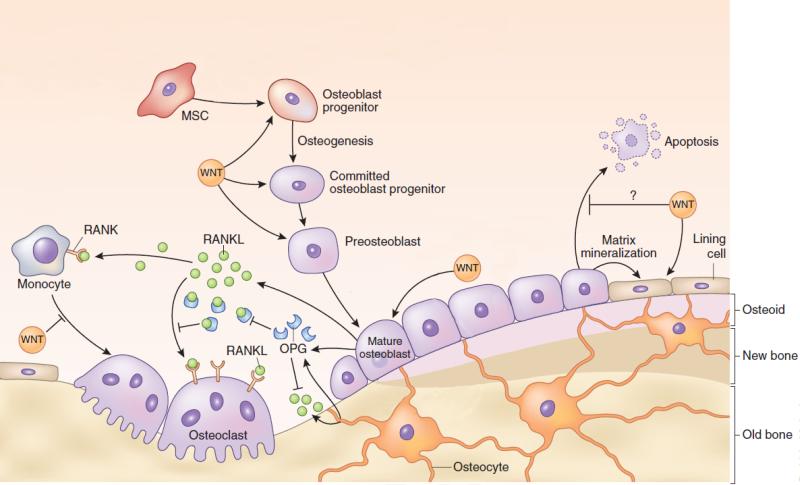
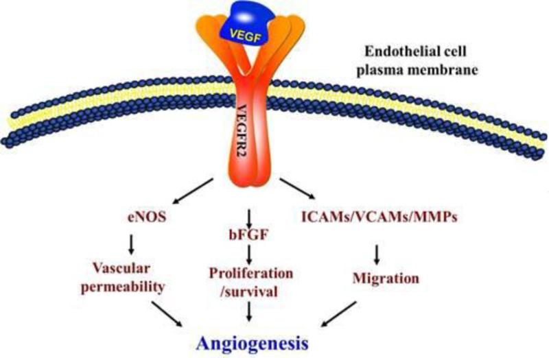
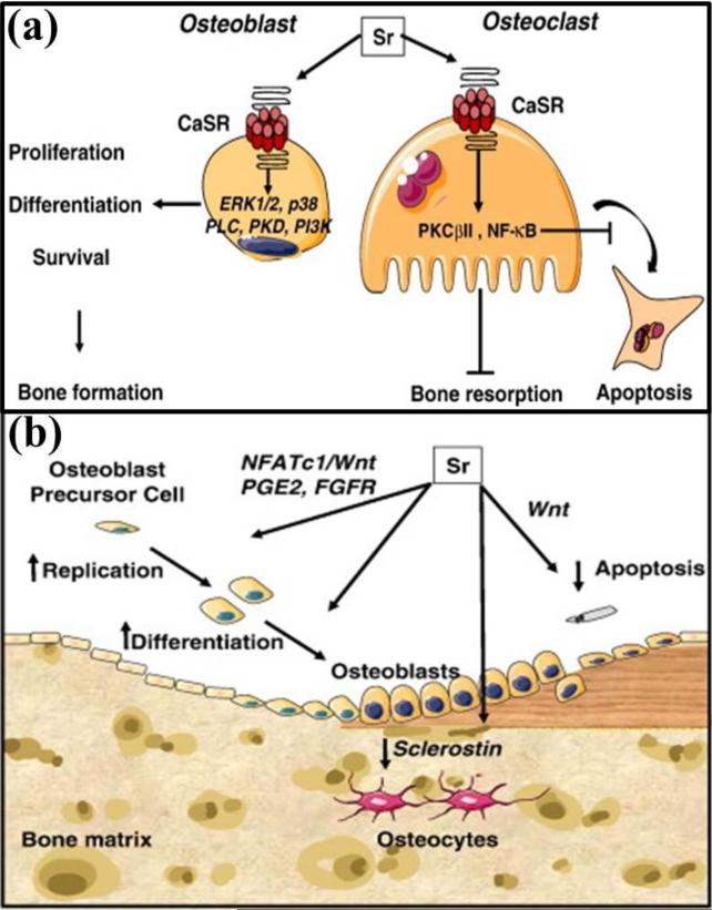
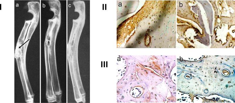
General trends in synthetic bone grafting materials are shifting towards approaches that can illicit osteoinductive properties. Pharmacologics and biologics have been used in combination with calcium phosphate (CaP) ceramics, however, they have recently become the target of scrutiny over safety. The importance of trace elements in natural bone health is well documented. Ions, for example, lithium, zinc, magnesium, manganese, silicon, strontium, etc., have been shown to increase osteogenesis and neovascularization. Incorporation of dopants (trace metal ions) into CaPs can provide a platform for safe and efficient delivery in clinical applications where increased bone healing is favorable. This review highlights the use of trace elements in CaP biomaterials, and offers an insight into the mechanisms of how metal ions can enhance both osteogenesis and angiogenesis.

Keywords: angiogenesis; bone remodeling; calcium phosphates; dopants or trace metal ions; osteogenesis.


Figures

Figure 1
Figure 1
Schematic demonstrating the important role of WNT in the various aspects of the bone formation process. This figure also defines the relationship between osteoblasts and osteoclasts in the bone remodeling process. Osteoblasts produce RANKL which stimulates osteoclastogenesis. At the same time, however, osteoblasts also produce OPG which competitively binds to RANKL to help decrease osteoclastogenesis. The balance of OPG:RANKL is a very important factor in healthy bone formation. This figure has been modified from its original version. Adapted with permission from reference [27].
Figure 2
Figure 2
Schematic representation of the mechanisms VEGF activates in endothelial cells to promote angiogenesis. VEGF released by osteoblastic (and other) cells will activate the transmembrane VEFGR2 receptors in endothelial cells, which in turn will activate several pathways responsible for angiogenesis, including eNOS, basic fibroblast growth factor (bFGF), intercellular adhesion molecules (ICAMS), vascular cell adhesion proteins (VCAM) and matrix metalloproteinases (MMPs). Adapted with permission from reference [39].
Figure 3
Figure 3
(a) A schematic showing the dual mechanism of action by strontium (Sr): stimulatory role on bone forming osteoblast cells, and inhibitory role on bone resorbing osteoclast cells. Sr demonstrates its stimulatory effect on osteoblast cells through the activation of calcium sensing receptor (CaSR) and downstream signalling pathways, which promotes osteoblast proliferation, differentiation and survival. While the same CaSR and downstream signalling pathways activation by Sr in osteoclast cells induce apoptosis resulting in decreased bone resorption. Adapted with permission from reference [47]; (b) A schematic showing how Sr activates osteoblastogenesis: Sr induces increased production of nuclear factor of activated Tc (NFATc)/Wnt signaling, prostaglandin E2 (PGE2), activation of fibroblast growth factor receptor (FGFR) in osteoblastic cells, and reduction of the sclerostin expression (a Wnt antagonist produced by osteocytes). Adapted with permission from reference [47].
Figure 4
Figure 4
I – (a) Shows arrow pointing to defect site in rabbit radiuses directly after implantation. Radiodensity is much greater than that of surrounding natural bone tissue. (b) Shows defect site after 16 weeks implantation with strontium doped calcium polyphosphate (SCPP). Implant is completely integrated into surrounding tissue as evidenced by similar radio opacity. (c) Shows defect site after 16 weeks implantation with a pure calcium polyphosphate (CPP) graft. Implantation site is much more radio dense than the surrounding bone tissue. II – (a) SCPP and (b) CPP tissue sections after 2 weeks stained for collagen I. Brown color indicates collagen formation. III – (a) SCPP and (b) CPP tissue sections after 2 weeks implantation stained for BMP-2. The red/brown color indicates BMP-2. Adapted with permission from reference [51].
Figure 5
Figure 5
(a) Schematic demonstrating the normal bone remodeling process (left) between osteoblasts and osteoclasts. Reactive oxide species inhibit OPG production in osteoblasts while increasing RANKL production allowing increased osteoclastogenesis and subsequent bone resorption. MnSOD acts to neutralize the formation of ROS. (b) A more detailed look at the signaling pathways ROS has been shown to influence in both osteoclasts and osteoblasts. Adapted with permission from reference [89].
Figure 6
Figure 6
(a) Effect of dopants on in vitro strength degradation rate of undoped and TCP doped with 1 wt. % MgO-1 wt. % SrO, 1 wt. % SrO-0.5 wt. % SiO2 and 1 wt. % MgO- 1 wt. % SrO- 0.5 wt. % SiO2 compacts soaked for 0, 2, 4, 8, 12 and 16 weeks in SBF( **P < 0.05, *P > 0.05; n = 6). Adapted with permission from reference [93]; (b) bone remodeling after 12 and 16 weeks in undoped TCP and SrO-MgO doped TCP. A uniform bone remodeling initiation was observed after 12-week with Sr/Mg doped TCP as compared to undoped TCP. A uniform and compact interface was observed between MgO/SrO doped implant and newly remodeled bone compared to undoped TCP. Adapted with permission from reference [94]; (c) Influence of metal ions on tartrate-resistant acid phosphatase (TRAP) expression by osteoclasts (Inset: Fluorescence microscopy images of cells after 14 days culture: red: actin cytoskeleton, green: vitronectin receptor αvβ3 integrin, and blue: nucleolus). Adapted with permission from reference [95]; (d) (i) Photograph of the microwave sintered 3D printed (3DP) TCP and Sr-Mg doped TCP scaffolds for in vivo implantation, (ii) Schematic of the distal femoral cortical defect model (anterior view); (e) Photomicrograph of 3DP pure TCP implants (i and iii), and Sr/Mg doped TCP implants (ii and iv) showing the new bone formation and bone remodeling inside the interconnected macro and intrinsic micro pores of the 3DP scaffolds after 12 and 16 weeks in rat distal femoral defect model. Modified Masson Goldner's trichrome staining of transverse section. OB: old bone, NB: new bone and BM: bone marrow. Color description: dark grey/black: scaffold; orange/red: osteoid; green/bluish: new mineralized bone (NMB)/ old bone; (f) Histomorphometric analysis of bone area fraction (total newly formed bone area/total area, %) showing the new bone formation induced by pure and Sr-Mg doped TCP at different time points (**p < 0.05, *p > 0.05, n=6).
Figure 7
Figure 7
I – (a) Shows the progression of new bone growth into either pure TCP or SiO2/ZnO doped TCP scaffolds over the course of 12 weeks using Goldner's Trichrome stain. (b) represents the histomorphological evaluation of same data. (c) Shows vonWillebrand factor staining to evaluate angiogenesis over the course of 16 weeks. (d) represents quantitative analysis of the same data. Adapted with permission from reference [95].

References

    1. Bose S, Tarafder S. Calcium phosphate ceramic systems in growth factor and drug delivery for bone tissue engineering: A review. Acta Biomater. 2012;8:1401–21. - PMC - PubMed
    1. Carano RAD, Filvaroff EH. Angiogenesis and bone repair. Drug Discov Today. 2003;8:980–9. - PubMed
    1. Gerber HP, Ferrara N. Angiogenesis and bone growth. Trends Cardiovasc Med. 2000;10:223–8. - PubMed
    1. Geiger M. Collagen sponges for bone regeneration with rhBMP-2. Adv Drug Deliv Rev. 2003;55:1613–29. - PubMed
    1. Tamai N, Myoui A, Hirao M, et al. A new biotechnology for articular cartilage repair: subchondral implantation of a composite of interconnected porous hydroxyapatite, synthetic polymer (PLA-PEG), and bone morphogenetic protein-2 (rhBMP-2). Osteoarthritis Cartilage. 2005;13:405–17. - PubMed

Publication types