. 2013 Sep 12;154(6):1342-55.
doi: 10.1016/j.cell.2013.08.025. Epub 2013 Sep 5.

Spatial organization of Hippo signaling at the plasma membrane mediated by the tumor suppressor Merlin/NF2

Affiliations

Spatial organization of Hippo signaling at the plasma membrane mediated by the tumor suppressor Merlin/NF2

Feng Yin et al. Cell. .

Abstract

Although Merlin/NF2 was discovered two decades ago as a tumor suppressor underlying Neurofibromatosis type II, its precise molecular mechanism remains poorly understood. Recent studies in Drosophila revealed a potential link between Merlin and the Hippo pathway by placing Merlin genetically upstream of the kinase Hpo/Mst. In contrast to the commonly depicted linear model of Merlin functioning through Hpo/Mst, here we show that in both Drosophila and mammals, Merlin promotes downstream Hippo signaling without activating the intrinsic kinase activity of Hpo/Mst. Instead, Merlin directly binds and recruits the effector kinase Wts/Lats to the plasma membrane. Membrane recruitment, in turn, promotes Wts phosphorylation by the Hpo-Sav kinase complex. We further show that disruption of the actin cytoskeleton promotes Merlin-Wts interactions, which implicates Merlin in actin-mediated regulation of Hippo signaling. Our findings elucidate an important molecular function of Merlin and highlight the plasma membrane as a critical subcellular compartment for Hippo signal transduction.


Figures

Figure 1
Figure 1. Merlin promotes Hippo signaling without stimulating the intrinsic kinase activity of Hpo/Mst. See also Figure S1
(A-C) Mer/Mer1-600 promotes Wts and Yki phosphorylation without affecting Hpo phosphorylation. S2R+ cell lysates expressing the indicated constructs were probed for P-Wts (A), P-Yki (B) or P-Hpo (C). Relative P-Wts and Wts levels were quantified in Figure S1B. (D) In vitro kinase assay. Myc-Hpo was expressed alone or together with HA-Mer1-600 in S2R+ cells, immunoprecipitated, and incubated with GST-Wts, and the reaction products were probed for P-Wts. (E) S2R+ cells transfected with the indicated plasmids along with control or hpo dsRNA were probed with the indicated antibodies. Note reduction of P-Wts levels by hpo RNAi. (F) RNAi of mer leads to decreased P-Yki and P-Wts but increased P-Hpo levels in S2R+ cells. Also note comparable levels of total Yki, Wts or Hpo in control and RNAimer cells. (G) NF2 promotes Lats1 phosphorylation without affecting Mst1 phosphorylation. HEK293 cells expressing the indicated constructs were probed for P-Lats and P-Mst. (H) Control (Nf2flox/flox) and Nf2 (Alb-Cre; Nf2flox/flox) livers from 2-month old littermates were probed with the indicated antibodies. Note the comparable levels of Lats1 and YAP but diminished P-Lats and P-YAP levels in Nf2 mutant livers. Also note the increased P-Mst levels in Nf2 mutants.
Figure 2
Figure 2. Merlin promotes plasma membrane association of Wts/Lats in Drosophila and mammalian cells. See also Figure S2
(A) Mer1-600, but not Mer or Ex, induces Wts membrane translocation. S2R+ cells expressing the indicated constructs were fractioned into membrane (M) and cytosolic (C) fraction and probed with the indicated antibodies. T: total cell lysates. (B) Mer1-600 induces membrane association of Wts and WtsT1077A in S2R+ cells. Subcellular fractions were analyzed as in (A). (C) RNAi of mer results in a significant increase of endogenous Wts in the cytoplasmic fraction relative to the membrane fraction. Subcellular fractions were probed with α–Wts antibody. Nervana, a plasma membrane-localized Na+/K+ ATPase, serves as a control for fractionation. (D) NF2 promotes membrane association of Lats1 and Lats1T1079A in HEK293 cells, as revealed by subcellular fractionation. (E) Loss of NF2 results in increased cytosolic accumulation of endogenous Lats1 and Lats2. Subcellular fractions of confluent FH-912 and FC-912 cells were probed. (F) FC-912 cells were transfected with empty vector (pcDNA), NF2 or NF2 mutants along with Myc-Lats1. Subcellular fractions were probed for Myc-Lats1. FC-912 cells expressing empty vector or NF2 mutants had higher Myc-Lats1 levels in cytosolic than membrane fraction, but FC-912 cells expressing wildtype NF2 showed similar cytosolic and membrane Myc-Lats1 levels. The cell lysates were also probed for the expression of NF2 mutants (bottom gel). (G) Liver sections from 8-day-old control and Nf2-deficient littermates, stained with Lats2 (red) and the plasma membrane marker Pan-cadherin (green). Lats2 is concentrated at the cell cortex immediately on the inner face of plasma membrane in wildtype hepatocytes, but shows a uniform cytoplasmic localization in Nf2-deficient hepatocytes.
Figure 3
Figure 3. Active Merlin binds to Wts/Lats through its FERM domain. See also Figure S3
(A) Wts associates with Mer1-600, but not Mer or Ex, as revealed by immunoprecipitation in S2R+ cells as indicated. (B) Mer1-600 exhibits an open conformation. S2R+ cells expressing Mer-N fragment (1-375) and wildtype (375-635) or truncated (375-600) Mer-C fragment were subjected to immunoprecipitation. (C) Lats1 associates with N-terminal half of NF2, as revealed by immunoprecipitation in HEK293 cells. (D) Co-immunoprecipitation between endogenous NF2 and Lats1/2 in HEK293 cells. Endogenous Lats1 or Lats2 was detected in α-NF2, but not control, immunoprecipitates. (E) NF2-Lats1 binding in vitro. HEK293 cell lysates containing Myc-Lats1 or Myc-Lats13m were incubated with glutathione Sepharose beads containing GST (as a control) or GST-NF2. GST-NF2 bound to Myc-Lats1 but not Myc-Lats13m. (F-H) C-terminal truncation of NF2 results in a more open conformation and enhances Lats1 binding and phosphorylation. HEK293 cells expressing the indicated constructs were probed for association between NF2-N and NF2-C fragments (F), NF2-Lats1 association (G) and Lats1 hydrophobic motif phosphorylation (H). (I) Wildtype NF2, but not disease-associated missense mutants, associates with Lats1, as revealed by immunoprecipitation in HEK293 cells. (J) Wildtype NF2, but not disease-associated missense mutants, promotes membrane association of Lats1 in HEK293 cells, as revealed by subcellular fractionation.
Figure 4
Figure 4. Identification of a conserved N-terminal motif in Wts/Lats that mediates Merlin-Wts/Lats interactions. See also Figure S4
(A) HEK293 cells expressing HA-NF2 and various truncations of Lats1 were subjected to immunoprecipitation as indicated. (B) Alignment of the conserved N-terminal motif (CNM) in Wts/Lats protein from different species. (C) Myc-Lats1, but not Myc-Lats13m, was co-immunoprecipitated with HA-NF2 in HEK293 cells. (D-E) The CNM is required for NF2-induced membrane association of Lats1 in HEK293 cells. Subcellular fractionation (D) and immunofluorescent staining (E) of Myc-Lats1 or Myc-Lats13m in the presence or absence of NF2. (F-G) The CNM is required for Mer1-600-mediated membrane association of Wts in Drosophila S2R+ cells. Subcellular fractionation (F) and immunofluorescent staining (G) of V5-Wts or V5-Wts3m in the presence or absence of Mer1-600. (H) UAS-Wts and UAS-Wts3m transgenes inserted at identical chromosomal locus were crossed to nub-Gal4 driver. Note the small-wing phenotype induced by Wts, but not Wts3m.
Figure 5
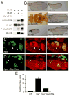
Figure 5. Constitutive membrane targeting promotes Hpo-mediated hydrophobic motif phosphorylation and tumor suppressor activity of Wts. See also Figure S5
(A) S2R+ cells expressing the indicated constructs were probed for P-Yki and P-Wts. (B) Enhanced growth-suppressive activity of membrane-targeted Wts and dominant negative activity of membrane-targeted WtsT1077A. All UAS Wts transgenes were inserted at the same locus. Note the complete loss of adult wings in nub: Myr-Wts flies, and the suppression of small-wing phenotype of nub:Hpo in flies that co-expressed Myr-WtsT1077A. (C-E) Partial rescue of hpo mutant clones by Myr-Wts. Eye discs containing clones of the indicated genotype (GFP+, arrowheads) were stained for Diap1 (red). Note elevated Diap1 levels in hpo clones without or with Myr-Wts expression. Also note the smaller clone size and less rounded shape of the latter genotype. Clones size is quantified in (E).
Figure 6
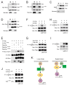
Figure 6. Sav-mediated membrane association of Hpo and regulation of Merlin-Wts binding by actin cytoskeleton. See also Figure S6
(A) S2R+ cells expressing Myc-Hpo together with HA-Mer1-600 or FLAG-Sav were fractioned into membrane (M) and cytosolic (C) fraction. T: total cell lysates. Sav, but not Mer1-600, promoted Hpo membrane association. (B) S2R+ cells expressing V5-Wts together with HA-Mer1-600 or FLAG-Sav were analyzed by subcellular fractionation. Mer1-600, but not Sav, promoted Wts membrane association. (C) RNAi of sav results in decreased membrane localization of endogenous Hpo. Subcellular fractions were probed with α–Hpo antibody. (D) Savshrp6 mimics a sav allele that deletes just the SARAH domain (Kango-Singh et al., 2002). Unlike wildtype Sav, Savshrp6 failed to induce membrane recruitment of Hpo. (E) S2R+ cells expressing the indicated constructs were analyzed for P-Wts. Wildtype Sav, but not Savshrp6 (Kango-Singh et al., 2002) or SavΔFBM (Yu et al., 2010), greatly potentiated Mer1-600-induced Wts phosphorylation. (F-G) S2R+ cells expressing the indicated constructs were treated with DMSO (0.1%, lane 1), LatB (1 μg/ml, lane 2) or Fosk (30 μM, lane 3) for 1 hr, followed by immunoprecipitation. LatB, but not Fosk, promoted Mer-Wts interaction (F). Neither drug affected Sav-Hpo interaction (G). (H) S2R+ cells expressing the indicated constructed were treated with DMSO (0.1%, lane 1), LatB (1 μg/ml, lane 2) or C3 (2 μg/ml, lane 3), followed by immunoprecipitation. (I) S2R+ cells were treated with DMSO (0.1%, lane 1), LatB (1 μg/ml, lane 2) or C3 (2 μg/ml, lane 3), followed by immunoprecipitation with anti-Mer antibody and detection with anti-Wts antibody. (J) S2R+ cells expressing HA-Mer1-600 and V5-Wts were treated with DMSO, LatB or C3 as in (H-I) followed by immunoprecipitation. Neither treatment affected interactions between Mer1-600 and Wts. (K) Schematic models of Merlin function in Hippo signaling. For simplicity, only proteins relevant to the current study are illustrated in the schematic diagrams. Left: the prevailing linear model placing Merlin upstream of Hpo activation. Right: a new model based on the current study highlighting a dual mechanism in which Merlin and Sav promotes the membrane association of Wts and Hpo, respectively.
Figure 7
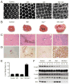
Figure 7. Loss of Mer/NF2 and Sav/Sav1 leads to synergistic defects in Hippo signaling in Drosophila and mice
(A) Synergistic effect of mer and sav mutations in Drosophila. Mid-pupal retina of the indicated genotypes was stained for Discs-Large to highlight cell outlines. Note the significantly increased number of interommatidial cells in mer; sav double mutant eyes. (B-E) Synergistic effect of Nf2 and Sav1 mutations in mice. Livers from 8 day-old pups of the following genotype were analyzed: control (Alb-Cre), Nf2 mutant (Alb-Cre; Nf2flox2/flox2), Sav1 mutant (Alb-Cre; Sav1flox/flox), and double mutant (Alb-Cre; Nf2flox2/flox2; Sav1flox/flox). Whole amount liver images (B), H&E staining (C) and Pan-CK staining (D) were shown, along with quantification of liver weight (E). While Nf2 and Sav1 livers displayed mild or no overproliferation of the CK-positive BECs, Nf2; Sav1 double mutant livers were predominantly occupied by the BECs. Also note the unique external appearance and overgrowth of Nf2; Sav1 double mutant livers. (F) Western blot analysis of liver lysates from mouse livers analyzed in (B-E). Liver lysates from three mice of each genotype were probed. Note the further decrease in YAP and Lats phosphorylation in Nf2; Sav1 double mutant livers compared to the single mutant. Also note the comparable level of Lats1 in control and Nf2 mutant livers.

Similar articles

Cited by

References

    1. Ahronowitz I, Xin W, Kiely R, Sims K, MacCollin M, Nunes FP. Mutational spectrum of the NF2 gene: a meta-analysis of 12 years of research and diagnostic laboratory findings. Hum Mutat. 2007;28:1–12. - PubMed
    1. Bischof J, Maeda RK, Hediger M, Karch F, Basler K. An optimized transgenesis system for Drosophila using germ-line-specific phiC31 integrases. Proc Natl Acad Sci U S A. 2007;104:3312–3317. - PMC - PubMed
    1. Bossuyt W, Chen CL, Chen Q, Sudol M, McNeill H, Pan D, Kopp A, Halder G. An evolutionary shift in the regulation of the Hippo pathway between mice and flies. Oncogene 2013 - PMC - PubMed
    1. Bretscher A, Edwards K, Fehon RG. ERM proteins and merlin: integrators at the cell cortex. Nat Rev Mol Cell Biol. 2002;3:586–599. - PubMed
    1. Cai J, Zhang N, Zheng Y, de Wilde RF, Maitra A, Pan D. The Hippo signaling pathway restricts the oncogenic potential of an intestinal regeneration program. Genes Dev. 2010;24:2383–2388. - PMC - PubMed

Publication types

MeSH terms

Substances