. 2013 Sep 12;4(5):1022-34.
doi: 10.1016/j.celrep.2013.07.048. Epub 2013 Sep 5.

Endothelial Jagged-1 is necessary for homeostatic and regenerative hematopoiesis

Affiliations

Endothelial Jagged-1 is necessary for homeostatic and regenerative hematopoiesis

Michael G Poulos et al. Cell Rep. .

Abstract

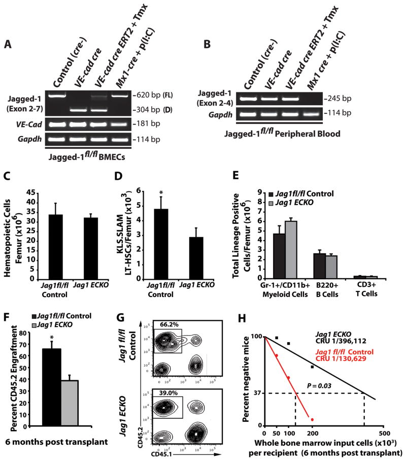
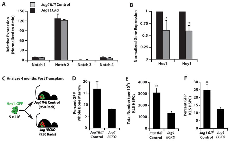
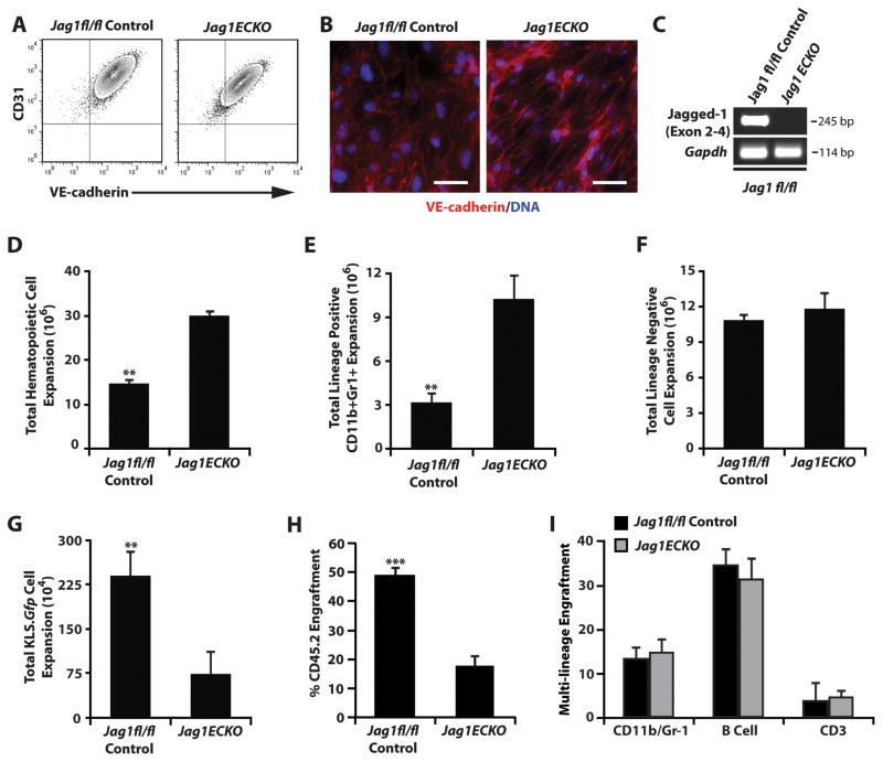
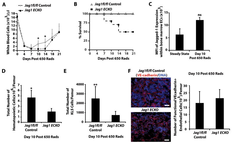
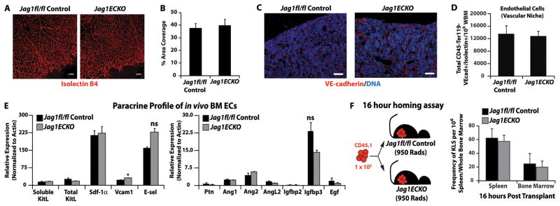
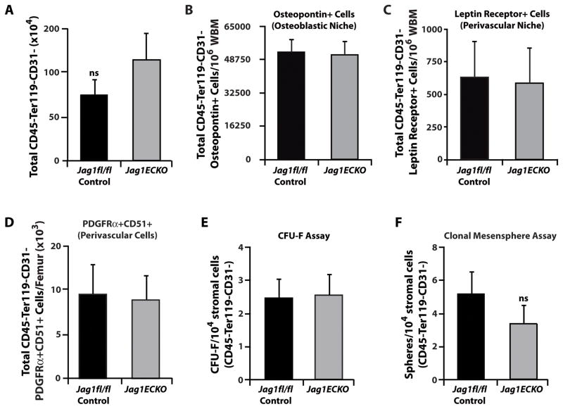
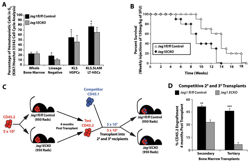
The bone marrow (BM) microenvironment is composed of multiple niche cells that, by producing paracrine factors, maintain and regenerate the hematopoietic stem cell (HSC) pool (Morrison and Spradling, 2008). We have previously demonstrated that endothelial cells support the proper regeneration of the hematopoietic system following myeloablation (Butler et al., 2010; Hooper et al., 2009; Kobayashi et al., 2010). Here, we demonstrate that expression of the angiocrine factor Jagged-1, supplied by the BM vascular niche, regulates homeostatic and regenerative hematopoiesis through a Notch-dependent mechanism. Conditional deletion of Jagged-1 in endothelial cells (Jag1((ECKO)) mice) results in a profound decrease in hematopoiesis and premature exhaustion of the adult HSC pool, whereas quantification and functional assays demonstrate that loss of Jagged-1 does not perturb vascular or mesenchymal compartments. Taken together, these data demonstrate that the instructive function of endothelial-specific Jagged-1 is required to support the self-renewal and regenerative capacity of HSCs in the adult BM vascular niche.


Figures

Figure 1
Figure 1. Maintenance of the HSC pool requires endothelial-specific expression of Jagged-1
Constitutive VE-cadherin cre transgenic mice promote efficient in vivo deletion of an exon 4–5 floxed Jag1 allele in BM endothelial cells. Jag 1fl/fl conditional mice were crossed with either constitutive VE-cadherin cre, tamoxifen (Tmx) inducible VE-cadherin creERT2, or Poly(I:C) inducible Mx1 cre mice and cDNA was synthesized from either BM endothelial cells sorted to purity (CD45, Ter119, VE-Cadherin+, Isolectin GS-IB4+) or peripheral blood. A)RT -PCR using primers spanning conditional Jag1 exons 4–5 were used to assay for the extent of full length (FL) and deleted (D) transcripts in BM ECs. VE-cadherin expression was used to assess for purity. B)RT -PCR analysis of peripheral blood using Jag1 specific primers located within floxed exon 4 demonstrate efficient elimination of exon 4 containing transcripts in Mx1 cre, but not constitutive VE-cadherin cre or inducible VE-cadherin creERT2 mice. C, D) Steady state analysis of control and Jag1(ECKO) mice demonstrates similar levels in BM cellularity C) but a significant increase in the frequency of phenotypic LT-HSC per 106 whole BM cells D) as compared to Jag1fl/fl controls (n=12/cohort). E) Analysis of lineage-committed progenitors shows no significant differences in Jag1(ECKO) mice as compare to Jag1fl/fl controls (n=8 control mice; n=10 Jag1(ECKO) mice). F) To test if whole BM from Jag1(ECKO) mice had decreased engraftment potential, we performed a competitive repopulation assay and demonstrated that whole BM from wild type donors were significantly more efficient at sustaining long-term repopulating ability as compared to Jag1(ECKO) mice. G) Representative contour plots of CD45.1 vs. CD45.2 engraftment 4 months post transplant (n=5/cohort, 3 independent experiments). H) Limiting dilution analysis of the frequency of long-term, multilineage reconstitution cells in Jag1(ECKO) mice as compared to Jag1fl/fl controls (n= 20/cohort, 3 independent experiments). All data represents mean ± s.d. (*P<0.05). See also Figure S1 and Figure S2.
Figure 2
Figure 2. Endothelial-specific deletion of Jagged-1 interferes with Notch signaling in HSPCs
A) KLS HSPCs were sorted from control and Jag1(ECKO) mice and analyzed for Notch receptors and B) downstream Notch target genes. There were no significant differences between A) Notch receptors but significant decrease in the B) Notch target genes Hes1 and Hey1 in the Jag1(ECKO) KLS cells. C) Whole BM (5 × 10⁵) from Hes1-GFP knock-in mice was transplanted into lethally irradiated control and Jag1(ECKO) mice and allowed to recover for 4 months. D) Percentage of GFP+ whole bone marrow. E) Total number of KLS cells and F) percentage of GFP+ KLS cells. There was a significant decrease in whole BM and KLS cells positive for Hes1 GFP in Jag1(ECKO) mice.
Figure 3
Figure 3. Endothelial-specific deletion of Jagged-1 impairs ex vivo expansion of Notch+ KLS cells
AC) Confirmation of endothelial cell phenotype of isolated endothelium from control and Jag1(ECKO) mice. A) Flow cytometric analysis of cultured endothelial cells based on the co-expression of endothelial-specific markers CD31 and VE-cadherin. B) Fluorescent images demonstrating junctional VE-cadherin staining of cultured endothelial cells. C) Confirmation of Jagged-1 deletion in cultured Jag1(ECKO) endothelium. DF) 50,000 Lineage-hematopoietic cells from Notch.GFP reporter mice were co-cultured with control and Jag1(ECKO) endothelial cells for 7 days in serum-free culture conditions supplemented with 50 ng/mL of stem cell factor. Hematopoietic cells co-cultured with Jag1(ECKO) endothelial cells resulted in a significant increase in expansion of the total number of hematopoietic cells D) with the majority of the cells being CD11b+Gr1+ Lineage cells E) but with no change in the expansion of Lineage cells F) when compared to control endothelial cells. However, there was a significant decrease in the total number of KLS cells (data not shown) and KLS cells that were actively signaling through the Notch pathway (GFP+) G). HI) Competitive repopulation of expanded hematopoietic cells demonstrates that Lineage hematopoietic cells that were co-cultured with control endothelial cells resulted in the expansion of HSCs with a greater H) engraftment potential with similar I) multilineage potential as compared to Jag1(ECKO) endothelial cells. All data represents mean ± s.d. (**P<0.01, **P<0.001); scale bar 50μm.
Figure 4
Figure 4. Endothelial-specific deletion of Jagged-1 interferes with hematopoietic regeneration
A) Analysis of white blood cell recovery in Jag1(ECKO) mice following sublethal irradiation of 650 Rads (n=10/cohort up to day 10, n=6 for duration of analysis, 3 independent experiments). B) Survival curve of control and Jag1(ECKO) mice post sublethal irradiation. All of the control mice fully recover and survived. However, Jag1(ECKO) mice begin succumbing to hematopoietic failure by day 10 and 50% of the cohort has died by day 18 post sublethal irradiation. C) Jagged-1 expression in BM endothelial cells at steady state and day 10 post sublethal irradiation. D, E) Analysis of the BM at day 10 post-sublethal irradiation demonstrated that Jag1(ECKO) mice have a significant decrease in BM cellularity D) and a decrease in KLS HSPCs E) as compared to controls (n=4/cohort, 3 independent experiments). F) Representative fluorescent images of the diaphysis and trabecular regions of Jag1fl/fl and Jag1(ECKO) mice as well as quantification of functional BM endothelial cells. Note the increase in BM cellular integrity in control mice as compared to Jag1(ECKO) mice without any significant changes in vascular density within the BM. All data represents mean ± s.d. (*P<0.05, **P<0.01); scale bar 50μm.
Figure 5
Figure 5. Jag1(ECKO) mice do not manifest postnatal vascular deficiencies that lead to hematopoietic dysfunction
A) Visualization of the retinal vasculature at P6 using isolectin B4 immunofluorescence shows no change in vascular density in Jag1(ECKO) mice, as compared to littermate controls. B) Quantification of vascular parameters at P6 in Jag1fl/fl controls and Jag1(ECKO) retinas. n=3 per group/2 independent experiments. C) Visualization of the BM vascular niche (VE-cadherin-Alexa647/DAPI nuclear counterstain). D) Quantification of the BM vascular niche shows no significant difference between the numbers of functionally patent CD45Ter119VEcadherin+Isolectin B4+ BM endothelial cells. N=5 for each experimental group for three independent experiments. E) Bone marrow endothelial cells were isolated and subjected to angiocrine profiling for known pro-hematopoietic angiocrine factors. The angiocrine profile was not significantly different between the Jag1fl/fl and Jag1(ECKO) cohorts except for Vcam1. N=3 for each experiment group. Due to the role of Vcam in homing of HSPCs, we performed a F) 16 hour homing assay where 105 whole BM from CD45.1 mice was transplanted into lethally irradiated Jag1fl/fl controls and Jag1(ECKO) mice and analyzed for donor engraftment 16 hours post transplant. Note that there were no significant differences in the homing of wild type whole BM into control and Jag1(ECKO) recipients. NS=non-significant, scale bar 50μm.
Figure 6
Figure 6. Jag1(ECKO) mice do not manifest alterations to the mesenchymal stem cell or perivascular niches in the BM microenvironment
A) Quantification of non-vascular stromal cells. BD) Within the non-vascular niche cell compartment we quantified B) Osteopontin+ osteoblast niche cells and C) Leptin Receptor+ and D) PDGFRα+/CD51+ perivascular niche cells. Note that endothelial-specific deletion of Jagged-1 does not result in dropout of other critical BM niche cells. n=9 per cohort/3 independent experiments. EF) MSC activity of CD45 Ter119 CD31 stromal cells in Jag1fl/fl and Jag1(ECKO) mice. No differences were detected in the E) number of CFU-Fs and F) clonal mesenspheres derived from 10,000 sorted CD45 Ter119 CD31 bone marrow stromal cells isolated from Jag1fl/fl and Jag1(ECKO) mice. Each functional assay was independently repeated three times with n=3 mice per test. These data suggest that the hematopoietic defect observed in Jag1(ECKO) mice is not an indirect result of alterations to the architectural integrity and geometry of the BM niche, rather a direct instructional role of endothelial cell-specific Jagged-1 expression. N=9 for each experimental group for three independent experiments. All data represents mean ± s.d., ns (statistically non significant).
Figure 7
Figure 7. Endothelial-specific deletion of Jagged-1 impairs the self-renewal potential of HSCs
A) Steady state cell cycle analysis of hematopoietic subpopulations from control and Jag1(ECKO) mice based on Ki67/Hoechst 33342 intracellular staining. Whole BM from both cohorts showed no significant difference in their cell cycle status. However, there was a significant decrease in the percentage of cells that were in G0/quiescence in Jag1(ECKO) mice in the hematopoietic subpopulations (Lineage Negative, KLS HSPCs, and KLS.SLAM) as compared to controls. B) Survival curve monitoring the morbidity/mortality of Jag1fl/fl and Jag1(ECKO) mice following weekly injections of a sublethal dose of 5-FU (150 mg/kg). Jag1(ECKO) mice succumbed to hematopoietic failure by 14 weeks post serial 5-FU injections, whereas, control mice had a significant increase in longevity; with the entire cohort dying at 19 weeks post serial 5-FU injections. C) Schematic depicting transplantation strategy. D) Control and Jag1(ECKO) mice were lethally irradiated (950 Rads) and were transplanted with 5 × 10⁵ whole BM from CD45.2 wild type mice. Four months post transplant, primary CD45.2 cells were competitively transplanted with CD45.1 whole BM into secondary and tertiary Jag1fl/fl and Jag1(ECKO) recipients. CD45.2 engraftment 4 months post secondary transplant demonstrate that endothelial-specific deletion of Jagged-1 results in a significant decrease in engraftment efficiency and self-renewal potential. All data represents mean ± s.d. (*P<0.05, ***P<0.001).

References

    1. Adams GB, Chabner KT, Alley IR, Olson DP, Szczepiorkowski ZM, Poznansky MC, Kos CH, Pollak MR, Brown EM, Scadden DT. Stem cell engraftment at the endosteal niche is specified by the calcium-sensing receptor. Nature. 2006;439:599–603. - PubMed
    1. Benedito R, Roca C, Sorensen I, Adams S, Gossler A, Fruttiger M, Adams RH. The notch ligands Dll4 and Jagged1 have opposing effects on angiogenesis. Cell. 2009;137:1124–1135. - PubMed
    1. Butler JM, Nolan DJ, Vertes EL, Varnum-Finney B, Kobayashi H, Hooper AT, Seandel M, Shido K, White IA, Kobayashi M, et al. Endothelial cells are essential for the self-renewal and repopulation of Notch-dependent hematopoietic stem cells. Cell stem cell. 2010;6:251–264. - PMC - PubMed
    1. Calvi LM, Adams GB, Weibrecht KW, Weber JM, Olson DP, Knight MC, Martin RP, Schipani E, Divieti P, Bringhurst FR, et al. Osteoblastic cells regulate the haematopoietic stem cell niche. Nature. 2003;425:841–846. - PubMed
    1. Ding L, Morrison SJ. Haematopoietic stem cells and early lymphoid progenitors occupy distinct bone marrow niches. Nature. 2013;495:231–235. - PMC - PubMed

Publication types