. 2013 Sep;64(4):653-64.
doi: 10.1016/j.yhbeh.2013.08.016. Epub 2013 Sep 5.

Endogenous oxytocin is necessary for preferential Fos expression to male odors in the bed nucleus of the stria terminalis in female Syrian hamsters

Affiliations

Endogenous oxytocin is necessary for preferential Fos expression to male odors in the bed nucleus of the stria terminalis in female Syrian hamsters

Luis A Martinez et al. Horm Behav. 2013 Sep.

Abstract

Successful reproduction in mammals depends on proceptive or solicitational behaviors that enhance the probability of encountering potential mates. In female Syrian hamsters, one such behavior is vaginal scent marking. Recent evidence suggests that the neuropeptide oxytocin (OT) may be critical for regulating this behavior. Blockade of OT receptors in the bed nucleus of the stria terminalis (BNST) or the medial preoptic area (MPOA) decreases vaginal marking responses to male odors; lesion data suggest that BNST, rather than MPOA, mediates this effect. However, how OT interacts with sexual odor processing to drive preferential solicitation is not known. To address this issue, intact female Syrian hamsters were exposed to male or female odors and their brains processed for immunohistochemistry for Fos, a marker of recent neuronal activation, and OT. Additional females were injected intracerebroventricularly (ICV) with an oxytocin receptor antagonist (OTA) or vehicle, and then tested for vaginal marking and Fos responses to sexual odors. Colocalization of OT and Fos in the paraventricular nucleus of the hypothalamus was unchanged following exposure to male odors, but decreased following exposure to female odors. Following injections of OTA, Fos expression to male odors was decreased in BNST, but not in MPOA or the medial amygdala (MA). Fos expression in BNST may be functionally relevant for vaginal marking, given that there was a positive correlation between Fos expression and vaginal marking for BNST, but not MPOA or MA. Together, these data suggest that OT facilitation of neuronal activity in BNST underlies the facilitative effects of OT on solicitational responses to male odors.

Keywords: Appetitive; Chemosensory; Olfaction; Precopulatory; Sexual motivation.


Figures

Figure 1
Figure 1
Timelines of experimental manipulations.
Figure 2
Figure 2
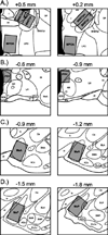
Counting domains for quantifying oxytocin- and Fos-positive cells. Domains (gray boxes) were fitted to the following areas: (A) the posteromedial (BNSTpm) and posterointermediate (BNSTpi) subdivisions of the bed nucleus of the stria terminalis, and the medial preoptic area (MPOA), (B) the paraventricular nucleus of the hypothalamus (PVH), (C) the anterior division of the medial amygdala (MeA), and (D) the posterior division of the medial amygdala (MeP). Atlas plates are modified from Morin and Wood (2001), and arranged in distances relative to bregma. ac, anterior commissure; ACo, anterior cortical amygdaloid nucleus; AH, anterior hypothalamus; BLA, anterior division of the basolateral amygdaloid nucleus; BLP, posterior division of the basolateral amygdaloid nucleus; BMA, anterior division of the basomedial amygdaloid nucleus; BMP, posterior division of the basomedial amygdaloid nucleus; BNSTpl, posterolateral subdivision of the bed nucleus of the stria terminalis; Ce, central amygdaloid nucleus; f, fornix; GP, globus pallidus; I, intercalated nuclei of the amygdala; ic, internal capsule; LH, lateral hypothalamus; LPO, lateral preoptic area; ot, optic tract; ox, optic chiasm; Pir, piriform cortex; PLCo, posterolateral cortical amygdaloid nucleus; SCN, superchiasmatic nucleus; SI, substantia innominata, sm, stria medullaris; SON, supraoptic nucleus; sox, supraoptic decussation; st, stria terminalis; VEn, ventral endopiriform nucleus.
Figure 3
Figure 3
Immunohistochemical staining for oxytocin and Fos. Representative photomicrographs of double-label immunohistochemistry in sections depicting (A) the paraventricular nucleus of the hypothalamus (PVH), (B) the medial preoptic area (MPOA), and (C) the anterior division of the medial amygdala (MeA). A higher magnification (40X objective) of the areas highlighted within the black boxes is provided on the right of each lower magnification (10X objective) image. White arrows indicate oxytocin-immunoreactive cells (brown cytoplasmic staining). Black arrows indicate Fos-immunoreactive cells (black nuclear staining). Gray arrows indicate cells immunoreactive for both oxytocin and Fos (brown cytoplasmic staining with black nuclear staining). Scale bars = 100 µm (lower magnification) and 25 µm (higher magnification).
Figure 4
Figure 4
Mean (± SEM) percentages of oxytocin/Fos double-labeled cells following stimulus exposures. In the paraventricular nucleus of the hypothalamus (PVH), a lower percentage of oxytocin-positive (OT+) cells were positive for Fos (Fos+) following exposure to female odors as compared to either male or clean odors. No differences were observed in the percentage of OT+/Fos+ cells across stimulus conditions in either the medial preoptic area (MPOA) or the anterior division of the medial amygdala (MeA). Few oxytocin-positive cells were observed in the bed nucleus of the stria terminalis (BNST) or posterior medial amygdala (MeP), and so double-labeled cells in those areas were not counted. Within each area, dissimilar letters = nonhomologous means (Tukey’s HSD post-hoc tests).
Figure 5
Figure 5
Mean (± SEM) number of vaginal marks following ICV drug injections. ICV injections of a selective oxytocin receptor antagonist (OTA) decreased vaginal marking in response to male odors, as compared to saline injections (A). OTA was most effective at the highest dose administered (900µM). There was no effect of OTA on vaginal marking in females exposed to female odors. V1aA injections did not affect vaginal marking in females exposed to either male or female odors (B). *p < .05 (Bonferroni correction), saline vs. 900 µM OTA in the male stimulus condition.
Figure 6
Figure 6
Mean (+/− SEM) densities of Fos-positive cells following ICV injections of OTA. In females exposed to male odors, ICV injections of a selective oxytocin receptor antagonist (OTA) reduced the density of Fos-positive cells in both the posteromedial (BNSTpm) and posterointermediate (BNSTpi) subdivisions of the bed nucleus of the stria terminalis, as compared to saline injections (A, B). This effect was not seen in females exposed to female odors. Although there was an overall increase in the density of Fos-positive cells in the medial preoptic area (MPOA) and the anterior (MeA) and posterior (MeP) divisions of the medial amygdala in response to male vs. female odors, there was no effect of OTA on Fos expression within these areas (C–E). *p < .05, OTA vs. saline in the male odor condition.
Figure 7
Figure 7
Relationships between the density of Fos-positive cells and the number of vaginal marks. When examining responses to male odors only (A), the density of Fos-positive cells was positively correlated with the number of vaginal marks for the posteromedial (BNSTpm) and posterointermediate (BNSTpi) subdivisions of the bed nucleus of the stria terminalis, but not the medial preoptic area (MPOA), the anterior division of the medial amygdala (MeA), or the posterior division of the medial amygdala (MeP). This pattern of results was also seen when looking at overall responses across odor stimulus condition, when first controlling for the effect of odor stimulus on Fos expression and vaginal marking (B).

Similar articles

Cited by

References

    1. Arakawa H, Arakawa K, Deak T. Oxytocin and vasopressin in the medial amygdala differentially modulate approach and avoidance behavior toward illness-related social odor. Neuroscience. 2010;171:1141–1151. - PubMed
    1. Beach FA. Sexual attractivity, proceptivity, and receptivity in female mammals. Horm. Behav. 1976;7:105–138. - PubMed
    1. Been LE, Bauman JM, Petrulis A, Chang YH. X-ray kinematics analysis of vaginal scent marking in female Syrian hamsters (Mesocricetus auratus) Physiol. Behav. 2012;105:1021–1027. - PMC - PubMed
    1. Been LE, Petrulis A. Chemosensory and hormone information are relayed directly between the medial amygdala, posterior bed nucleus of the stria terminalis, and medial preoptic area in male Syrian hamsters. Horm. Behav. 2011;59:536–548. - PMC - PubMed
    1. Bennett AL, Greco B, Blasberg ME, Blaustein JD. Response to male odours in progestin receptor- and oestrogen receptor-containing cells in female rat brain. J. Neuroendocrinol. 2002;14:442–449. - PubMed

Publication types