Review
. 2013 Dec 1;531(2):150-7.
doi: 10.1016/j.gene.2013.08.061. Epub 2013 Sep 5.

Structure and function of the DNA ligases encoded by the mammalian LIG3 gene

Affiliations
Review

Structure and function of the DNA ligases encoded by the mammalian LIG3 gene

Alan E Tomkinson et al. Gene. .

Abstract

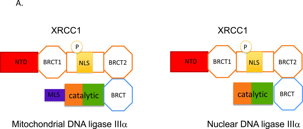
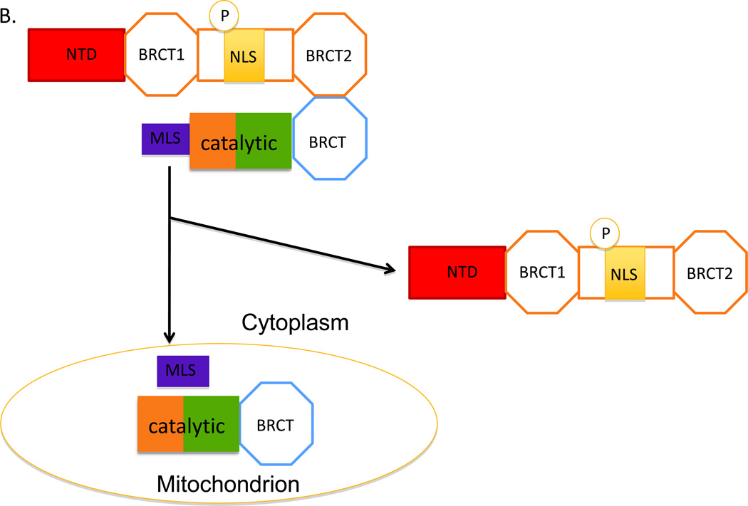
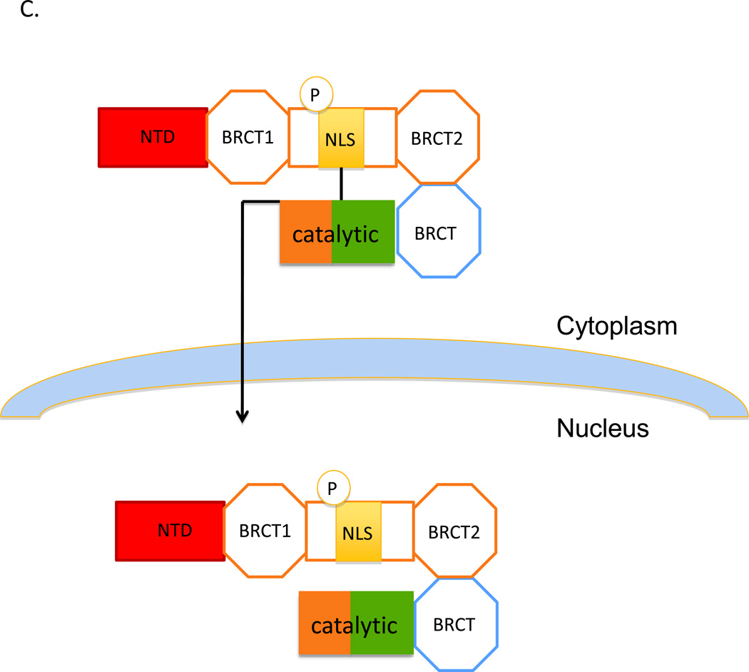
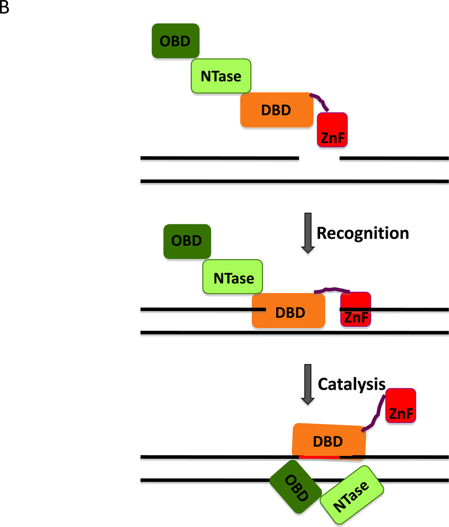
Among the mammalian genes encoding DNA ligases (LIG), the LIG3 gene is unique in that it encodes multiple DNA ligase polypeptides with different cellular functions. Notably, this nuclear gene encodes the only mitochondrial DNA ligase and so is essential for this organelle. In the nucleus, there is significant functional redundancy between DNA ligase IIIα and DNA ligase I in excision repair. In addition, DNA ligase IIIα is essential for DNA replication in the absence of the replicative DNA ligase, DNA ligase I. DNA ligase IIIα is a component of an alternative non-homologous end joining (NHEJ) pathway for DNA double-strand break (DSB) repair that is more active when the major DNA ligase IV-dependent pathway is defective. Unlike its other nuclear functions, the role of DNA ligase IIIα in alternative NHEJ is independent of its nuclear partner protein, X-ray repair cross-complementing protein 1 (XRCC1). DNA ligase IIIα is frequently overexpressed in cancer cells, acting as a biomarker for increased dependence upon alternative NHEJ for DSB repair and it is a promising novel therapeutic target.

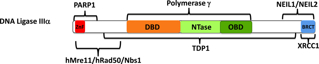
Keywords: BRCT; Cancer; DBD; DNA PK; DNA binding domain; DNA double-strand break; DNA ligase encoding gene; DNA ligases; DNA single-strand break; DNA-dependent protein kinase; DSB; LIG; LIG3; MLS; Mitochondria; NEIL; NHEJ; NLS; NTase; Nei endonoclease VIII-like protein; Neurodegeneration; Nuclear DNA repair; OBD; PARP1; PNKP; SSB; Tdp1; X-ray cross-complementing protein 1; XRCC1; ZnF; breast cancer susceptibility protein 1-related C-terminal; mitochondrial leader sequence; non-homologous end joining; nuclear localization signal; nucleotidyl transferase domain; oligonucleotide/oligosaccharide-fold binding domain; poly(ADP-ribose) polymerase 1; polynucleotide kinase phosphatase; tyrosyl phosphodiesterase 1; zinc finger.


Figures

Figure 1
Figure 1. DNA ligases encoded by the LIG3 gene
The four DNA ligase polypeptides encoded by the mammalain LIG3 gene are shown schematically. The positions of the zinc finger (ZnF, red) and the catalyic region, which contains the DNA binding domain (DBD, light brown), nucleotidyl transferase domain (NTase, light green) and oligonucleotide/oligosaccharide-fold binding domain (OBD, dark green), are indicated. Mitochondrial and nuclear versions of DNA ligase IIIα are generated by alternative translation initiation. The position of the mitochondrial leader sequence (MLS, purple) is indicated. An alterative splicing event that occurs in male germ cells replaces the C-terminal BRCT domain (BRCT, blue) of DNA ligase IIIα with short amino acid sequence that functions as a NLS (NLS, grey) in DNA ligase IIIβ.
Figure 2
Figure 2. Mitochondrial and nuclear versions of DNA ligase IIIα
A. Two polypeptides are generated from DNA ligase IIIα mRNA by alternative translation. Mitochondrial DNA ligase IIIα has an additional N-terminal mitochondrial leader sequence (MLS, purple) in a addition to the common catalytic region (brown/green) and C-terminal BRCT domain (BRCT). Since this domain mediates the interaction with the C-terminal BRCT2 domain of XRCC1, it is assumed that both the mitochondrial and nuclear versions of DNA ligase IIIα will form complexes with XRCC1, which also contains an N-terminal domain (NTD, red), a second BRCT domain (BRCT1) and a nuclear localization domain (NLS, yellow) whose activity appears to be dependent upon phosphorylation (P) of adjacent residues. B. The complex containing mitochondrial DNA ligase IIIα is targed to the mitochondria by the MLS. It is assumed that the activity of the MLS is much greater than the activity of XRCC1 NLS. The MLS initiates passage of the DNA ligase IIIα polypeptide through the mitochondrial membrane and is then removed by proteolysis. Unfolding of the DNA ligase IIIα polypeptide as it passes through the mitochondrial membrane disrupts the interaction with XRCC1, leaving free XRCC1 in the cytoplasm. C. The transport of DNA ligase IIIα lacking the MLS into the nucleus is mediated by the NLS of XRCC1 which binds to the nuclear pore complex, resulting in passage of the XRCC1 into the nucleus.
Figure 3
Figure 3. Interaction of DNA ligase III with nicked DNA
A. Space filling model showing the DNA binding (DBD, light brown), nucleotidyl transferase (NTase, light green) and oligonucleotide/oligosaccharide-fold binding domains (OBD, dark green) of DNA ligase III enaging a short oligonucleotide (Black) containing a non-ligatable nick (Cotner-Gohara et al., 2010). The image was made using PyMol (http://www.pymol.org). B. A fragment of DNA ligase III encompassing the zinc finger (ZnF, red) and the catalytic region (DNA binding domain, DBD; nucleotidyl transferase domain, NTase; oligonucleotide/oligosaccharide-fold binding domain, OBD) and a nicked DNA duplex are shown schematically (upper panel). The single strand interruption is initially recognized by a combination of the ZnF and the DBD (middle panel). If the nick is ligatable, the ZnF is displaced by a combination of the NTase and OBD. The DBD, NTase and OBD encirle and ligate the nicked DNA.
Figure 4
Figure 4. Protein partners of DNA ligase IIIα
The regions of DNA ligase IIIα involved in interactions with hMre11/hRad50/Nbs1, NEIL1 and NEIL2, PARP1, TDP1, XRCC1 and mitochondrial DNA polymerase γ are indicated.

Similar articles

Cited by

References

    1. Ahel I, Rass U, El-Khamisy SF, Katyal S, Clements PM, McKinnon PJ, Caldecott KW, West SC. The neurodegenerative disease protein aprataxin resolves abortive DNA ligation intermediates. Nature. 2006;443:713–716. - PubMed
    1. Ame J, Spenlehauer C, de Murcia G. The PARP superfamily. Bioessays. 2004;26:882–893. - PubMed
    1. Arakawa H, Bednar T, Wang M, Paul K, Mladenov E, Bencsik-Theilen AA, Iliakis G. Functional redundancy between DNA ligases I and III in DNA replication in vertebrate cells. Nucleic Acids Res. 2012;40:2599–2610. - PMC - PubMed
    1. Barnes DE, Tomkinson AE, Lehmann AR, Webster AD, Lindahl T. Mutations in the DNA ligase I gene of an individual with immunodeficiencies and cellular hypersensitivity to DNA-damaging agents. Cell. 1992;69:495–503. - PubMed
    1. Barzilai A. The contribution of the DNA damage response to neuronal viability. Antioxid Redox Signal. 2007;9:211–218. - PubMed

Publication types