. 2013 Oct;20(10):1191-8.
doi: 10.1038/nsmb.2666. Epub 2013 Sep 8.

A cancer-associated BRCA2 mutation reveals masked nuclear export signals controlling localization

Affiliations

A cancer-associated BRCA2 mutation reveals masked nuclear export signals controlling localization

Anand D Jeyasekharan et al. Nat Struct Mol Biol. 2013 Oct.

Abstract

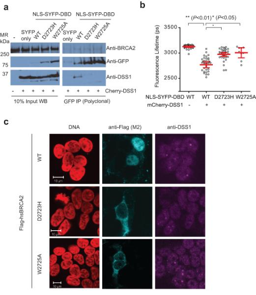
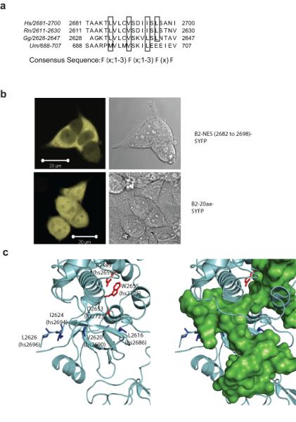
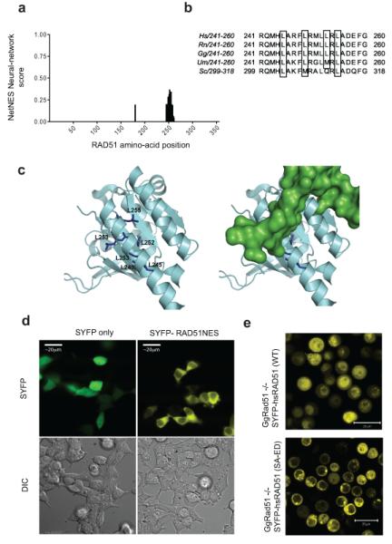
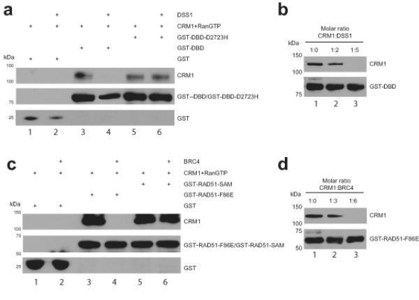
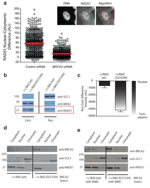
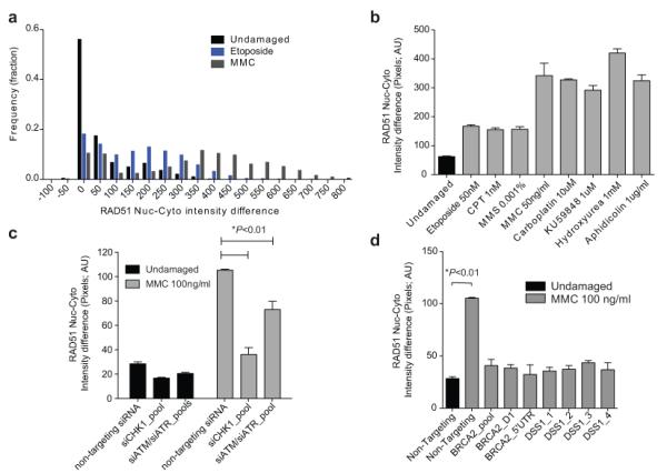
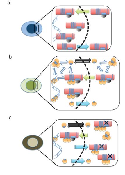
Germline missense mutations affecting a single BRCA2 allele predispose humans to cancer. Here we identify a protein-targeting mechanism that is disrupted by the cancer-associated mutation, BRCA2(D2723H), and that controls the nuclear localization of BRCA2 and its cargo, the recombination enzyme RAD51. A nuclear export signal (NES) in BRCA2 is masked by its interaction with a partner protein, DSS1, such that point mutations impairing BRCA2-DSS1 binding render BRCA2 cytoplasmic. In turn, cytoplasmic mislocalization of mutant BRCA2 inhibits the nuclear retention of RAD51 by exposing a similar NES in RAD51 that is usually obscured by the BRCA2-RAD51 interaction. Thus, a series of NES-masking interactions localizes BRCA2 and RAD51 in the nucleus. Notably, BRCA2(D2723H) decreases RAD51 nuclear retention even when wild-type BRCA2 is also present. Our findings suggest a mechanism for the regulation of the nucleocytoplasmic distribution of BRCA2 and RAD51 and its impairment by a heterozygous disease-associated mutation.


Figures

Figure 1
Figure 1. DSS1 binding correlates with localization of BRCA2
a) is an immunoprecipitation experiment of YFP tagged NLS-DBD fragments with Cherry-DSS1 in 293T cells, using an anti-GFP polyclonal serum. The left panels are of the whole cell lysate (10% of the input), and the panels on the right are of the immunoprecipitate. Transfection of free SYFP with cherry-DSS1 serves as the negative control for the western blot as well as for the immunoprecipitation, and endogenous BRCA2 as the loading control for the experiment. Endogenous DSS1 is difficult to visualize on standard Western blots due to its acidity and small size, requiring unique gel and transfer conditions sub-optimal for the relatively large DBD fragment. Uncropped blots are in Supplementary Fig 8. b) is a dot plot of the mean SYFP lifetime per cell from a FRET-FLIM experiment with NLS-SYFP-DBD and Cherry-DSS1. Each dot represents a single cell; the lines and error bars represent the mean and 95% confidence interval for the population studied in the experiment. U2OS cells co-transfected with the indicated constructs were analyzed by TCSPC. NLS-SYFP DBD forms have a lifetime of about 3200ps, which drops to about 2900ps when mCherry-DSS1 is co-transfected (p<0.01 by two-tailed Student’s t-test, n~30), in contrast to the mutant forms of the DBD. c) shows representative immunofluorescent confocal micrographs of 293T cells nucleofected with Flag-tagged full-length versions of BRCA2, WT or mutant D2723H and W2725A. DNA is colored in red, BRCA2 (anti-Flag) in Cyan, and endogenous DSS1 (anti-DSS1) in Magenta.
Figure 2
Figure 2. DSS1 regulates a functional NES in BRCA2
a) is a clustal W alignment of the nuclear exclusion signal within the DBD of BRCA2, with the critical hydrophobic residues that potentially contribute to the NES consensus marked by black boxes. b) is a representative fluorescent micrograph of 293T cells transfected with a BRCA2 NES-SYFP construct (upper panel) and SYFP tagged to a similarly sized sequence within BRCA2 (lower panel), which serves as a negative control. c) is a rendering of the 1YIJ structure of rnBRCA2 DBD. The DBD is shown in grey, and the critical hydrophobic NES residues highlighted in blue. Mutations that affect DSS1 binding are depicted in red. The right panel shows a superimposition of DSS1 (green).
Figure 3
Figure 3. BRCA2 regulates a functional NES in RAD51
a) is a histogram of the NES scores for amino acids in the HsRAD51 sequence, as determined by the NetNES algorithm, showing the potential NES sequence in the C-terminus of the protein. b) is a sequence alignment of the residues comprising the RAD51 NES from yeast to human. The hydrophobic residues are marked by black boxes. c) is a rendering of the crystal structure 1N0W, in the same color scheme as Figure 2c. NES residues in RAD51 are shown in blue. The right panel shows a superimposition of the BRC repeat (green). d) shows representative micrographs of 293T cells transfected with a RAD51-NES tagged version of SYFP (right), in comparison to free SYFP alone. e) is a fluorescence micrograph of SYFP-tagged wildtype RAD51, compared with a mutant form lacking BRCA2 binding (SYFP-RAD51 (SA-ED)). SYFP-tagged proteins were expressed in Rad51−/−DT40 cells that conditionally express Tet-regulated untagged RAD51. Micrographs were taken 12h after the depletion of untagged RAD51 using doxycycline .
Figure 4
Figure 4. CRM1 binding to NESs in BRCA2 or RAD51 is masked by DSS1 or BRC4 respectively
a) Immunoblots of GST pull-down assessing binding of CRM1 to GST-DBD and GST-DBD-D2723H immobilized on a glutathione sepharose matrix in the absence (lanes 3 and 5) or presence (lanes 4 and 6) of DSS1 at the 5-fold molar excess optimized from the representative titration shown in panel b). GST was used as a specificity control. DSS1 inhibits the binding of CRM1 to DBD (lane 4) but not to DBD-D2723H (lane 6). c) Immunoblots of GST pull-down assessing binding of CRM1 to GST-RAD51-F86E and GST-RAD51-SAM immobilized on a glutathione sepharose matrix in the absence (lanes 3 and 5) and presence of (lanes 4 and 6) of BRC4 peptide at the 6-fold molar excess optimized from the representative titration shown in panel d). GST was used as a specificity control. BRC4 peptide disrupts the binding of CRM1 to RAD51-F86E (lane 4) but not to RAD51-SAM (lane 6). Proteins were visualized with antibodies against His or GST. CRM1 protein was His-tagged. Uncropped blots are in Supplementary Fig 8.
Figure 5
Figure 5. RAD51 mislocalization by BRCA2 depletion or mutation
a) is a dot plot of the mean nucleo-cytoplasmic difference of RAD51 per cell, determined for a population of cells with and without exposure to BRCA2 siRNA. The figures in the inset represent the algorithm used for determining the nucleo-cytoplasmic difference by automated microscopy. The left image is of the nucleus as defined by DAPI, the middle image is that of RAD51 staining. The algorithm for nucleo-cytoplasmic intensity difference calculation (see Methods) is overlayed on RAD51 in the right panel. b) is a cell fractionation experiment from heterozygous mouse ES cells, with Western blotting for RAD51 to assess localization. MEK2 (predominantly cytoplasmic) and SCC1 (predominantly nuclear) serve as loading controls and controls for the efficiency of fractionation. c) is a bar graph quantitating the results from three independent fractionation experiments. The nucleo-cytoplasmic intensity difference for RAD51 in each experiment was obtained by generating densitometric profiles for each band (identical exposure) using ImageJ, and then subtracting the cytoplasmic value from the nuclear value. Lower (more negative) values indicate more cytoplasmic protein (n=3, error bars represent SEM). d) shows a cell fractionation experiment of ES cells carrying BACs expressing either the WT or D2723H mutant of BRCA2, in the absence or presence (E) of DNA damage induced by exposure to 100 ng/ml of MMC for 20 hrs. SCC1 is used as a control for fractionation. One experiment representative of 3 independent repeats is shown. Uncropped blots are in Supplementary Fig 8.
Figure 6
Figure 6. RAD51 nuclear enrichment is a BRCA2-dependent DNA damage response
a) represents the frequency distribution of the RAD51 nuclear-cytoplasmic pixel intensity difference (NC-difference) from different populations of MCF10A cells analyzed by automated immunofluorescence microscopy. Results for untreated cells are in black, and for cells treated with etoposide or MMC in blue or grey respectively (n=2000 each). b) shows the mean NC-difference for RAD51 in MCF10A cells treated with the indicated forms of DNA damage. Each bar represents the mean ± standard error of 4 wells in a 96-well plate, with 500 cells counted per well. c) shows the mean NC-difference for RAD51 in MCF10A cells pre-treated with the indicated siRNAs, with (grey) or without (black) exposure to MMC. The increase in the NC-difference after DNA damage is significantly affected (Student’s two tailed t-test, p<0.05, n=500 cells, 3 wells) by depletion of ATM, ATR or CHK1. d) shows the RAD51 NC-difference in MCF10A cells following knockdown of BRCA2 and DSS1 using multiple independent siRNA’s. The black bar on the left represents undamaged cells. The nuclear enrichment seen after DNA damage is significantly affected (p<0.01, Student’s two tailed t-test, n=500 cells, 3 wells) by depletion of both BRCA2 and DSS1. Supplementary Fig 6a-d shows the depletion efficiency with the siRNAs used.
Figure 7
Figure 7. A hypothetical model for BRCA2 and RAD51 nuclear localization through masking of nuclear export sequences
a) depicts the proposed mechanism wherein the nuclear retention of BRCA2 (red oblong) is allowed when its binding to DSS1 (black semi-circle) obscures an NES motif. Nuclear transport (green arrow) is presumably directed by NLSs previously identified in the C-terminal region of BRCA2, which are not shown. Dissociation of BRCA2 from DSS1 may permit nuclear export (blue arrow). b) depicts the proposed mechanism wherein the nuclear retention of RAD51 (yellow circle) is allowed when its binding to the BRC repeats of BRCA2 obscures an NES motif. Nuclear transport (green arrow) of the BRCA2-RAD51 complex is presumably directed by the NLSs in BRCA2. In addition, cytosolic RAD51 may exist in an equilibrium (thin blue arrows) between free monomers and oligomers. Momomeric RAD51 is small enough to diffuse freely across the nuclear membrane (black arrows), whereas oligomers are not. Dissociation of RAD51 from BRCA2 may permit nuclear export (blue arrow). The net result of these different processes is to localize RAD51 predominantly in the nucleus. c) depicts how the processes depicted in the preceding panels may be affected by the BRCA2 D2723H mutation, which prevents DSS1 binding (black X). Mutant BRCA2 is exported from the nucleus (blue arrows) resulting in predominant cytoplasmic localization, and this is proposed to shift the balance towards cytoplasmic localization of RAD51, despite the presence of wildtype BRCA2.

Similar articles

Cited by

References

    1. The Breast Cancer Linkage Consortium Cancer risks in BRCA2 mutation carriers. The Breast Cancer Linkage Consortium. J Natl Cancer Inst. 1999;91:1310–6. - PubMed
    1. Collins N, et al. Consistent loss of the wild type allele in breast cancers from a family linked to the BRCA2 gene on chromosome 13q12-13. Oncogene. 1995;10:1673–5. - PubMed
    1. King TA, et al. Heterogenic loss of the wild-type BRCA allele in human breast tumorigenesis. Ann Surg Oncol. 2007;14:2510–8. - PubMed
    1. Knudson AG., Jr. Mutation and cancer: statistical study of retinoblastoma. Proc Natl Acad Sci U S A. 1971;68:820–3. - PMC - PubMed
    1. Skoulidis F, et al. Germline Brca2 heterozygosity promotes Kras(G12D) -driven carcinogenesis in a murine model of familial pancreatic cancer. Cancer Cell. 2010;18:499–509. - PubMed

References for Methods

    1. Hattori H, Skoulidis F, Russell P, Venkitaraman AR. Context dependence of checkpoint kinase 1 as a therapeutic target for pancreatic cancers deficient in the BRCA2 tumor suppressor. Mol Cancer Ther. 2011;10:670–8. - PMC - PubMed
    1. Jeyasekharan AD, et al. DNA damage regulates the mobility of Brca2 within the nucleoplasm of living cells. Proc Natl Acad Sci USA. 2010;107:21937–42. - PMC - PubMed
    1. Yu DS, et al. Dynamic control of Rad51 recombinase by self-association and interaction with BRCA2. Mol Cell. 2003;12:1029–41. - PubMed
    1. Guttler T, et al. NES consensus redefined by structures of PKI-type and Rev-type nuclear export signals bound to CRM1. Nat Struct Mol Biol. 2010;17:1367–1376. - PubMed
    1. Frey S, Richter RP, Gorlich D. FG-rich repeats of nuclear pore proteins form a three-dimensional meshwork with hydrogel-like properties. Science. 2006;314:815–7. - PubMed