. 2014 Jun 26;33(26):3383-91.
doi: 10.1038/onc.2013.327. Epub 2013 Sep 9.

High incidence of female reproductive tract cancers in FA-deficient HPV16-transgenic mice correlates with E7's induction of DNA damage response, an activity mediated by E7's inactivation of pocket proteins

Affiliations

High incidence of female reproductive tract cancers in FA-deficient HPV16-transgenic mice correlates with E7's induction of DNA damage response, an activity mediated by E7's inactivation of pocket proteins

J W Park et al. Oncogene. .

Abstract

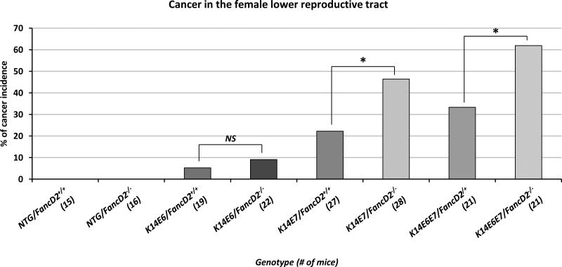
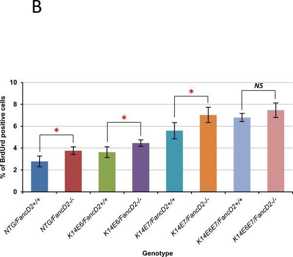
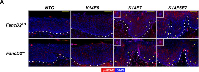
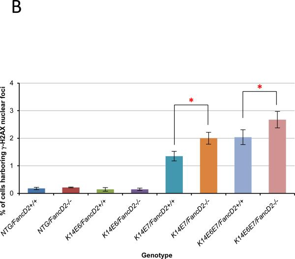
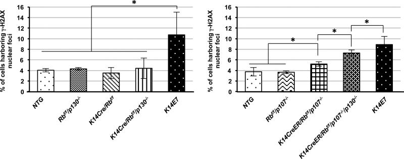
Fanconi anemia (FA) is a rare genetic disorder caused by defects in a DNA damage repair system, the FA pathway. FA patients frequently develop squamous cell carcinoma (SCC) at sites that are associated with human papillomavirus (HPV)-driven cancer including the female reproductive tract. To assess experimentally whether FA deficiency increases susceptibility to HPV-associated cervical/vaginal cancer, we monitored cancer incidence in the female lower reproductive tract of FA-deficient mice expressing HPV16 oncogenes, E6 and/or E7. FA deficiency specifically increased the incidence of cancers in mice expressing E7; but this effect was not observed in mice just expressing E6. We also observed that E7, but not E6, induced DNA damage as scored by induction of γ-H2AX and 53BP1 (p53 binding protein 1) nuclear foci, and this induction was heightened in FA-deficient tissue. Finally, we discovered that this induction of DNA damage responses was recapitulated in mice deficient in expression of 'pocket' proteins, pRb, p107 and p130, which are established targets of E7. Our findings support the hypothesis that E7 induces cancer by causing DNA damage at least in part through the inactivation of pocket proteins. This hypothesis explains why a deficiency in DNA damage repair would increase susceptibility to E7-driven cancer.


Figures

Figure 1
Figure 1. HPV-associated cancer incidence in the lower reproductive tract
Mice were treated with 17β-estradiol which is predominant estrogen for a period of 6 months to achieve a state of persistent estrus. After the estrogen treatment, the female lower reproductive tract was harvested, fixed and sectioned. Every tenth, 5 μm section was stained with hematoxylin and eosin (H&E) and histopathologically evaluated the worst lesion scored as the final diagnosis. Cervical and/or vaginal cancers were scored in each genotype of mice. The cancer incidence in K14E7/FancD2+/+ is significantly higher than it in NTG/FancD2+/+ (P=0.03) or K14E6/FancD2+/+ mice (P=0.07). fancD2 deficiency significantly increased cancer incidence in K14E7 and K14E6E7 mice. Asterisk (*) means significant difference (P<0.05). NS means no significant difference. All statistical analyses were performed using a one-sided Barnard's exact test.
Figure 2
Figure 2. Expression of two biomarkers, MCM7 and p16, in cervical carcinomas derived from K14E6, K14E7, K14E6E7 mice on both fancD2-sufficient and -deficient backgrounds
Shown are representative immunohistochemistry with anti-MCM7 and anti-p16. Brown-stained nuclear cells are positive for the both markers. Hematoxylin (Blue) is used for nuclear counter staining. Scale bar, 50 μm. *K14E6/FancD2+/+ tumor was a vaginal cancer due to no cervical cancer.
Figure 3
Figure 3. Cell proliferation modulated by HPV oncogenes and fancD2 deficiency
A, to monitor the newly synthesized DNA at the cervical epithelia in each genotype of mice, mice were intraperitoneally injected with BrdUrd 1 hour before euthanized. Immunofluorescence with anti-BrdUrd (Red) was performed. DAPI (Blue) was used for a nuclear counterstaining. Scale bar, 50 μm. The white dot line indicates basal membrane. B, at least three mice from NTG, K14E6, K14E7, and K14E6E7 mice in the presence and absence of fancD2 expression were randomly selected and more than eight image frames of cells at the epithelia of cervix were quantified for each genotype of mice. The amount of BrdUrd-positive cells over total number of cells was plotted in each case (columns); bars, Standard deviation (SD). Asterisk (*) means significant difference (P<0.05). NS means no significant difference (P>0.05). In the epithelial layer of the cervical tissues, HPV oncogene expression significantly induced the number of BrdUrd-positive cells (NTG/FancD2+/+ vs. K14E6/FancD2+/+, K14E7/FancD2+/+, or K14E6E7/FancD2+/+; P<0.05). fancD2 deficiency statistically increased proliferation in NTG, K14E6, and K14E7 mice (NTG/FancD2+/+ vs. NTG/FancD2−/−, K14E6/FancD2+/+ vs. K14E6/FancD2−/−, and K14E7/FancD2+/+ vs. K14E7/FancD2−/−; P<0.05), not in K14E6E7 mice (K14E6E7/FancD2+/+ vs. K14E6E7/FancD2−/−; P=0.15). All statistical analyses were performed using a two-sided Wilcoxon Rank sum test.
Figure 4
Figure 4. FancD2 deficiency induced E7-dependent DNA damage response via β-H2AX
A, to detect DNA damage response, tissue sections from each group were stained for anti-γ-H2AX (Red) antibody. DAPI (Blue) is used for a nuclear counterstaining. Scale bar, 50 μm. γ-H2AX nuclear-foci positive cells are highlighted by yellow arrows in the images. Insets provide magnified views of cells with γ-H2AX positive nuclear-foci. The white dot line indicates basal membrane. B, at least three mice from NTG, K14E6, K14E7, and K14E6E7 mice in the presence and absence of fancD2 expression were randomly selected and more than eight image frames of cells at the epithelia of cervix were quantified for each mouse. The amount of γ-H2AX nuclear-foci positive cells over total number of cells was plotted in each case (columns); bar, Standard deviation (SD). In the epithelial layer of the cervical tissues, E7 expression and E6/E7 double expression significantly induced the number of γ-H2AX nuclear-foci positive cells on fancD2-sufficient background (NTG/FancD2 or K14E6/FancD2+/+ vs. K14E7/FancD2+/+ or K14E6E7/FancD2+/+; P<0.05). fancD2 deficiency statistically increased proliferation in K14E7 and K14E6E7 mice (K14E7/FancD2+/+ vs. K14E7/FancD2−/− and K14E6E7/FancD2+/+ vs. K14E6E7/FancD2−/−; P<0.05), not in NTG and K14E6 mice. Asterisk (*) means significant difference (P<0.05). NS means no significant difference (P>0.05). All statistical comparisons were performed using a two-sided Wilcoxon Rank sum test.
Figure 5
Figure 5. Deficiency of Pocket protein family members Increased DNA damage via γ-H2AX in the cervical epithelia
A and B. At least three mice from each genotype were randomly selected and more than eight image frames of cells at the epithelia of cervix were quantified for each mouse. The amount of γ-H2AX nuclear-foci positive cells over total number of cells was plotted in each case (columns); bar, Standard deviation (SD). A. In the epithelial layer of the cervical tissues, K14Cre/pRbf/f, pRbf/f/p130−/−, and K14Cre/pRbf/f/p130−/− mice failed to increase the number of γ-H2AX nuclear-foci positive cells compared with NTG mice (P>0.05). E7 expression cause a significant increase in DNA damage compared with NTG, K14Cre/pRbf/f, pRbf/f/p130−/−, or K14Cre/pRbf/f/p130−/− mice (P<0.05). B. Single deficiency of p107 failed to increase DNA damage via γ-H2AX foci (pRbf/f/p107−/− vs. NTG; P>0.05). Double deletion of pRb and p107 significantly induced the number of cells harboring with γ-H2AX foci compared with single deletion of p107 (K14CreER/pRbf/f/p107−/− vs. pRbf/f/p107−/−; P<0.05). The frequency of γ-H2AX foci positive cells in the triple knockout mice was significantly higher than it in pRb and p107 double knockout mice, but was significantly lower than it in K14E7 mice. (K14CreER/pRbf/f/p130f/f/p107−/− vs. K14CreER/pRbf/f/p107−/−; P<0.05 and K14CreER/pRbf/f/p130f/f/p107−/− vs. K14E7; P<0.05). Asterisk (*) means significant difference (P<0.05). NS means no significant difference (P>0.05). All statistical comparisons were performed using a two-sided Wilcoxon Rank sum test. Note that In Fig. 4, E7 expression induced γ-H2AX in only ~1.5% of cells, but in the data presented in this figure, it induces it in ~10% of cells. This difference is solely attributed to the difference in genetic backgrounds between the mice used in the studies reported in these two figures, not due to differences in staining efficiency, because the same low % of cells with γ-H2AX positive foci was observed in sections of K14E7 transgenic mice taken from the study reported in Fig. 4, when they were stained alongside the samples from the experiment reported in this figure.

Similar articles

Cited by

References

    1. Kutler DI, Singh B, Satagopan J, Batish SD, Berwick M, Giampietro PF, et al. A 20-year perspective on the International Fanconi Anemia Registry (IFAR). Blood. 2003 Feb 15;101(4):1249–56. - PubMed
    1. Garner E, Smogorzewska A. Ubiquitylation and the Fanconi anemia pathway. FEBS Lett. [Research Support, N.I.H., Extramural Research Support, Non-U.S. Gov't Review] 2011 Sep 16;585(18):2853–60. - PMC - PubMed
    1. Joenje H, Patel KJ. The emerging genetic and molecular basis of Fanconi anaemia. Nat Rev Genet. 2001 Jun;2(6):446–57. - PubMed
    1. Crossan GP, Patel KJ. The Fanconi anaemia pathway orchestrates incisions at sites of crosslinked DNA. J Pathol. [Research Support, Non-U.S. Gov't Review] 2012 Jan;226(2):326–37. - PubMed
    1. Kennedy RD, D'Andrea AD. The Fanconi Anemia/BRCA pathway: new faces in the crowd. Genes Dev. 2005 Dec 15;19(24):2925–40. - PubMed

Publication types

MeSH terms

Substances