. 2013:4:2368.
doi: 10.1038/ncomms3368.

RecG and UvsW catalyse robust DNA rewinding critical for stalled DNA replication fork rescue

Affiliations
Free PMC article

RecG and UvsW catalyse robust DNA rewinding critical for stalled DNA replication fork rescue

Maria Manosas et al. Nat Commun. 2013.
Free PMC article

Erratum in

  • Nat Commun. 2014;5:4210. Bianco, Piero [corrected to Bianco, Piero R]

Abstract

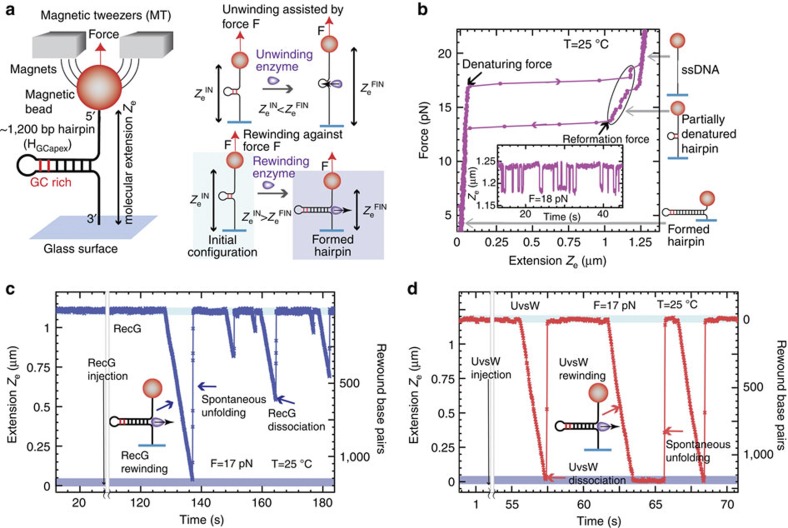
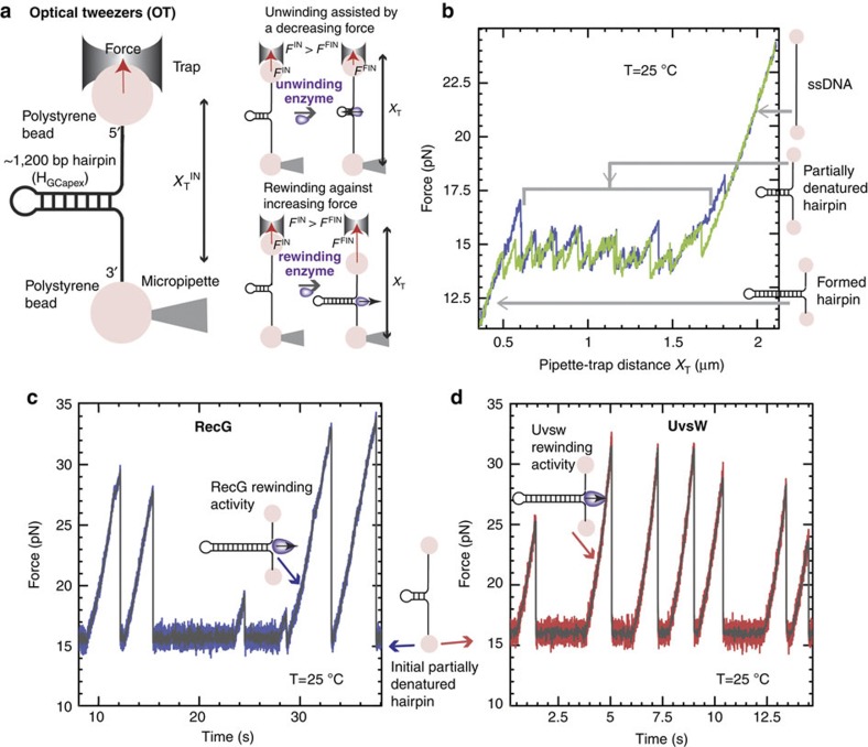
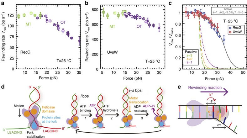
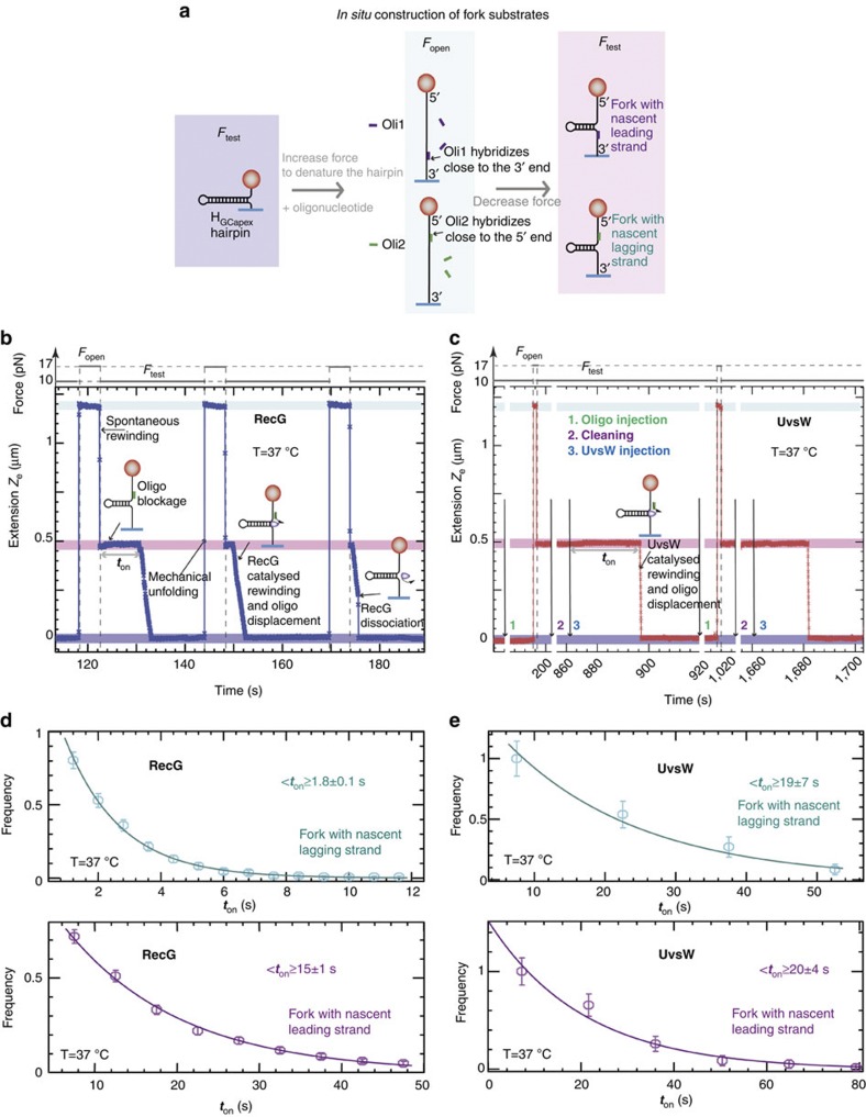
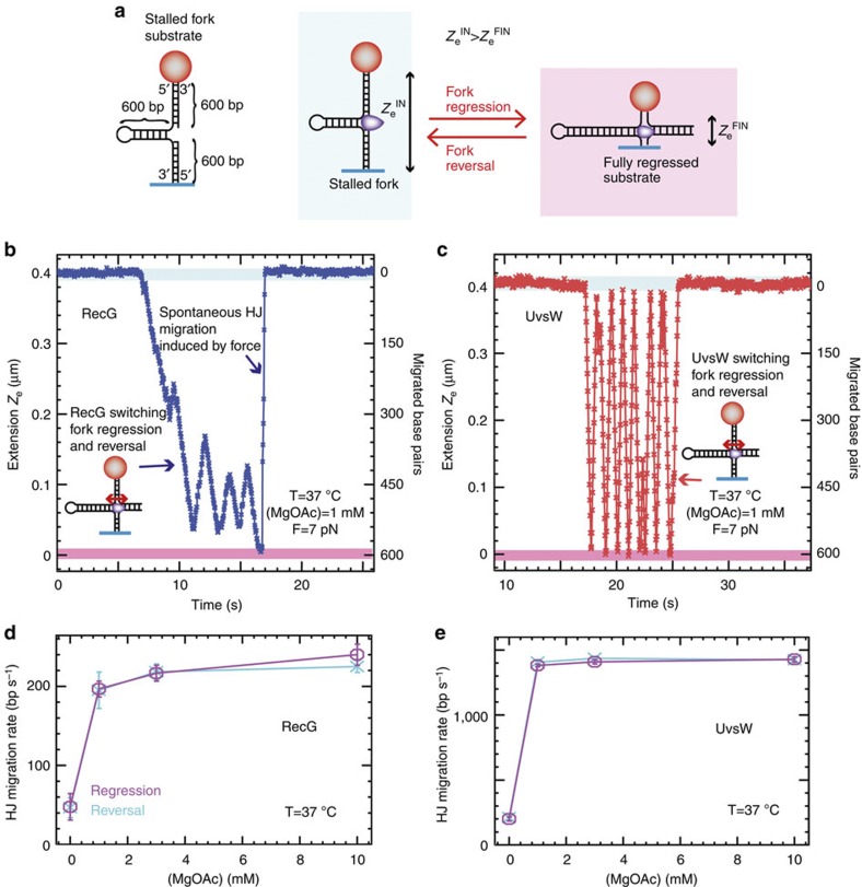
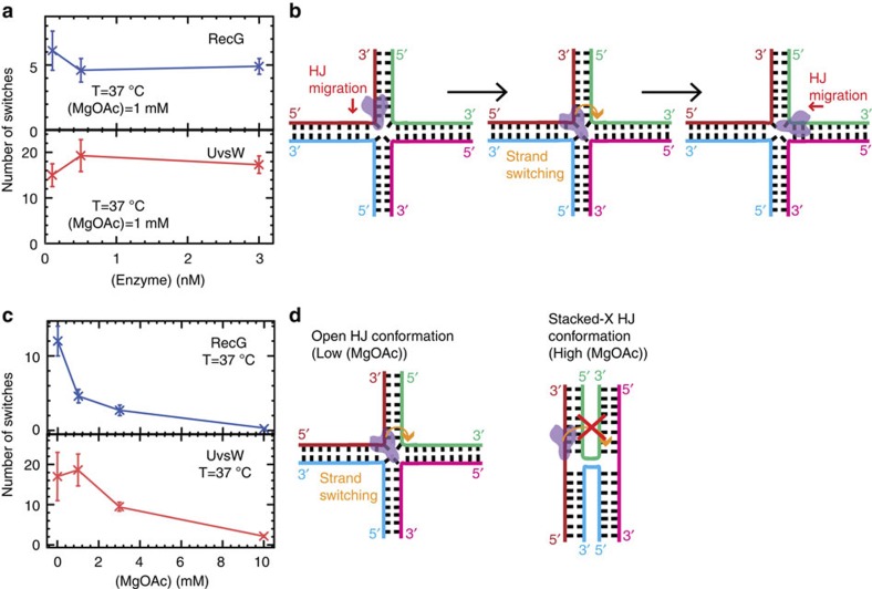
Helicases that both unwind and rewind DNA have central roles in DNA repair and genetic recombination. In contrast to unwinding, DNA rewinding by helicases has proved difficult to characterize biochemically because of its thermodynamically downhill nature. Here we use single-molecule assays to mechanically destabilize a DNA molecule and follow, in real time, unwinding and rewinding by two DNA repair helicases, bacteriophage T4 UvsW and Escherichia coli RecG. We find that both enzymes are robust rewinding enzymes, which can work against opposing forces as large as 35 pN, revealing their active character. The generation of work during the rewinding reaction allows them to couple rewinding to DNA unwinding and/or protein displacement reactions central to the rescue of stalled DNA replication forks. The overall results support a general mechanism for monomeric rewinding enzymes.


Figures

Figure 1
Figure 1. Efficient rewinding of DNA is monitored in real time using magnetic tweezers.
(a) Schematic of the MT experimental setup and progress of the reaction. A DNA hairpin substrate was tethered between a glass surface and a magnetic bead held in a magnetic tweezers. The GC-rich region of the HGCapex hairpin is shown in red. The DNA rewinding or unwinding reaction was followed by monitoring changes in the distance between the bead and the glass surface (Ze). (b) Hairpin configurations can be precisely followed using MT, and typical data in the absence of protein are shown. The force as a function of Ze for the HGCapex hairpin at 25 °C demonstrated stable hairpin folding below 15 pN and mechanical unfolding above 17 pN. Inset shows the Ze(t) at 18 pN where the molecule hops between the fully open (that is, completely denatured) and partially denatured hairpin configurations (c,d). Representative traces of rewinding reactions catalysed by RecG (c) and UvsW (d) enzymes. Reactions were carried out as described in Methods. Rewinding bursts were detected as decreases in Ze. The molecular extensions corresponding to the initial partially denatured hairpin and the final fully formed hairpin are highlighted in light and dark blue, respectively.
Figure 2
Figure 2. Efficient rewinding of DNA at very large forces is monitored in real time using optical tweezers.
(a) Schematic of the OT experimental setup and progress of the reaction. A DNA hairpin substrate was tethered between two beads—one trapped in the optical trap and the other fixed in a tip of a micropipette. In this passive configuration, the DNA rewinding or unwinding reaction was followed by monitoring changes in force. (b) Hairpin states can be precisely followed using OT, and typical progress curves in the absence of protein are shown. The force as a function of XT for the HGCapex hairpin at 25 °C shows a broad region resulting in a partially unzipped hairpin configuration. (c,d) Representative traces of rewinding reactions catalysed by RecG (c) and UvsW (d) proteins. Experimental traces showing force as a function of time starting with the initial partially denatured hairpin configuration. Rewinding bursts were detected as force rips.
Figure 3
Figure 3. RecG and UvsW catalyse efficient rewinding reactions against large opposing forces.
(a,b) Mean rate of rewinding as a function of force for RecG (a, number n of experimental traces analysed from 56 to 428 depending on the conditions) and UvsW (b, n from 37 to 214 depending on the conditions) at 25 °C. Reactions were carried out using MT (green squares) for forces ≤15 pN and OT (purple circles and crosses) for opposing forces ≥15 pN. Rates at high forces are computed as described in Methods both from OT-passive data (Fig. 2, purple circles) and from OT force-jump data (Supplementary Fig. S3, purple crosses). Error bars are s.e.m. (c) The experimentally measured rate of rewinding for RecG (blue circles) and UvsW (red diamonds) normalized to the maximum rate of rewinding as a function of the force. Error bars are s.e.m. The results are compared with the predictions obtained from the extended model for a passive enzyme with different step sizes and for an active enzyme model that agrees very well with the experimental results (s=1, ΔGa=5.5 kBT, m=3). (d) Rewinding scheme based on that previously proposed for RecG fork regression. Helicase domains are marked in yellow, whereas the protein region that makes contact with the fork is shown in blue (for example, wedge domain of RecG). Along the ATP cycle at least three different states can be considered: protein without nucleotide bound (1), protein with ATP bound (2) and protein with ADP bound (3). (e) Schematics of the extended model used to describe the UvsW and RecG rewinding behaviours: α and β are the base pair opening and closing rates; k+ is the forward translocation rate; s is the enzyme step size and m is the range of the protein–DNA interaction potential (in the figure s=1 and m=3).
Figure 4
Figure 4. Substrate requirements for RecG- and UvsW-catalysed fork regression are distinct.
(a) Schematics of the forked-DNA substrate construction with nascent lagging or leading strand from the HGCapex hairpin. (b,c) Experimental traces showing the cyclic fork regression assay for RecG (b) and UvsW (c) proteins at 37 °C. The molecular extensions corresponding to the initially formed hairpin, the totally denatured substrate and the partially denatured hairpin are highlighted in dark blue, light blue and pink, respectively. For RecG, a single injection of oligonucleotide and enzyme is sufficient, whereas for UvsW measurements three separate injections are needed: first oligonucleotide injection, second buffer injection and third UvsW injection. (d,e) The distributions of enzyme-binding times for RecG (d), (number n of experimental traces analysed from 427 to 566 depending on the conditions) and UvsW (e), (n from 55 to 86 depending on the conditions) and different fork geometries. Error bars are inversely proportional to the square root of the number of points for each bin.
Figure 5
Figure 5. RecG and UvsW catalyse the Holliday junction formation and migration from a stalled fork intermediate.
(a) Schematic of the stalled fork substrate and the fork regression and HJ branch migration reaction. (b,c) RecG and UvsW branch migration traces performed in a buffer containing 1 mM MgOAc at 37 °C. The molecular extensions corresponding to the initial stalled fork and the final fully regressed configurations are highlighted in light blue and pink, respectively. (d,e) RecG (d, number of experimental traces analysed n from 64 to 126 depending on the conditions) and UvsW (e, n from 53 to 209 depending on the conditions) mean branch-migration rate during fork regression (magenta) and fork reversal (cyan) at 7 pN as a function of the MgOAc concentration. Error bars are s.e.m.
Figure 6
Figure 6. Switches in the direction of HJ branch migration catalysed by single RecG and UvsW enzymes are dependent on the ionic strength.
(a) The mean number of switches in the HJ branch-migration direction catalysed by RecG (number n of experimental traces analysed from 25 to 76 depending on the conditions) and UvsW (n from 17 to 68 depending on the conditions) at 37 °C, 1 mM MgAc and 7 pN as a function of the enzyme concentration. Error bars are s.e.m. The average number of switches remains constant when changing the enzyme concentration (from 0.1 to 3 nM), showing that the HJ branch-migration switching is mediated by a single enzyme (RecG or UvsW). (b) Schematics of the enzyme strand-switching mechanism, which can lead to changes in direction of the HJ branch migration. (c) The mean number of switches in the HJ branch-migration direction catalysed by RecG (n from 33 to 74 depending on the conditions) and UvsW (n from 17 to 41 depending on the conditions) at 7 pN and 37 °C as a function of the MgOAc concentration. Error bars are s.e.m. (d) Schematics of the open and X-stacked HJ conformations favoured at low and high ionic strength, which, respectively, facilitate and hinder the enzyme strand-switching transition.

Similar articles

Cited by

References

    1. Lohman T. M. & Bjornson K. P. Mechanisms of helicase-catalyzed DNA unwinding. Annu. Rev. Biochem. 65, 169–214 (1996). - PubMed
    1. Matson S. W., Bean D. W. & George J. W. DNA helicases: enzymes with essential roles in all aspects of DNA metabolism. Bioessays 16, 13–22 (1994). - PubMed
    1. Nelson S. W. & Benkovic S. J. The T4 phage UvsW protein contains both DNA unwinding and strand annealing activities. J. Biol. Chem. 282, 407–416 (2007). - PubMed
    1. McGlynn P. & Lloyd R. G. Rescue of stalled replication forks by RecG: simultaneous translocation on the leading and lagging strand templates supports an active DNA unwinding model of fork reversal and Holliday junction formation. Proc. Natl Acad. Sci. USA 98, 8227–8234 (2001). - PMC - PubMed
    1. Cheok C. F., Wu L., Garcia P. L., Janscak P. & Hickson I. D. The Bloom's syndrome helicase promotes the annealing of complementary single-stranded DNA. Nucleic Acids Res. 33, 3932–3941 (2005). - PMC - PubMed

Publication types

MeSH terms