. 2013 Sep 1;27(17):1903-16.
doi: 10.1101/gad.219899.113.

Regulation of constitutive and alternative splicing by PRMT5 reveals a role for Mdm4 pre-mRNA in sensing defects in the spliceosomal machinery

Affiliations

Regulation of constitutive and alternative splicing by PRMT5 reveals a role for Mdm4 pre-mRNA in sensing defects in the spliceosomal machinery

Marco Bezzi et al. Genes Dev. .

Abstract

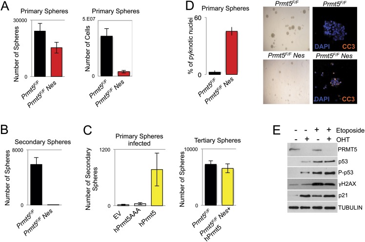
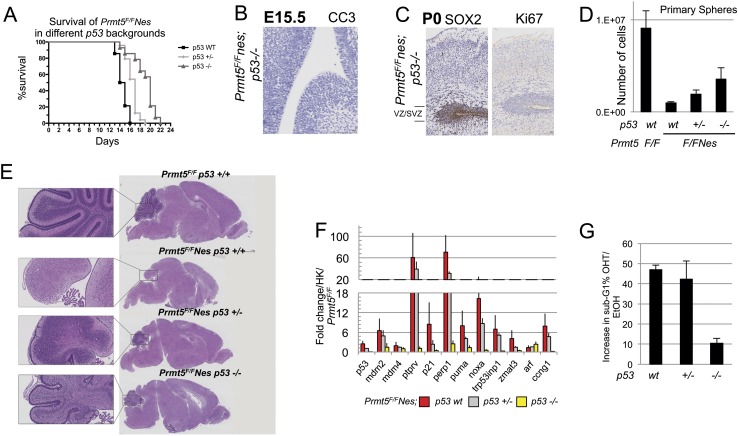
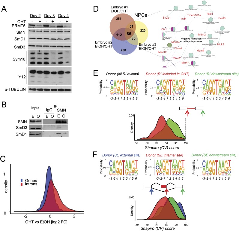
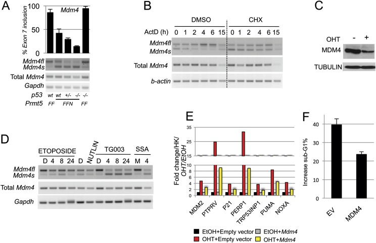
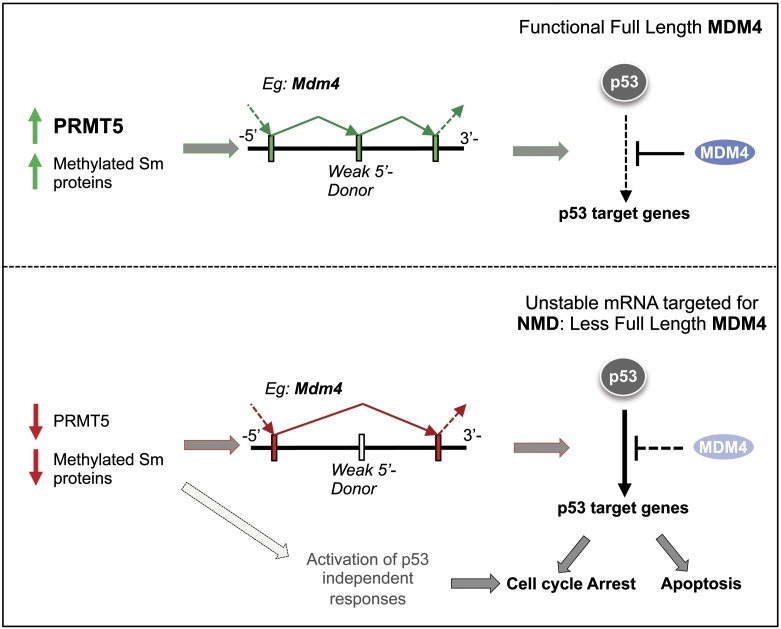
The tight control of gene expression at the level of both transcription and post-transcriptional RNA processing is essential for mammalian development. We here investigate the role of protein arginine methyltransferase 5 (PRMT5), a putative splicing regulator and transcriptional cofactor, in mammalian development. We demonstrate that selective deletion of PRMT5 in neural stem/progenitor cells (NPCs) leads to postnatal death in mice. At the molecular level, the absence of PRMT5 results in reduced methylation of Sm proteins, aberrant constitutive splicing, and the alternative splicing of specific mRNAs with weak 5' donor sites. Intriguingly, the products of these mRNAs are, among others, several proteins regulating cell cycle progression. We identify Mdm4 as one of these key mRNAs that senses the defects in the spliceosomal machinery and transduces the signal to activate the p53 response, providing a mechanistic explanation of the phenotype observed in vivo. Our data demonstrate that PRMT5 is a master regulator of splicing in mammals and uncover a new role for the Mdm4 pre-mRNA, which could be exploited for anti-cancer therapy.

Keywords: MDM4; PRMT5; arginine methylation; development; p53; splicing.


Figures

Figure 1.
Figure 1.
PRMT5 deficiency in the CNS results in early postnatal lethality. Nestin-Cre-induced deletion of the PRMT5 gene in the CNS. (A) Weight in milligrams of wild-type (Prmt5F/F) and Prmt5-deleted (Prmt5F/FNes) brains at three different time points (E17.5, P0, and P10). Brain sizes of P10 Prmt5F/F and Prmt5F/FNes mice are shown as an example in the right panel. (B) Hematoxylin and eosin (H&E)-stained sagittal and coronal sections of P10 cerebrum (Cr) and the cerebellum (Cb) from Prmt5F/F (right) and Prmt5F/FNes (left). (C) Coronal sections of P0 brains. Cellularity of the cortical plate (CP) and VZ/SVZ are indicated in wild-type (wt; black) and mutant (red) brains. (MZ) Marginal zone; (SP) subplate; (IZ) intermediate zone. (D) SOX2 and Ki67 immunohistochemistry (IHC) staining in P0 brains. (E) Cleaved Caspase 3 (CC3) staining is shown in both the cortex and the gangliomic eminence.
Figure 2.
Figure 2.
PRMT5 is required for NPC homeostasis. (A) Number of primary neurospheres and total number of cells from cultures of E14.5 dorsal telencephalon NPCs derived from Prmt5F/F and Prmt5F/FNes embryos. Each bar represents an average of at least three experiments. (B) Number of secondary neurospheres, as in A. (C) Primary neurospheres (left panel) from Prmt5F/FNes mice infected with empty vector (EV), wild-type PRMT5 (hPRMT5), or a catalytically inactive PRMT5 mutant (hPRMT5AAA) and passaged to derive secondary and tertiary neurospheres (right panel). (D) Neurospheres derived from Prmt5F/F or Prmt5F/FNes NPCs were stained with DAPI and CC3, and the percentage of pyknotic nuclei was counted. (E) Protein levels upon treatment with OHT and subsequent PRMT5 depletion for 4 d. The antibodies used are indicated on the right of each panel. As a positive control, p53 and the DDR were induced by treating cells with 10 μM etoposide for 2 h.
Figure 3.
Figure 3.
p53 deletion partially rescues Prmt5F/FNes developmental defects. (A) Kaplan-Meier survival analysis of Prmt5F/FNes mice in a p53wt (n = 14), p53+/− (n = 24), or p53−/− (n = 14) background. (B) Nestin-Cre-induced deletion of the PRMT5 gene in the CNS of p53−/− embryos. Coronal sections of E15.5 brains stained for CC3 (B) and P0 brains stained for SOX2 and Ki67 (C) to identify stem cells and assess their proliferation status. The antibodies used are indicated for each panel. (D) Total number of NPC cells grown as primary neurospheres derived from Prmt5F/FNes;p53wt, Prmt5F/FNes;p53+/−, and Prmt5F/FNes;p53−/− as indicated. (E) H&E-stained coronal brain sections of PRMT5F/FNes mice with different p53 backgrounds. The cerebellum is shown at a higher magnification in the inset. (F) Expression of p53 up-regulated target genes in NPCs from different genotypes as indicated. The activation of the genes is expressed as the average fold change of three embryos/NPCs, normalized against Prmt5F/F;p53wt and HK. (G) NPCs treated with OHT to delete Prmt5 were stained with propidium iodide and subjected to FACS. Bars indicate the increase in sub-G1/apoptotic cell populations, normalized to EtOH-treated cells. P53 genotypes are indicated.
Figure 4.
Figure 4.
PRMT5 loss leads to malfunction of the constitutive splicing machinery and to alternative splicing events. (A) PRMT5, SmD1, SmD3, and SMN1 levels were assessed in Prmt5F/FER NPC cells depleted of PRMT5 after 2, 3, and 4 d after OHT treatment. Levels of symmetric arginine dimethylation were assessed by staining SmB/B′, SmD1, and SmD3 with SYM10 and Y12 antibodies. (B) Coimmunoprecipitation between SMN, SmD3, and SmD1, as indicated, in the presence ([E] EtOH) or absence ([O] OHT) of PRMT5. (C) Total number of reads in introns (red) or genes (blue) expressed as fold change of the events in NPCs lacking PRMT5 over control (wild-type PRMT5). A smooth density estimate is drawn as calculated by a Gaussian kernel. (D) Number of genes affected by alternative splicing events in each NPC population (derived from independent embryos). (Right panel) (Snapshot; full figure is in Supplemental Fig. S4B.) Network representation of the differentially spliced genes upon Prmt5 deletion in NPCs. The gene ontology (GO) terms are represented as nodes based on their κ scores. The edges represent the relationships between the GO terms and the shared genes. (E) Shapiro (CV) score of 5′ donor sites of the RI events in NPCs identified by MATS. A smooth density estimate is drawn as calculated by a Gaussian kernel. The top panels depict the sequence logo of the 5′ donor of all RI events (left) and the 5′ donor of the RI events detected upon PRMT5 deletion (right, indicated by the red arrow). The CV score of the downstream donor site is displayed for direct comparison. (F) Same as in E. Shapiro (CV) score of 5′ donor sites of the SE events (in red). The CV scores of the exclusion site (left, indicated by the blue arrow) and the downstream donor site (right, indicated by the green arrow) are displayed for direct comparison.
Figure 5.
Figure 5.
Mdm4 alternative splicing event is a sensor of PRMT5 depletion and defects in the constitutive splicing machinery. (A) PCR validation and relative quantification of the alternative splicing event taking place on the Mdm4 mRNA upon PRMT5 deletion in different p53 genetic backgrounds. (B) Semiquantitative PCR of the indicated transcripts upon CHX (100 μg/mL) treatment to block NMD. Cells were pretreated for 3 h and then for the indicated time with 5 μg/mL Actinomycin D to block transcription. (C) MDM4 full-length protein levels are reduced upon PRMT5 deletion. (O) OHT. Tubulin was used as a loading control. (D) PCR detecting both Mdm4 and Mdm4s in wild-type (wt) and mutant NPCs upon inhibition of the core splicing machinery, 100 μM TG003, and 30 ng/mL Spliceostatin A (SSA), or p53 stabilization (Nutlin and 5 μM etoposide). (D) DMSO; (M) MetOH. (E) Full-length Mdm4, re-expressed in PRMT5-depleted NPCs, is able to partially rescue the activation of the p53 response. PCR quantification of p53 target genes upon PRMT5 deletion in cells re-expressing full-length Mdm4 (gray and blue bars) or negative control, empty vector plasmid (black and red bars). A representative experiment of three is shown as an example. (F) NPCs infected with a retroviral vector stably expressing MDM4 or empty vector (EV) control. Prmt5 was deleted (OHT), and cells were stained with propidium iodide and subjected to FACS. Bars indicate the increase in sub-G1/apoptotic cell populations, normalized to EtOH-treated cells.
Figure 6.
Figure 6.
PRMT5 depletion triggers Mdm4 alternative splicing and p53 activation in multiple organs. (A) Experimental strategy used to delete PRMT5 at mid-gestation (E10.5). Embryos were analyzed at E15.5 and E17.5. Upon TAM injection, no pups were born alive. (Bottom panel) Efficiency of CRE recombination taking place in different organs. (B) Weight of PRMT5 wild-type (EtOH) or PRMT5-deleted (TAM) whole embryos at E15.5 and E17.5. (Right panel) Representative example of E15.5 embryos with wild-type (left) or deleted PRMT5 (right). (C) Quantitative PCR (qPCR) quantification of p53 targets in the indicated organs upon PRMT5 deletion. (Bottom panel) PCR validation and relative quantification of the alternative splicing event taking place on the Mdm4 mRNA upon PRMT5 deletion in the same organs. (D) H&E staining of wild-type and knockout E15.5 lung and liver sections. In the liver, light-purple-stained hepatocytes and dark-purple-stained hematopoietic precursor cells are easily detectable. Note the dramatic loss of the latter and the corresponding loss of ki67 staining. Below each H&E staining are the IHC stainings of lung and liver sections from a representative embryo. CC3 was used to detect apoptotic cells, and Ki67 was used to detect proliferating cells. Mdm4 pre-mRNA senses defects in the spliceosomal machinery in cancer lines. (E) PCR quantification of the alternative splicing event taking place on the Mdm4 mRNA upon PRMT5 knockdown (KD). Scramble shRNA was used as a control (Scr). Treatment with the splicing inhibitor TG003 (or with DMSO vehicle control) was used as an alternative way of perturbing the splicing machinery. The experiments were performed in the indicated human cancer cells (shown at the top). GAPDH was used as a loading control. (F) Quantification of Mdm4fl/Mdm4s splicing levels 4 d after infection and 2-d selection in 1 mg/mL puromycin upon knockdown with three different shRNA lentiviral constructs (Sh1–Sh3) in U2OS cells. (Scr) Scrambled control shRNA.
Figure 7.
Figure 7.
Mdm4 pre-mRNA senses defects in the spliceosomal machinery in cancer lines. Schematic model of the data presented in the study: Upon PRMT5 deletion (or reduction), we observed a loss of symmetric arginine dimethylation at key components of the splicing machinery (SmB/B′, SmD1, SmD3, and possibly others). This leads to aberrant snRNP maturation. The consequence is the activation of a sensing mechanism, which is linked to alternative splicing of key mRNAs (mainly RIs and SEs). As an example, we show Mdm4, which induces a potent p53 transcriptional activation. (Bottom) Other alternative splicing events might be equally important and will ultimately result in a p53-independent cell cycle arrest.

Similar articles

Cited by

References

    1. Akhtar RS, Geng Y, Klocke BJ, Latham CB, Villunger A, Michalak EM, Strasser A, Carroll SL, Roth KA 2006. BH3-only proapoptotic Bcl-2 family members Noxa and Puma mediate neural precursor cell death. J Neurosci 26: 7257–7264 - PMC - PubMed
    1. Allende-Vega N, Dayal S, Agarwala U, Sparks A, Bourdon JC, Saville MK 2013. p53 is activated in response to disruption of the pre-mRNA splicing machinery. Oncogene 32: 1–14 - PubMed
    1. Barbosa-Morais NL, Irimia M, Pan Q, Xiong HY, Gueroussov S, Lee LJ, Slobodeniuc V, Kutter C, Watt S, Colak R, et al. 2012. The evolutionary landscape of alternative splicing in vertebrate species. Science 338: 1587–1593 - PubMed
    1. Bedford MT, Clarke SG 2009. Protein arginine methylation in mammals: Who, what, and why. Mol Cell 33: 1–13 - PMC - PubMed
    1. Bindea G, Mlecnik B, Hackl H, Charoentong P, Tosolini M, Kirilovsky A, Fridman WH, Pages F, Trajanoski Z, Galon J 2009. ClueGO: A Cytoscape plug-in to decipher functionally grouped gene ontology and pathway annotation networks. Bioinformatics 25: 1091–1093 - PMC - PubMed

Publication types

MeSH terms