. 2013 Nov 15;126(Pt 22):5132-42.
doi: 10.1242/jcs.127555. Epub 2013 Sep 6.

Activation of the Ca²+-sensing receptor induces deposition of tight junction components to the epithelial cell plasma membrane

Affiliations

Activation of the Ca²+-sensing receptor induces deposition of tight junction components to the epithelial cell plasma membrane

François Jouret et al. J Cell Sci. .

Abstract

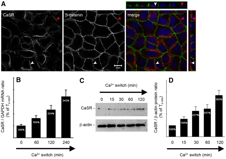
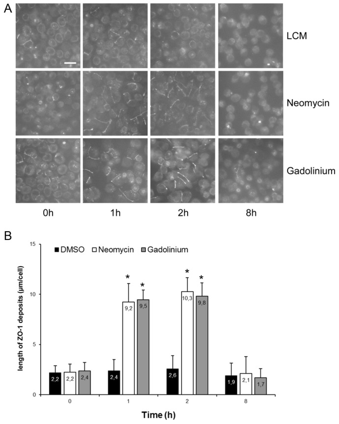
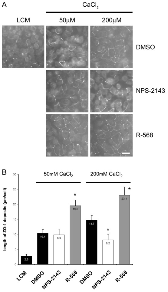
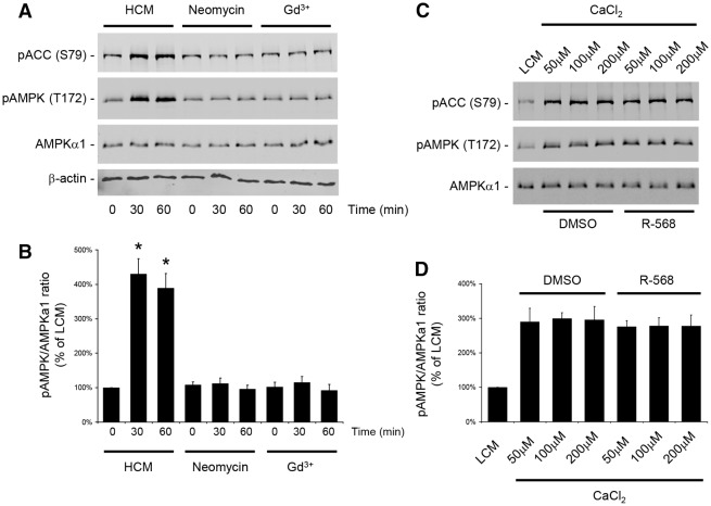
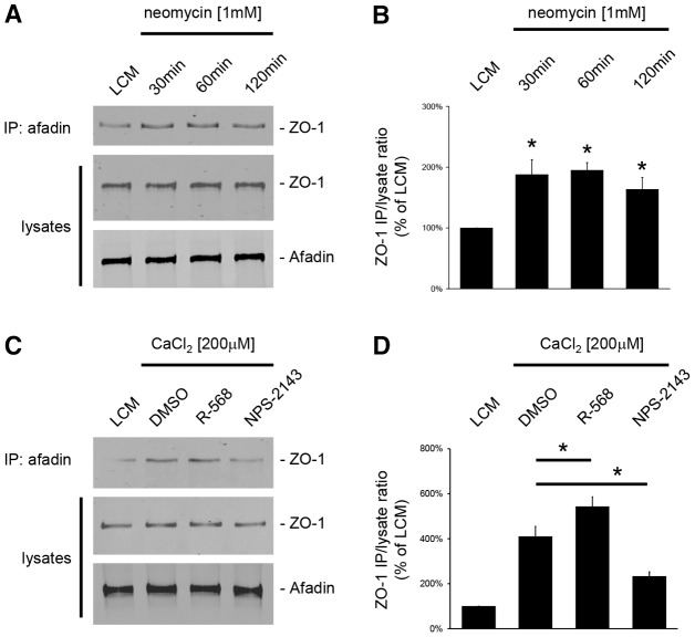
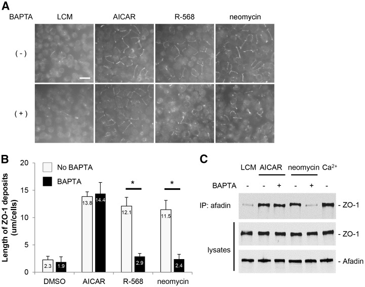
The Ca(2+)-sensing receptor (CaSR) belongs to the G-protein-coupled receptor superfamily and plays essential roles in divalent ion homeostasis and cell differentiation. Because extracellular Ca(2+) is essential for the development of stable epithelial tight junctions (TJs), we hypothesized that the CaSR participates in regulating TJ assembly. We first assessed the expression of the CaSR in Madin-Darby canine kidney (MDCK) cells at steady state and following manipulations that modulate TJ assembly. Next, we examined the effects of CaSR agonists and antagonists on TJ assembly. Immunofluorescence studies indicate that endogenous CaSR is located at the basolateral pole of MDCK cells. Stable transfection of human CaSR in MDCK cells further reveals that this protein co-distributes with β-catenin on the basolateral membrane. Switching MDCK cells from low-Ca(2+) medium to medium containing a normal Ca(2+) concentration significantly increases CaSR expression at both the mRNA and protein levels. Exposure of MDCK cells maintained in low-Ca(2+) conditions to the CaSR agonists neomycin, Gd(3+) or R-568 causes the transient relocation of the tight junction components ZO-1 and occludin to sites of cell-cell contact, while inducing no significant changes in the expression of mRNAs encoding junction-associated proteins. Stimulation of CaSR also increases the interaction between ZO-1 and the F-actin-binding protein I-afadin. This effect does not involve activation of the AMP-activated protein kinase. By contrast, CaSR inhibition by NPS-2143 significantly decreases interaction of ZO-1 with I-afadin and reduces deposition of ZO-1 at the cell surface following a Ca(2+) switch from 5 µM to 200 µM [Ca(2+)]e. Pre-exposure of MDCK cells to the cell-permeant Ca(2+) chelator BAPTA-AM, similarly prevents TJ assembly caused by CaSR activation. Finally, stable transfection of MDCK cells with a cDNA encoding a human disease-associated gain-of-function mutant form of the CaSR increases the transepithelial electrical resistance of these cells in comparison to expression of the wild-type human CaSR. These observations suggest that the CaSR participates in regulating TJ assembly.

Keywords: Calcium-sensing receptor; Epithelia; Tight junction.


Figures

Fig. 1.
Fig. 1.
Expression of the CaSR in MDCK cells at steady-state and following Ca2+ switch. (A) Representative co-immunofluorescence using rabbit polyclonal antibodies directed against the CaSR and mouse monoclonal antibodies directed against β-catenin, a marker of the basolateral membrane, in confluent MDCK cells. Merge panels show the xyz planes, with the CaSR labeled in red and β-catenin in green. Arrowheads indicate the basolateral side. Scale bar: 10 µm. (B–D) Comparative quantification of mRNA (B) and protein (C,D) expression of the CaSR in MDCK cells following Ca2+ switch. MDCK cells were lysed and processed at the indicated times after Ca2+ switch. Real-time RT-PCR was performed using specific primers directed against canine CaSR (target gene) and canine GAPDH (housekeeping gene), and the quantification was performed using the MxPro QPCR software (Stratagene). Immunoblotting quantification was performed using ImageJ software after normalization to β-actin expression levels. Data represent means ± s.d.
Fig. 2.
Fig. 2.
Deposition of ZO-1 at the plasma membrane following activation of the CaSR in MDCK cells. (A) Confluent MDCK cells were incubated in low-Ca2+ medium (LCM) for 16 hours, exposed to fresh LCM supplemented with DMSO or with the CaSR agonists neomycin (1 mM) or Gd3+ (100 µM) for the indicated time points, fixed in ice-cold methanol, and immunostained for ZO-1. Scale bar: 50 µm. (B) Quantification of ZO-1 relocation to the cell membrane in A. Data represent means ± s.d., and are representative of four independent experiments. ZO-1 length per cell is measured within each of six randomly selected fields of view. *P≤0.05 versus incubation with DMSO by Student's t-test.
Fig. 3.
Fig. 3.
Deposition of ZO-1 at the plasma membrane after modulation of the CaSR by R-568 in MDCK cells. (A) Confluent MDCK cells were incubated in low-Ca2+ medium (LCM) for 16 hours, exposed to fresh LCM supplemented with 50 µM or 200 µM CaCl2 and DMSO, the CaSR antagonist NPS-2143 (1 µM) or the CaSR agonist R-568 (800 nM) for 2 hours, fixed in ice-cold methanol and immunostained for ZO-1. Scale bar: 50 µm. (B) Quantification of ZO-1 relocation to cell membrane in A. Data are means ± s.d., and are representative of four independent experiments. ZO-1 length per cell is measured within each of six randomly selected fields of view. *P≤0.05 versus incubation with DMSO by Student's t-test.
Fig. 4.
Fig. 4.
CaSR activation does not stimulate AMPK phosphorylation or activation in MDCK cells. (A) Confluent MDCK cells were incubated in low-Ca2+ medium (LCM) for 16 hours, exposed to high-Ca2+ medium (HCM) or fresh LCM supplemented with the CaSR agonists neomycin (1 mM) or Gd3+ (100 µM) for 60 minutes, lysed in the presence of protease and phosphatase inhibitors, and probed with the indicated antibodies in a western blot analysis. (B) The quantification of the immunoreactive signal for phospho-AMPK was performed using the Odyssey Infrared Scanner (Li-Cor Biosciences) after normalization to the AMPKα1 expression level. Data represent mean percentages ± s.d., with the LCM level used as a reference (100%). *P≤0.05 versus LCM level by Student's t-test. (C) Confluent MDCK cells were incubated in LCM for 16 hours, exposed to fresh LCM supplemented with 50, 100 or 200 µM CaCl2, with or without R-568 (800 nM) for 60 minutes, lysed in the presence of protease and phosphatase inhibitors and probed with the indicated antibodies in a western blot analysis. (D) The quantification of the immunoreactive signal for phospho-AMPK was performed as in B. No significant difference was observed between the lysates exposed to R-568 versus DMSO for any Ca2+ concentration.
Fig. 5.
Fig. 5.
Increased interaction between ZO-1 and I-afadin following activation of the CaSR in MDCK cells. (A) Confluent MDCK cells were incubated in low-Ca2+ medium (LCM) for 16 hours, exposed to fresh LCM supplemented with the CaSR agonist neomycin (1 mM) for the indicated time intervals, and lysed in presence of protease inhibitors. Cell lysates were immunoprecipitated using rabbit polyclonal antibodies directed against I-afadin and Protein-A agarose beads. Equal amounts of immunoprecipitates were then separated on SDS-PAGE and probed with mouse monoclonal anti-ZO-1 antibodies. Total cell lysates were simultaneously subjected to immunoblotting using anti-I-afadin and anti-ZO1 antibodies. (B) The quantification of the immunoreactive signal for ZO-1 in immunoprecipitates was performed using the Odyssey Infrared Scanner (Li-Cor Biosciences) and normalized to ZO-1 expression in total cell lysates. Data represent mean percentages ± s.d., with the LCM level used as a reference (100%). *P≤0.05 versus LCM level by Student's t-test. (C) Confluent MDCK cells were incubated in low-Ca2+ medium (LCM) for 16 hours, exposed to fresh LCM supplemented with 200 µM CaCl2 and DMSO or the CaSR agonist R-568 (800 nM) or the CaSR antagonist NPS-2143 (1 µM) for 120 minutes and lysed in presence of protease inhibitors. Immunoprecipitation was performed as described in A. (D) The quantification of immunoreactive signals was performed as described in B. Data represent mean percentages ± s.d., with the LCM level used as a reference (100%). *P≤0.05 between indicated pairs using Student's t-test.
Fig. 6.
Fig. 6.
Effects of the cell-permeant Ca2+ chelator, BAPTA-AM, on ZO-1 relocation to the plasma membrane after CaSR stimulation or AMPK activation in MDCK cells. (A) Confluent MDCK cells were incubated in low-Ca2+ medium (LCM) for 16 hours, exposed to fresh LCM supplemented or not with BAPTA-AM (50 µM) for 30 minutes, incubated with the CaSR agonists neomycin (1 mM) or R-568 (800 nM), or with the AMPK activator AICAR (1 mM) for 2 hours, fixed in ice-cold methanol and immunostained for ZO-1. Scale bar: 50 µm. (B) Quantification of ZO-1 relocation to cell membrane in A. Data represent means ± s.d. and are representative of three independent experiments. ZO-1 length per cell is measured within each of six randomly selected fields. *P≤0.05 between indicated pairs using Student's t-test. (C) Confluent MDCK cells were incubated in LCM for 16 hours, exposed to fresh LCM supplemented or not with BAPTA-AM (50 µM) for 30 minutes, incubated with the CaSR agonist neomycin (1 mM) or with the AMPK activator AICAR (1 mM) for 2 hours. Cells were lysed in the presence of protease inhibitors and immunoprecipitation was performed using rabbit polyclonal antibodies directed against I-afadin and Protein-A agarose beads. Equal amounts of immunoprecipitates were then separated on SDS-PAGE and probed with mouse monoclonal anti-ZO-1 antibodies. Total cell lysates were simultaneously subjected to immunoblotting using anti-I-afadin and anti-ZO1 antibodies. These data are representative of three independent experiments.
Fig. 7.
Fig. 7.
Expression of human wild-type and the P221L active mutant of the CaSR in MDCK cells. (A,B) Representative co-immunofluorescence using rabbit polyclonal antibodies directed against FLAG and mouse monoclonal antibodies directed against β-catenin, a marker of the basolateral membrane, in MDCK cells stably transfected with wild-type human CaSR-FLAG (A) or with the human disease-associated P221L active mutant of the CaSR-FLAG (B). Merge panels show xyz planes, with the CaSR-FLAG labeled in red and β-catenin in green. Scale bars: 10 µm. (C) MDCK cells stably expressing human wild-type or P221L active mutant of the CaSR-FLAG were lysed in the presence of protease inhibitors and immunoprecipitated using mouse monoclonal antibodies directed against FLAG and protein-A agarose beads. Equal amounts of immunoprecipitates (IP) were then separated on SDS-PAGE and probed with rabbit polyclonal antibodies directed against β-catenin. Total cell lysates (L) were simultaneously subjected to immunoblotting using anti-FLAG and anti-β-catenin antibodies. These data are representative of three independent experiments. (D) MDCK cells stably expressing human wild-type or P221L active mutant of the CaSR-FLAG were lysed in the presence of protease inhibitors, incubated with the PGNase F and endo H deglycosylation enzymes for 60 minutes at 37°C, and probed with anti-FLAG antibodies in a western blot analysis. The arrowhead indicates the endo-H-resistant upper band of the CaSR-FLAG.

References

    1. Bai M., Quinn S., Trivedi S., Kifor O., Pearce S. H., Pollak M. R., Krapcho K., Hebert S. C., Brown E. M. (1996). Expression and characterization of inactivating and activating mutations in the human Ca2+o-sensing receptor. J. Biol. Chem. 271, 19537–19545 10.1074/jbc.271.32.19537 - DOI - PubMed
    1. Balda M. S., González-Mariscal L., Contreras R. G., Macias-Silva M., Torres-Marquez M. E., García-Sáinz J. A., Cereijido M. (1991). Assembly and sealing of tight junctions: possible participation of G-proteins, phospholipase C, protein kinase C and calmodulin. J. Membr. Biol. 122, 193–202 10.1007/BF01871420 - DOI - PubMed
    1. Boller K., Vestweber D., Kemler R. (1985). Cell-adhesion molecule uvomorulin is localized in the intermediate junctions of adult intestinal epithelial cells. J. Cell Biol. 100, 327–332 10.1083/jcb.100.1.327 - DOI - PMC - PubMed
    1. Bräuner-Osborne H., Wellendorph P., Jensen A. A. (2007). Structure, pharmacology and therapeutic prospects of family C G-protein coupled receptors. Curr. Drug Targets 8, 169–184 10.2174/138945007779315614 - DOI - PubMed
    1. Brennan S. C., Conigrave A. D. (2009). Regulation of cellular signal transduction pathways by the extracellular calcium-sensing receptor. Curr. Pharm. Biotechnol. 10, 270–281 10.2174/138920109787847484 - DOI - PubMed

Publication types